Introduction
The snow crab (Chionoecetes opilio) is a recent addition to the Barents Sea fauna. It was first discovered in 1996 in Russian waters, but the range has expanded to include the eastern Norwegian parts of the Barents Sea where a fishery has developed (Kuzmin, Akhtarin, and Menis 1999; Alvsvåg, Agnalt, and Jørstad 2008; Holt et al. 2021). The origin of the Barents Sea snow crab is unclear. One hypothesis is that the species has been introduced by ballast water from Canadian western Atlantic waters (Kuzmin, Akhtarin, and Menis 1999). Recent genetic studies are not consistent with this (Dahle, Agnalt, and Farestveit 2014; Dahle et al. 2022). The Barents Sea population appears to be genetically distinct from populations in both Canadian Atlantic waters and Alaskan waters, more so from the former. There is also no evidence of a recent bottleneck in the Barents Sea population, which would be expected in the case of an accidental introduction of a limited number of individuals (Dahle, Agnalt, and Farestveit 2014; Dahle et al. 2022). Therefore, a recent range expansion from a small Arctic refugial population could be a possibility (Sundet and Bakanev 2014).
Hematodinium is a parasitic dinoflagellate infecting marine decapod crustaceans, often found in the haemolymph (blood) of the host. Infections result in severe pathologies, such as organ failure and respiratory dysfunction, which can lead to death. Infected crustaceans exhibit systemic signs such as lethargy and cloudy or milky haemolymph that does not clot in the periphery or around the internal viscera (Stentiford and Shields 2005). In addition, there are other macroscopic signs, such as discoloured carapace, although the results are inconsistent. The complete Hematodinium life cycle remains unknown, but transmission is thought to be direct, with no evidence of the involvement of an intermediate host (Alimin et al. 2024).
Hematodinium spp. infections have been detected in snow crabs in most areas of their distribution, from the banks off Newfoundland and Labrador (Dawe 2002; Gaudet et al. 2015), western Greenland (Eigemann, Burmeister, and Skovgaard 2010) and in the Bering Sea (Jensen et al. 2010; Morado 2011). The parasite may cause bitter crab disease (BCD) in Chionoecetes spp., including snow crabs. This can have commercial consequences, as a parasite-related biochemical alteration of the meat products from BCD cause a bitter taste (Small 2012).
In Canadian Atlantic waters prevalence of Hematodinium spp. has been shown to be low, max. 16% but usually less than 5%, based on haemolymph cloudy- or milkiness (Dawe 2002). Off western Greenland the prevalence has been found to be 46%, and in the Bering Sea as high as 82% (including subclinical infections), based on PCR analyses of haemolymph samples (Eigemann, Burmeister, and Skovgaard 2010; Jensen et al. 2010).
If the origin of the invading snow crab in the Barents Sea is a range expansion, i.e., of an enzootic crab population, the available studies cited suggests that Hematodinium spp. could be present. However, if, despite the genetic evidence (Dahle, Agnalt, and Farestveit 2014; Dahle et al. 2022), the recent invasion is because of anthropogenic introduction of a small number of crabs or larvae, then it is more likely that a host specific parasite is absent (e.g., Torchin et al. 2003). The aim of the present study was to search for evidence of Hematodinium infection in the invading snow crabs of the Barents Sea.
Materials and methods
Snow crabs were collected during a research cruise with RV “Helmer Hansen” from 13 to 27 November 2016 in the eastern Barents Sea between 75.03°N and 77.52°N and 28.06°E and 34.60°E. Haemolymph samples were taken from 142 crabs (Table 1). Leeches occurring on the crabs were registered and identified morphologically. A subsample was also identified genetically by using COI sequencing within the framework of the Norwegian Barcode of Life (NorBOL) network (see http://www.norbol.org/), in a Barcoding of Life Database (BOLD; see http://boldsystems.org/) project dedicated to Norwegian leeches (project NOHIR).
Haemolymph samples were taken using sterile disposable 1 mm syringes inserted at the junction of the basis and ischium base of the crab’s 2nd swimming leg, as it was the largest appendage. Presence of Hematodinium spp. infection was assessed using microscopic examination of fresh haemolymph smear samples prepared onboard and PCR. Smears were prepared immediately after sampling and examined by an experienced parasitologist using bright field microscopy at x 400 magnification. For PCR analyses, a further haemolymph sample was ethanol preserved (96%) and stored at -20°C. Ethanol preserved haemolymph was mixed well, and 1-1.5 ml taken out and centrifuged at 15 700 x g for 5-10 min. The ethanol supernatant was then removed, and the pellet dried at 60°C. DNA was extracted from the dried pellet using the DNeasy® Tissue Kit protocol for animal tissues (Qiagen, Hilden, Germany). DNA concentration was measured using NanoDrop. The DNA concentration (average concentration at this step 114 ng/µL) was then adjusted to 100 ng/µL if higher. A positive control sample was provided by Rose Kerr and Grant D. Stentiford (Cefas), representing DNA from hepatopancreas tissue from Hematodinium sp. infected Carcinus maenas.
Two PCR assays were used. The first was the Haemat-assay of Gruebl et al. (2002) and Jensen et al. (2010), with primers Hemat-F-1487 and Hemat-R-1654 with annealing temperature (TA) of 56°C (expected product size 197 bp). When this did not produce any results in the samples tested, a nested PCR was performed following Eigemann, Burmeister, and Skovgaard (2010), employing in the first round the Hemat-F-1487/ Hsp2r primers (TA 58; 692 bp product) and in the second round 18SF2/Hem3R (TA 58, 467 bp product). Some selected samples were also analysed with the Hsp1f and Hsp2R primers (Hudson and Adlard 1994), using a range (50°C -60°C with 2 degrees interval) of annealing temperatures. Some products of the wrong size (<618 bp) were sequenced, using the amplification primers. The PCR amplifications were performed in a total volume of 25 μl using 4 μl of template DNA and a reaction mixture consisting of following final concentration: 1× PCR buffer, 1X Q-solution, 0.625 U HotStarTaq DNA polymerase (Qiagen cat nr 203203) 0.2 mM of each dNTP, 0.5mM of both the reverse and forward primer and 10.375 μl dH2O.
The PCR conditions were as follows: after an initial 15 min denaturation step at 95°C, samples were taken through 35 amplification cycles, each consisting of a 1 min denaturation step at 94ºC, a 1 min primer annealing step at a temperature dependent of the primer combination used, and a 1 min extension step at 72ºC. A prolonged extension step of 10 min at 72°C completed each reaction. Products were visualized on 1% agarose gels.
Results
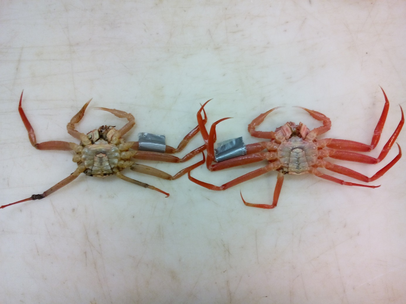
The haemolymph was clear in most crabs (97.2%). Only 4 (2.8%) showed milky to opaque haemolymph appearance. In 18 crabs (12.7%), pale brownish exoskeletal lesions were seen on the ventral side (Figure 1). Leeches occurred on 11 crabs (prevalence 7.7%, intensity of 1-8 (mean 2.5, SD 2.1)). Leeches examined (N=11) were determined morphologically as Johanssonia arctica. COI sequencing of three specimens showed that they all belong to a barcode cluster recognized by the Barcode Index Number (BIN) BOLD:AAF9634, which confirms the identity as J. arctica. The sequences are numbered NOHIR029-19, NOHIR030-19 and NOHIR031-19, respectively.
_exhibiting_carapace_discolouration_(left)_.jpeg)
Microscopic examination of the fresh haemolymph smear samples examined did not reveal any evidence of Hematodinium spp. infection. The PCR testing on haemolymph DNA also revealed no positive samples, except the positive controls. Therefore, the Hematodinium spp. prevalence was 0%, with 95% confidence interval 0-2.6%. Using the Hsp1f/2r primers with low annealing temperature, 4 weak bands of the wrong size were obtained. Sequences of these products gave in BLAST searches decapod genomic hits, and two protist sequences, a ciliate (324 nt, 98.1% identity to FR874823 18S) and a diatom (564nt, 99.8% identity to MT796594 18S-ITS1) (see sequences before the reference section).
Discussion
PCR screening on haemolymph samples from Chionoecetes spp. has previously met problems. Using the ITS1 primers Hsp 1f/2r of Hudson and Adlard (1994), Jensen et al. (2010) got clear bands at expected size from Hematodinium - infected crab, but also noted unspecific bands of varying size in both positive and negative controls. Our preliminary testing on 27 samples, including those from crabs with opaque haemolymph also gave some bands of the “wrong” size. Sequencing of the products showed that some were likely host (genomic decapod hits) while two were protists. These, a diatom and a ciliate, could represent contaminations from the syringe penetration site (diatom), or possibly other blood infections (ciliate) (see Stentiford and Shields 2005). These observations show that amplification of suitable template DNA occurred in these samples, meaning amplifiable Hematodinium spp. DNA was absent. Due to the problems with the Hsp 1f/2r assay, we followed Jensen et al. (2010) in instead using the Hemat-F-1487/Hemat-R-1654 primer pair (Gruebl et al. 2002), amplifying a short (~190 bp) product from 18S. This testing of all crabs gave no positive samples except the positive controls. We therefore also tested all samples with a nested PCR successfully employed by Eigemann, Burmeister, and Skovgaard (2010) on Greenlandic snow crab blood samples. Again, all proved negative, except the positive controls.
Our results therefore suggest that the sampled crabs were negative for Hematodinium. Hematodinium spp. infections often show seasonality in prevalence and intensity (e.g., Stentiford, Neil, and Atkinson 2001; Sheppard et al. 2003; Hamilton, Shaw, and Morritt 2009; Smith et al. 2015). We only had samples from November and do not know if this is a season of low prevalence in Arctic decapods. However, there are no seasonal trends in prevalence of bitter crab disease in snow crab in the Newfoundland and Labrador area (Dawe 2002). In the eastern Bering Sea, annual overall prevalence of Hematodinium spp. in snow crabs has been shown to be low, around 3-5% (Morado 2011). Still, with the sample size of 142 used in this study, and a prevalence of 3%, the probability of detecting at least one infected individual is high (0.99 assuming the probability of infection follows the same binomial distribution for all individuals). The sampled crabs were taken offshore in the central parts of the Barents Sea. Eigemann, Burmeister, and Skovgaard (2010) noted a 3-times higher prevalence of Hematodinium spp. positive C. opilio in inshore compared to offshore stations in western Greenland. However, prevalence was still 10% or higher in the offshore stations. Dawe (2002), who examined a much larger area on the Newfoundland and southern Labrador Continental Shelf, noted no effect of distance from the coast on Hematodinium spp. prevalence. Also, a higher prevalence has been noted with depth in C. opilio (Pestal et al. 2003), possibly relating to substrate type rather than depth since prevalence has been found to be higher in crabs from mud/sand bottoms compared to other habitats (Shields et al. 2007). The lack of findings of Hematodinium spp. in the present study is therefore not likely a consequence of having sampled areas far from the coast.
Although admittedly speculative, a lack of Hematodinium spp. in the Barents Sea snow crabs could be taken as support for an introduction hypothesis, since such an event could have represented inadvertent spread of juveniles such as larvae not carrying the parasites occurring in the native range. However, this infection status could eventually change, since snow crabs are susceptible, and the apparent same genotypes occur in native Arctoboreal or Arctic decapod species (Eigemann, Burmeister, and Skovgaard 2010). In such a scenario, the invasive crabs in the Barents Sea may be expected to gradually develop a parasite or symbiont fauna (symbiome) more like that in the native range, as they encounter parasitic taxa to which they are susceptible. However, there are many unknown factors related to these interpretations. This includes the native range of the Barents Sea snow crabs, the occurrence of Hematodinium spp. in the native range and whether Hematodinium spp. occur in other decapod species in the Barents Sea or adjacent areas (e.g., the Arctic Ocean north of Western Russia and Siberia).
eDNA searches have revealed the presence of Hematodinium in sediments and in the water column (Pitula et al. 2012; Hanif et al. 2013; Davies et al. 2019), but no evidence of an intermediate host has been found. It thus seems likely that transmission is direct, although the existence of an intermediate host cannot yet be excluded (Stentiford and Shields 2005; Lohan et al. 2012; Meyers 2014; Li, Li, and Huang 2021). A relationship between Hematodinium spp. prevalence or bitter crab disease and crab density has not been observed in snow crab (Dawe 2002; Shields et al. 2007).
Another finding is, as has been observed by others, that Hematodinium spp. infections cannot be reliably diagnosed by presence of opaque haemolymph, which was observed in 2.8 % of the crabs sampled here. Thus, Davies et al. (2022) found that opaque haemolymph can be caused by other micro-eukaryotes and bacterial infections and occurred in only one in three Hematodinium infected shore crabs.
Acknowledgements
This project was funded through the internal IMR project SnowMan (Project No: 14862). We thank Rose Kerr and Grant D. Stentiford at Cefas for providing Hematodinium DNA for the positive controls.
Sequences
>Diatom-Snowcrab-24
GTTCCCCTTGAACGAGGAATTCCTAGTAAACGCACATCATCAATGTGCATTGATTACGTCCCTGCCCTTTGTACACACCGCCCGTCGCACCTACCGATTGAGTGGTCCGGTGAACCCTCGAGATTGTGATTAATTTCTTTTATTAGAATTTGATTGTGAGAACTTGTGTAAACCTTATCACTTAGAGGAAGGTGAAGTCGTAACAAGGTTTCCGTAGGTGAACCTGCGGAAGGATCATTAACACACCGATCTAAGATCTCAACTCCCGTGAAGAAGCAGTACAGCCTGGCTTTCCCCGCAGCTATCATGCTCGGCCGCCTCGCAGTCTGTGACTATCAACATAATACCGTAGCGTAGAGCGCAAGCTCGAAGCTACAGGTTGGCAAGCAATTGTCAACCACCCAATACCAAACAATAACATATAACCAGAAGCCTAAATGAGTTGACGAGCCTGTCGTGCACTTGTGCGCTTCAGGCACTCCTCATACAAGTATATAAATGTATACAACTTTCAGCGATGGATGTCTAGGCTCCCACAACGATGAAGAACGCAGCGAAATGCGA
99.8% identity to MT796594 Chaetoceros gelius 18S-ITS
>Ciliate-Snowcrab-18
AGGAATTCCTAGTAAGCCTGGGTCATCAACCCATGTTGATTACGTCCCTGCCCTTTGTACACACCGCCCGTCGCTCCTACCGATTCGAGTGCCTCGGCGAATGCTTCGGATTGGGTTCCCTTAGGAACTCGAAAAGTTGTGTAAGCCATGTCACTTAGAGGAAGGATAAGGCGTAACAAGGTTTCCGTAGGTGAACCTGCGGAAGGATCATTCTCGCCAATATAACAACTTAGTTAGTCTTCGGGCTAACTTTGTAAAAAATTAAAAAAAAAAAAAAAAAGAAAATTTTCAACGGTGGATATCTGGGTTCTCATACCGAGAAAAAACG
98.1% identity to FR874823, uncultured marine picoeukaryote (Ciliophora) partial 18S