Loading [Contrib]/a11y/accessibility-menu.js
Skip to main content
null
Bulletin of the EAFP
  • Menu
  • Articles
    • Case study
    • Expert Opinion
    • Method
    • Note
    • Research article
    • Review
    • Workshop Report
    • All
  • For Authors
  • Editorial Board
  • About
  • Issues
  • search

RSS Feed

Enter the URL below into your favorite RSS reader.

http://localhost:39917/feed
Research article
Vol. 44, Issue 2, 2024June 08, 2024 CEST

Phenotypic, genotypic and virulence traits analysis of aeromonads causing massive mortality in farmed Oreochromis niloticus

Mamdouh Y. Elgendy, Professor, PhD Fish diseases and management, Mohamed Abdelsalam, Professor, Amany Kenawy, professor, Nehal A. Younis, Assistant professor,
AeromonadsAntibiotic resistanceNile tilapiaPathogenicitySummer mortality
Copyright Logoccby-4.0 • https://doi.org/10.48045/001c.118913
Photo by National Cancer Institute on Unsplash
Bulletin of the EAFP
Elgendy, Mamdouh Y., Mohamed Abdelsalam, Amany Kenawy, and Nehal A. Younis. 2024. “Phenotypic, Genotypic and Virulence Traits Analysis of Aeromonads Causing Massive Mortality in Farmed Oreochromis Niloticus.” Bulletin of the European Association of Fish Pathologists 44 (2). https:/​/​doi.org/​10.48045/​001c.118913.
Save article as...▾
Download all (3)
  • Fig. 1. (a) naturally infected tilapia showing hemorrhages on the external body surface and at the base of fins (circle); (b) naturally infected tilapia showing congestion and enlargement of the liver (circle). (c, d, e, f): Fish used in the pathogenicity testing (c) Nile tilapia injected with saline showing normal appearance of external body surface (square) and caudle fin (square); (d, e, & f) tilapia injected with aeromonads showing: (d) erosions and tail rot (circle); (e) hemorrhages on the caudal fin (circle); and (f) skin darkening (square).
    Download
  • Fig. 2. Phylogenetic tree based on the gyrB gene of Aeromonas spp.
    Download
  • Fig. 3. Histopathological lesions in Nile tilapia experimentally infected with aeromonads, haematoxylin and eosin (H&E) stained sections. (A) Skin showing degenerative changes in epidermal cells (d) edema in dermis and underlying muscular layer (o), Bar = 50 μm. (B) Gills showing (h) severe hyperplasia, (c) congestion and (n) mononuclear inflammatory cells infiltration, Bar = 50 μm. (C) Spleen showing severe hemorrhages (h) and lymphocytic depletion (p), Bar = 20 μm. (D): Liver showing severe congestion (c), hemorrhages (h), diffuse vacuolar degeneration of hepatocytes (d), necrosis (n) and mononuclear inflammatory cells infiltration in between the hepatic parenchyma (m), Bar = 50 μm. (E) kidneys showing congestion (c), degenerative and necrotic changes in the tubular epithelium and in the endothelial lining the glomerular tufts (d &n), Bar = 50 μm. (F): Brain showing marked neuronal degeneration (nd), congestion (c) and hemorrhages (h), Bar = 50 μm.
    Download

Sorry, something went wrong. Please try again.

If this problem reoccurs, please contact Scholastica Support

Error message:

undefined

View more stats

Abstract

Motile aeromonads were identified in earthen-pond-farmed Oreochromis niloticus that suffered massive mortalities in Egypt during the summer of 2020. The fish showed hemorrhagic septicemic signs. Poor management practices and inadequate water quality measures were observed in the affected earthen ponds. Motile aeromonads (n = 31 isolates) were identified from 70 fish specimens. Based on their phenotypic and molecular characteristics, isolates were identified as: Aeromonas hydrophila (n = 12), A. veronii (n = 10), A. caviae (n = 5), and A. sobria (n = 4). Bacteriological examination of farm water samples also revealed aeromonads (n=9) and some fish-nonpathogenic bacteria. The aeromonad isolates recovered from fish exhibited lipase (52.5%) and protease (47.5%) activities and harboured some virulence genes: Ser (62.5%), Aer (55%), ela (37.5%), gcaT (32.5%), Hyl (25%), laf-A (22.5%), and Act (20%). They also harboured numerous antibiotic-resistance genes, including aadA (37.5%), tetC (32.5%), tetA (27.5%), sul 1 (20%), and blaTEM (10%). Virulence and antibiotic resistance genes were also noted in some of the Aeromonas spp. isolates obtained from farm water. Aeromonads were highly resistant to ampicillin, amoxicillin, and gentamicin but highly susceptible to ciprofloxacin and florfenicol antibiotics. Aeromonas spp pathogenicity was confirmed by the experimental infection of Oreochromis niloticus. Our results indicate a positive correlation between excessive tilapia mortalities, motile Aeromonas septicemia and adverse water quality parameters measured during the summer. This study provides data on the virulence, pathogenicity, and antibiotic resistance of motile aeromonads affecting fish and humans, which will be useful for developing efficient therapies.

Introduction

Motile Aeromonas septicemia (MAS) is one of the most important bacterial diseases in aquaculture caused by various Aeromonas species and results in severe economic losses (Elgendy, Moustafa, et al. 2015). Aeromonads are ubiquitous in aquatic environment, but under stressful conditions, they can cause serious infections and high levels mortality in farmed fish (El-Gohary, Zahran, and Abd El-Gawad 2020). Aeromonas hydrophila, A. sobria, A. caviae, and A. veronii are the most pathogenic Aeromonas spp. in aquaculture (Abdelsalam et al. 2021).

The pathogenesis of MAS is multifactorial and requires the interaction of several variables for the disease to develop. Aeromonas species secrete various extracellular enzymes and possess numerous virulence genes that enable bacteria to overcome the host’s immune system and initiate severe infections (Hu et al. 2012; El-Gohary, Zahran, and Abd El-Gawad 2020).

Nile tilapia (Oreochromis niloticus) aquaculture constitutes an integral part of the Egyptian aquaculture industry (Elgendy et al. 2022). Farmed tilapia are susceptible to numerous bacterial diseases causing massive mortalities and colossal economic losses (Abdelsalam et al. 2022). The excessive use of antibiotics by fish farmers to control such infections have detrimental impacts on the aquatic ecosystem and poses a threat to human health due to the emergence of resistant strains (Elgendy, Shaalan, et al. 2021; Ali et al. 2022; Algammal et al. 2022). This study was conducted to analyze Aeromonas spp. isolates recovered from farmed O. niloticus that suffered massive mortalities during summer. The molecular characteristics, virulence profile, antibiotic resistance and pathogenicity of aeromonads isolates were investigated.

Materials and methods

Case history and sampling

Abnormal mortalities (250 dead fish every day/earthen pond) were recorded in O. niloticus (110–200 g) within five earthen ponds in a fish farm in Beheira Governorate, Egypt, in June 2020. Fish were stocked at a density of 30,000 fish/acre. The farm owner used untreated poultry droppings to promote natural food production in the ponds and had rear-Pecking ducks (190 ducks per/acre). Cattle and sheep grazed at the banks between the earthen ponds. There were several dogs straying on the farm. Moribund fish exhibited signs of hemorrhagic septicemia and respiratory distress. The average temperature, dissolved oxygen level, and unionized ammonia level were 27°C, 4 mg/L, and 0.53 mg/L, respectively. A total of 70 moribund fish were collected and transferred in an ice box to the hydrobiology department laboratory NRC. Water samples (n = 15) (3 samples from each affected pond) were collected and analyzed according to Newaj-Fyzul et al. (2008).

Bacteriological examination

Bacteriological smears were obtained from fish hematopoietic organs (kidneys and liver) and subcultured onto tryptic soy agar. Water samples were collected and examined according to Newaj-Fyzul et al. (2008). Bacterial strains were purified and identified using the Vitek 2 compact device (bio-Merieux).

Molecular identification of Aeromonas spp.

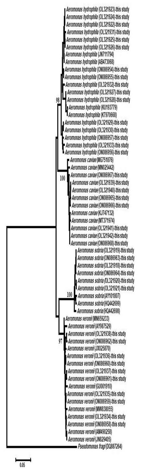
DNA was extracted from purified bacterial strains according to the PrepMan® Ultra Sample Preparation Reagent protocol. Polymerase chain reaction was conducted using gyrB-specific primers as described by Hu et al. (2012). The amplified gyrB was directly sequenced in two directions using the Sanger DNA sequencer ABI 3730xl DNA sequencer (Applied Biosystems™, USA) at Sigma (Cairo, Egypt), assembled using Bio Edit version 7.0 (Hall 1999) and aligned to other interrelated sequences in the GenBank database using BLASTN search (National Center for Biotechnology Information). The neighbor-joining phylogenetic tree was constructed using MEGA version X (Kumar et al. 2018).

Virulence characteristics of Aeromonas spp.

Extracellular enzymes

Lipase and protease activities were analyzed according to Carrazco-Palafox et al. (2018) and Sokol, Ohman, and Iglewski (1979).

Detection of virulence and antibiotic resistance genes

The presence of aerolysin (Aer), hemolysin (Hyl), lateral flagella A (laf-A), elastase (ela), serine protease (Ser), cytotoxic enterotoxin (Act), and glycerophospholipid-cholesterol acyltransferase (gcaT) genes in the recovered aeromonads strains was investigated according to Sun et al. (2016) using specific primers (Table 1). The strains were also analyzed for the presence of the following antibiotics resistance genes: aminoglycosides (aadA), β-lactams (blaTEM), tetracycline (tetC & tetA), and sulphonamide (sul 1) according to Ndi & Barton (2011) using specific primers (Table 1).

Table 1.Primers used in the study
Genes Primers size/bp References
The gyrB gene F: TCCGGCGGTCTGCACGGCGT
R TTGTCCGGGTTGTACTCGTC
1100 Hu et al. (2012)
Cytotoxic enterotoxin (Act) F: GAGAAGGTGACCACCAAGAACA
R: AACTGACATCGGCCTTGAACTC
232
Aerolysin (Aero) F: GAGCGAGAAGGTGACCACCAAGC
R:TTCCAGTCCCACCACTTCACTTCC
417 Nam and Joh (2007)
Serine protease (Ser) F: ACGGAGTGCGTTCTTCCTACTCCAG
R: CCGTTCATCACACCGTTGTAGTCG
211
Cholesterol
acyltransferase (gcaT)
F: CATGTCTCCGCCTATCACAACAAGC
R: CCAGAACATCTTGCCCTCACAGTTG
339
Elastase gene (ela) F: ACACGGTCAAGGAGATCAAC
F: CGCTGGTGTTGGCCAGCAGG
513 Sen and Rodgers (2004)
Lateral flagella A
(Laf-A)
F: GGTCTGCGCATCCAACTC
R: 5GCTCCAGACGGTTGATG
550 Merino et al. (2003)
Hemolysin (hly) F: GGCCGGTGGCCCGAAGATACGGG
R: GGCGGCGCCGGACGAGACGGG
579 Heuzenroeder, Wong, and Flower 1999)
Aminoglycoside resistance gene (aadA) F:GAGAACATAGCGTTGCCTTGG
R:TCGGCGCGATTTTGCCGGTTAC
198 Sunde and Norstrom (2005)
β-lactamase resistance gene (blaTEM) F: ATG AGT ATT CAA CAT TTC CG
R: CTG ACA GTTACC AATGCT TA
867 Rasheed et al. (1997)
Tetracycline resistance gene TetA F: GTA ATT CTG AGC ACT GTC GC
R: CTG CCT GGA CAA CAT TGC TT
956 Schmidt et al. (2001)
Tetracycline resistance gene (tetC) F: TCT AAC AAT GCG CTC ATC GT
R: GGT TGA AGG CTC TCA AGG GC
588
Sulphonamides resistance gene (sul1) F: CTT CGA TGA GAC CCG GCG GC
R: GCA AGG CGG AAA CCC GCG CC
436 Sundstrom et al. (1988)

Antibiotic susceptibility testing

The susceptibility of the Aeromonas spp. isolates to ampicillin 10 µg, amoxicillin 30 μg, gentamicin 10 μg, trimethoprim/sulfamethoxazole 1.25/23.75 μg, florfenicol 30 µg, ciprofloxacin 5 μg, and tetracycline 30 μg was evaluated using the disk diffusion method according to CLSI (2010).

Pathogenicity

A total of 720 healthy Nile tilapia with an average weight of 55 g were collected and acclimatized in aerated aquaria (50 L each) at 25°C ± 1°C. The fish were divided into groups of 10 fish/aquarium in duplicates. They were anesthetized using MS-222 (Sigma) (150 μgL-1). One isolate representative for each Aeromonas spp. was chosen at random and used to determine the LD50 value. Bacterial cultures were serially diluted, and each fish group was injected intraperitoneally (I/P) with 0.1 ml of the relevant bacterial culture suspension at concentrations ranging from 102 to 109 CFU/ml/fish (one isolate and one challenge dose per tank). Control fish were injected with 0.1 ml of sterile phosphate buffered saline (PBS). All fish were monitored for 13 days. The LD50 value was calculated according to Reed J. and Muencha H. (1938).

Histopathology

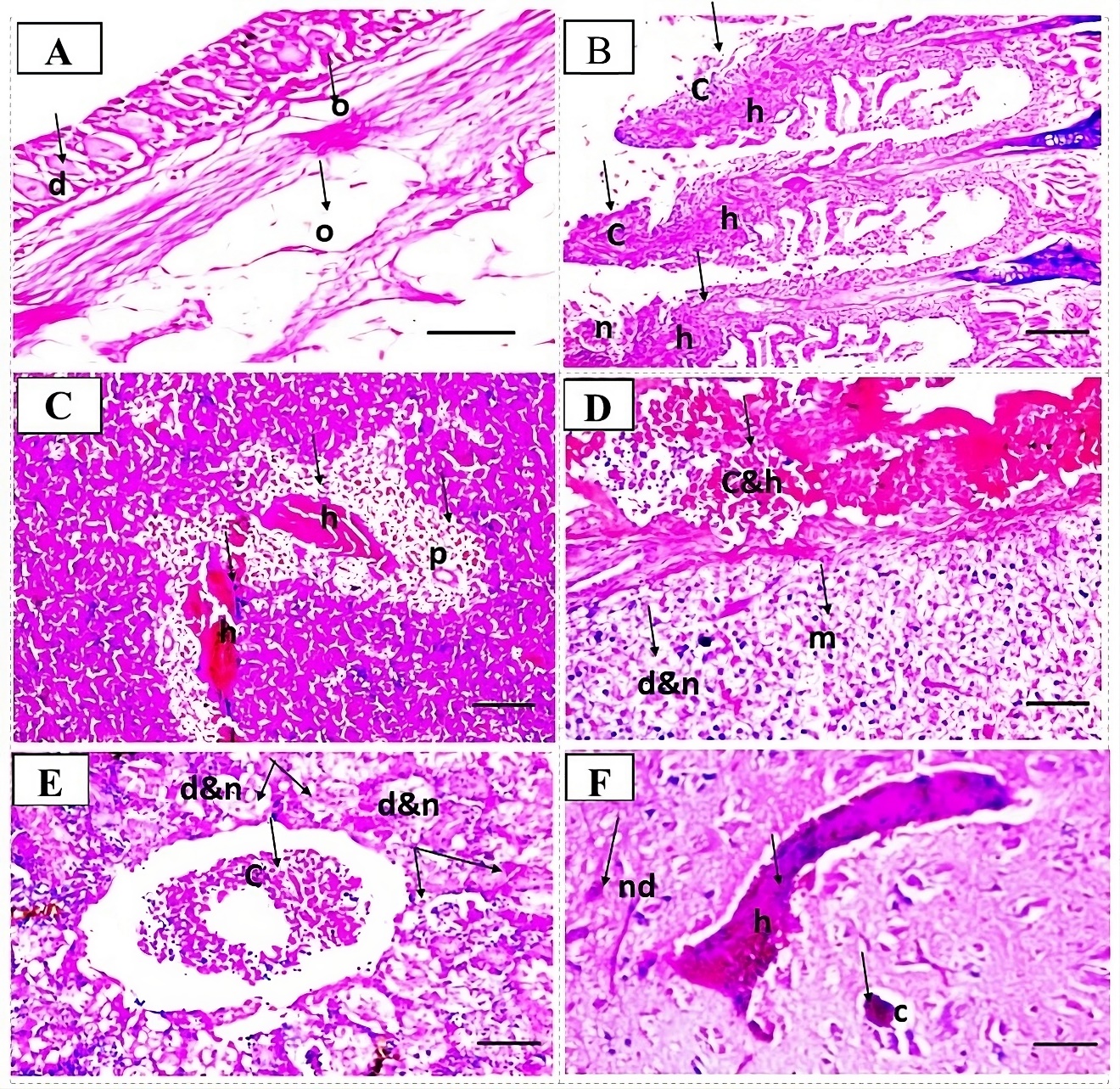
Haematoxylin and eosin (H&E) stained histopathological sections were prepared from tissue specimens collected from experimentally infected fish following the methods described by Bancroft and Gamble (2008).

Results

Clinical examination

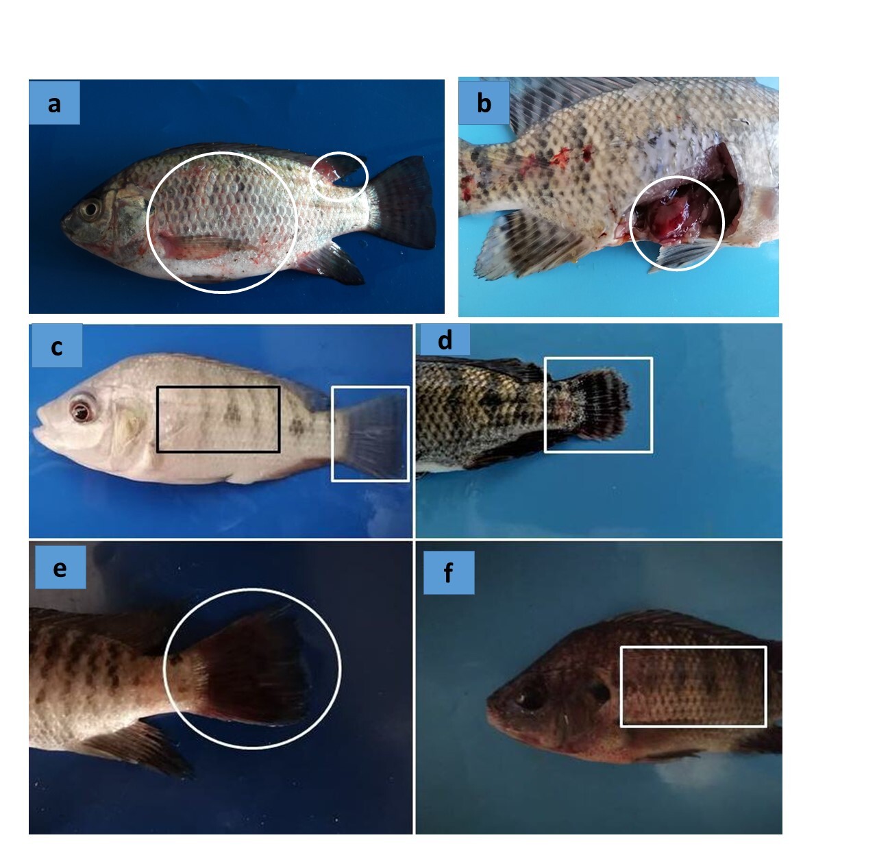
Moribund tilapia demonstrated signs of hemorrhagic septicemia. Petechial hemorrhages, fin rot, and skin erosions were widely distributed on the external body surface. Internally, the liver, spleen, and kidney were congested and enlarged. Ascites and exophthalmia were noted in some fish (Fig. 1).

Fig. 1
Fig. 1.(a) naturally infected tilapia showing hemorrhages on the external body surface and at the base of fins (circle); (b) naturally infected tilapia showing congestion and enlargement of the liver (circle). (c, d, e, f): Fish used in the pathogenicity testing (c) Nile tilapia injected with saline showing normal appearance of external body surface (square) and caudle fin (square); (d, e, & f) tilapia injected with aeromonads showing: (d) erosions and tail rot (circle); (e) hemorrhages on the caudal fin (circle); and (f) skin darkening (square).

Bacteriological examination

In total, 31 Aeromonas spp. isolates were recovered from the investigated fish specimens and identified as A. hydrophila (n = 12), A. veronii (n = 10), A. caviae (n = 5), and A. sobria (n = 4). Nine Aeromonas spp. isolates were discovered in the farm water samples, including A. hydrophila (n = 4), A. sobria (n = 2), and A. caviae (n = 3). A. veronii was not detected in the water samples. Some nonpathogenic bacteria (Proteus spp., Citrobacter freundii, and Shigella spp.) were also detected in some water and fish samples.

Molecular identification of Aeromonas spp.

A comparison of gyrB sequences revealed that all isolates (n = 40) belonged to the genus Aeromonas, the identity of which was confirmed as A. hydrophila (n = 16), A. caviae (n = 8), A. veronii (n = 10), and A. sobria (n = 6).

A. hydrophila isolated from water samples (OL321924, OL321926, OL321928, and OL321930) and that isolated from fish specimens (OL321922, OL321923, OL321925, OL321927, OL321929, OL321931–OL321933, and ON086954–ON086957) exhibited 99.72%–98.05% similarity to the following A. hydrophila strains: JN711793T, JN711794T, JN711795T, AB436660T, and JN711793T (typing strains) and HQ701864.1, GQ471011.1, JQ805070.1, AB473095.1, KF873661.1, MK484180.1, JN711794.1, and AB473068.1. The intraspecies similarity was 97.74%–100% for 12 isolates of A. hydrophila recovered from tilapia and water, with nucleotide differences ranging from 10 to 24 bp.

A. caviae recovered from fish (OL321939, OL321941, and ON086965–ON086967) and that recovered from water (OL321940, OL321942, and ON086968) showed 98.92%–98.22% similarity to the following A. caviae strains: AJ868400T, JN829530T, and KC924126T (typing strains) and MT371974.1, LC003106.1, MN855498.1, KJ747132.1, MN855516.1, KR140073.1, KC924174.1, and MN855511.1. The intraspecies similarity was 99.19%–100% for four A. caviae isolates recovered from fish and water, with eight nucleotide differences.

The sequence alignment of A. sobria isolated from fish (OL321918, OL321920, ON086963, and ON086964) and that recovered from water (OL321919 and OL321921) exhibited 98.91–98.15% similarity to the following A. sobria strains: AB473084T, HQ442698T, AF417631T (typing strains) and MG263589.1, AB473086.1, JN829516.1, MG263541.1, KP115770.1, KJ743530.1, AY101781.1, and HQ442698.1. The intraspecies similarity was 99.20%–100% for four A. sobria isolates recovered from Nile tilapia and water, with eight nucleotide differences.

The sequence alignment of A. veronii isolated from fish (OL321934–OL321938 and ON086958–ON086962) showed 99.34%–98.84% similarity to the following A. veronii strains: FN796748T, HM584508T, and AF417626T (typing strains) and MN659233.1, LC644255.1, AB829112.1, JF938686.1, KR140071.1, MN025464.1, LC003119.1, and JX025899.1. The intraspecies similarity was 98.56%–100% for five A. veronii isolates recovered from tilapia, with nucleotide differences ranging from 6 to 16 bp. The phylogenetic analysis confirmed the identity of the isolates (Fig. 2).

Fig. 2
Fig. 2.Phylogenetic tree based on the gyrB gene of Aeromonas spp.

Virulence characteristics of Aeromonas spp.

Proteolytic and lipolytic activities

Proteolytic and lipolytic activities were noted in (52.5%) and (47.5%) of isolates recovered from fish, respectively, and in (20%) and (17.5%) of isolates from water, respectively.

Detection of virulence and antibiotic resistance genes

Analysis of Aeromonas spp. isolates recovered from fish showed that Ser was the most commonly detected gene (62.5%), followed by Aer (55%), ela (37.5%), gcaT (32.5%), Hyl (25%), laf-A (22.5%), and Act (20%) gene. laf-A was missing in A. veronii and A. caviae isolates. Act and Hyl were absent in A. sobria isolates. The Aeromonas spp. isolates recovered from water also harboured Aer (17.5%), Ser (12.5%), laf-A (12.5%), ela (5%), Hyl (2.5%), and gcaT (2.5%) genes, while Act gene was missing in these isolates.

The aadA resistance gene was the most detected among Aeromonas spp. isolates obtained from fish (37.5%), followed by tetC (32.5%%), tetA (27.5%), sul 1 (20%), and blaTEM (10%). Aeromonas spp. isolates recovered from water harbored aadA (20%), tetC (15%), tetA (7.5%), and sul 1 (10%) genes, while blaTEM was not detected in any of these isolates.

Antibiotic susceptibility testing

All Aeromonas spp. isolates (100%) recovered from both fish and water were resistant to ampicillin 10 μg, amoxicillin 30 μg, and gentamicin 10 μg. Isolates were highly susceptible to ciprofloxacin 5 µg (47.5%), florfenicol (40%) and trimethoprim 1.25 μg/sulfamethoxazole 23.75 μg (32.5%).

Pathogenicity of Aeromonas spp.

Tilapia injected with Aeromonas spp. exhibited skin darkening, and hemorrhages on the external body surfaces, while some fish showed erosions and fin rot (Fig. 1). Control fish showed normal clinical signs. The LD50 value determined for the four tested aeromonad isolates was as follows: A. veronii (5.5 × 10⁵), A. hydrophila (4.21 × 10⁶), A. sobria (5.6 × 10⁶), and A. caviae (8.48 × 10⁶). Congestion and enlargement of the liver, spleen, and kidneys were the most common post-mortem lesions. Bacterial strains were reisolated from dead fish, and no mortalities were recorded in the control fish.

Histopathological observation

Degenerative, necrotic changes and mononuclear inflammatory cell infiltrations were the most commonly seen histopathological alterations (Fig. 3)

Fig. 3
Fig. 3.Histopathological lesions in Nile tilapia experimentally infected with aeromonads, haematoxylin and eosin (H&E) stained sections. (A) Skin showing degenerative changes in epidermal cells (d) edema in dermis and underlying muscular layer (o), Bar = 50 μm. (B) Gills showing (h) severe hyperplasia, (c) congestion and (n) mononuclear inflammatory cells infiltration, Bar = 50 μm. (C) Spleen showing severe hemorrhages (h) and lymphocytic depletion (p), Bar = 20 μm. (D): Liver showing severe congestion (c), hemorrhages (h), diffuse vacuolar degeneration of hepatocytes (d), necrosis (n) and mononuclear inflammatory cells infiltration in between the hepatic parenchyma (m), Bar = 50 μm. (E) kidneys showing congestion (c), degenerative and necrotic changes in the tubular epithelium and in the endothelial lining the glomerular tufts (d &n), Bar = 50 μm. (F): Brain showing marked neuronal degeneration (nd), congestion (c) and hemorrhages (h), Bar = 50 μm.

Discussion

Bacteriological and molecular examinations confirmed the identity of motile aeromonads involved in Nile tilapia mortality. The phenotypic and molecular characteristics of the isolates were consistent with previous findings (Abu-Elala et al. 2015). Outbreaks of MAS in the investigated farm was due to inadequate management and lack of biosecurity practices that negatively impacted the physiological and immunological status of fish, rendering them more susceptible to infections (Elgendy, Moustafa, et al. 2015; Elgendy, Soliman, et al. 2015). Untreated poultry manure acts as a source of infections (Abu-Elala et al. 2015).

The motile aeromonads isolated from fish and water displayed numerous virulence factors, including the production of extracellular enzymes that enable aeromonads to adapt to unfavourable environmental conditions and facilitated their invasion into fish (Chuang et al. 1997). The virulence genes of the aeromonads have an important role in their pathogenesis. A relatively similar virulence gene profile to the one seen here was reported by El-Gohary, Zahran, and Abd El-Gawad (2020).

The recovered isolates were resistant to most of the antibiotics tested and harbored numerous resistance genes similar to those reported by Ndi and Barton (2011). This dilemma is exacerbated by the contamination of fish farms with agricultural discharges and the use of untreated poultry manure. Aeromonas spp. are intrinsically resistant to ampicillin, amoxicillin/clavulanic acid, and cefazoline antibiotics (CLSI 2010). Application of good biosecurity measures, use of medicinal plants as alternative to chemotherapeutics, prudent usage of antimicrobials and performing antibiotic sensitivity testing are critical issues to reduce antibiotic resistance in aquaculture (Elgendy, Awad, et al. 2021; Elgendy et al. 2022).

The challenge experiment confirmed the pathogenicity of Aeromonas spp. isolates. The extracellular components and virulence genes of Aeromonas spp. are among the primary determinants of their pathogenicity (El-Gohary, Zahran, and Abd El-Gawad 2020).

Conclusion

Stressed fish lose their physiological condition and become vulnerable to motile aeromonad infections. Motile aeromonads harbour numerous virulence and antibiotic-resistance genes. Appropriate management and biosecurity practices are essential in aquaculture for protecting the health of farmed tilapia.


Conflicts of interest/Competing interests

All authors declare that they have no conflict of interest.

Authors’ contributions

This study was conducted in cooperation between all authors.

Funding

No fund

Ethics approval

The study followed the guidelines of the Institutional Animal Care and Use Committee, National Research Centre, Egypt under the number (231192022).

Submitted: July 03, 2022 CEST

Accepted: March 24, 2023 CEST

References

Abdelsalam, M., M.Y. Elgendy, M.R. Elfadadny, S.S. Ali, A.H. Sherif, and S.K. Abolghait. 2022. “A review of molecular diagnoses of bacterial fish diseases.” Aquaculture International 30 (1): 1–22. https:/​/​doi.org/​10.1007/​s10499-022-00983-8.
Google Scholar
Abdelsalam, M., M. A. Z. Ewiss, H. S. Khalefa, M. A. Mahmoud, M. Y. Elgendy, and D. A. Abdel-Moneam. 2021. “Coinfections of Aeromonas spp., Enterococcus faecalis, and Vibrio alginolyticus isolated from farmed Nile tilapia and African catfish in Egypt, with an emphasis on poor water quality.” Microb Pathog 160 (2021): 105213. https:/​/​doi.org/​10.1016/​j.micpath.2021.105213.
Google Scholar
Abu-Elala, N., M. Abdelsalam, S. H. Marouf, and A. Setta. 2015. “Comparative analysis of virulence genes, antibiotic resistance and gyrB-based phylogeny of motile Aeromonas species isolates from Nile tilapia and domestic fowl.” Lett Appl Microbiol 61:429–36. https:/​/​doi.org/​10.1111/​lam.12484.
Google Scholar
Algammal, A. M., R. A. Ibrahim, K. J. Alfifi, H. Ghabban, S. Alghamdi, A. Kabrah, A. R. Khafagy, et al. 2022. “A First Report of Molecular Typing, Virulence Traits, and Phenotypic and Genotypic Resistance Patterns of Newly Emerging XDR and MDR Aeromonas veronii in Mugil seheli.” Pathogens 11:1262. https:/​/​doi.org/​10.3390/​pathogens11111262.
Google Scholar
Ali, S. E., O. Mahana, C. V. Mohan, J. Delamare-Deboutteville, and M. Y. Elgendy. 2022. “Genetic characterization and antimicrobial profiling of bacterial isolates collected from Nile tilapia (Oreochromis niloticus) affected by summer mortality syndrome.” Journal of Fish Diseases 2022:1–15. https:/​/​doi.org/​10.1111/​jfd.13710.
Google Scholar
Bancroft, J.D., and M. Gamble. 2008. Theory and practice of histological techniques. 6th Edition. London: Churchill Livingstone.
Google Scholar
Carrazco-Palafox, J., B. E. Rivera-Chavira, N. Ramírez-Baca, L. I. Manzanares-Papayanopoulos, and G. V. Nevárez-Moorillón. 2018. “Improved method for qualitative screening of lipolytic bacterial strains.” MethodsX 5:68–74. https:/​/​doi.org/​10.1016/​j.mex.2018.01.004.
Google ScholarPubMed CentralPubMed
Chuang, Y. C., S. F. Chiou, J. H. Su, M. L. Wu, and M. C. Chang. 1997. “Molecular analysis and expression of the extracellular lipase of Aeromonas hydrophila MCC-2.” Microbiology 143:803–12. https:/​/​doi.org/​10.1099/​00221287-143-3-803.
Google Scholar
Elgendy, M. Y., M. Abdelsalam, S. A. Mohamed, and S. E. Ali. 2022. “Molecular characterization, virulence profiling, antibiotic susceptibility, and scanning electron microscopy of Flavobacterium columnare isolates retrieved from Nile tilapia (Oreochromis niloticus).” Aquac Int 30:845–62. https:/​/​doi.org/​10.1007/​s10499-021-00819-x.
Google Scholar
Elgendy, M. Y., E. S. Awad, D. A. Darwish, T. B. Ibrahim, W. S. E. Soliman, A. M. Kenawy, I. M. K. Abumourad, H. H. Abbas, and W. T. Abbas. 2021. “Investigations on the influence of moringa oleifera on the growth, haematology, immunity and disease resistance in Oreochromis niloticus with special reference to the analysis of antioxidant activities by PAGE electrophoresis.” Aquaculture Research 52:4938–95. https:/​/​doi.org/​10.1111/​are.15370.
Google Scholar
Elgendy, M. Y., M. Moustafa, A. Y. Gaafar, and T. B. Ibrahim. 2015. “Impacts of extreme cold-water conditions and some bacterial infections on earthen-pond cultured Nile tilapia, Oreochromis niloticus.” RJPBCS 6:136–45.
Google Scholar
Elgendy, M. Y., M. Shaalan, M. Abdelsalam, A. E. Eissa, M. M. El-Adawy, and A. A. Seida. 2021. “Antibacterial activity of silver nanoparticles against antibiotic-resistant Aeromonas veronii infections in Nile tilapia, Oreochromis niloticus (L.), in vitro and in vivo assay.” Aquaculture Research 35:901–20. https:/​/​doi.org/​10.1111/​are.15632.
Google Scholar
Elgendy, M. Y., W. S. Soliman, H. A. Hassan, A. M. Kenawy, and A. M. Liala. 2015. “Effect of abrupt environmental deterioration on the eruption of vibriosis in mari-cultured shrimp, Penaeus indicus, in Egypt.” Fish Aquat Sci 10:146–58. https:/​/​doi.org/​10.3923/​jfas.2015.146.158.
Google Scholar
El-Gohary, F. A., E. Zahran, and E. A. Abd El-Gawad. 2020. “Investigation of the Prevalence, Virulence Genes, and Antibiogram of Motile Aeromonads Isolated from Nile Tilapia Fish Farms in Egypt and Assessment of their Water Quality.” Animals. https:/​/​doi.org/​10.3390/​ani10081432.
Google Scholar
Hall, T. A. 1999. “Bioedit: A user friendly biological sequence alignment editor and analysis program for Windows 95/98/NT.” Nucleic Acids Symposium Series 41:95–98.
Google Scholar
Heuzenroeder, M. W., C. Y. Wong, and R. L. Flower. 1999. “Distribution of two hemolytic toxin genes in clinical and environmental isolates of Aeromonas spp. correlation with virulence in a suckling mouse model.” FEMS Microbiol Lett 174:131–36. https:/​/​doi.org/​10.1111/​j.1574-6968.1999.tb13559.x.
Google Scholar
Hu, M., N. Wang, Z. H. Pan, C. O. Lu, and Y. J. Liu. 2012. “Identity and virulence properties of Aeromonas isolates from diseased fish, healthy controls and water environment in China.” Lett Appl Microbiol 55:224–33. https:/​/​doi.org/​10.1111/​j.1472-765X.2012.03281.x.
Google Scholar
Kumar, S., G. Stecher, M. Li, C. Knyaz, and K. Tamura. 2018. “MEGA X: molecular evolutionary genetics analysis across computing platforms.” Mol Biol Evol 35:1547–49. https:/​/​doi.org/​10.1093/​molbev/​msy096.
Google ScholarPubMed CentralPubMed
Merino, S., R. Gavín, S. Vilches, J. Shaw, and J.M. Tomás. 2003. “A colonization factor (production of lateral flagella) of mesophilic Aeromonas spp. Is inactive in Aeromonas salmonicida strains.” Appl Environ Microbiol 69:663–67. https:/​/​doi.org/​10.1128/​AEM.69.1.663-667.2003.
Google ScholarPubMed CentralPubMed
Nam, I.-Y., and K. Joh. 2007. “Rapid detection of virulence factors of Aeromonas isolated from a trout farm by hexaplex-PCR.” J Microbiol 45:297–304.
Google Scholar
Ndi, O. L., and M. D. Barton. 2011. “Incidence of class 1 integron and other antibiotic resistance determinants in Aeromonas spp. from rainbow trout farms in Australia.” J Fish Dis 34:589–99. https:/​/​doi.org/​10.1111/​j.1365-2761.2011.01272.x.
Google Scholar
Newaj-Fyzul, A., A. Mutani, A. Ramsubhag, and A. Adesiyun. 2008. “Prevalence of Bacterial Pathogens and their Anti-microbial Resistance in Tilapia and their Pond Water in Trinidad.” Zoonoses Public Health 55:206–13. https:/​/​doi.org/​10.1111/​j.1863-2378.2007.01098.x.
Google Scholar
Rasheed, J. K., C. Jay, B. Metchock, F. Berkowitz, L. Weigel, J. Crellin, C. Steward, B. Hill, A. A. Medeiros, and F. C. Tenover. 1997. “Evolution of extended-spectrum beta-lactam resistance (SHV-8) in a strain of Escherichia coli during multiple episodes of bacteremia.” Antimicrob Agents Chemother 41:647–53. https:/​/​doi.org/​10.1128/​AAC.41.3.647.
Google ScholarPubMed CentralPubMed
Reed J. and Muencha H. 1938. “Simple method of estimating fifty per cent endpoints.” American Journal of Hygiene 27:493–97.
Google Scholar
Schmidt, A. S., M. S. Bruun, I. Dalsgaard, and J. L. Larsen. 2001. “Incidence, distribution and spread of tetracycline resistance determinants and integron-associated antibiotic resistance genes among motile aeromonads from fish farming environment.” Appl Environ Microbiol 67:5675–82. https:/​/​doi.org/​10.1128/​AEM.67.12.5675-5682.2001.
Google ScholarPubMed CentralPubMed
Sen, K., and M. Rodgers. 2004. “Distribution of six virulence factors in Aeromonas species isolated from US drinking water utilities: a PCR identification.” Appl Microbiol 97:1077–86. https:/​/​doi.org/​10.1111/​j.1365-2672.2004.02398.x.
Google Scholar
Sokol, P. A., D. E. Ohman, and B. H. Iglewski. 1979. “A more susceptible plate assay for detection of protease production by Pseudomonas aeruginosa.” J Clin Microbiol 9:538–40. https:/​/​doi.org/​10.1128/​jcm.9.4.538-540.1979.
Google ScholarPubMed CentralPubMed
Sun, J., X. Zhang, X. Gao, Q. Jiang, Y. Wen, and L. Lin. 2016. “Characterization of virulence properties of Aeromonas veronii isolated from diseased Gibel Carp (Carassius gibelio).” Int J Mol Sci 17:496–456. https:/​/​doi.org/​10.3390/​ijms17040496.
Google ScholarPubMed CentralPubMed
Sunde, M., and M. Norstrom. 2005. “The genetic background for streptomycin resistance in Escherichia coli influences the distribution of MICs.” J Antimicrob Chemother 56:87–90. https:/​/​doi.org/​10.1093/​jac/​dki150.
Google Scholar
Sundstrom, L., P. Radstrom, G. Swedberg, and O. Skold. 1988. “Site-specific recombination promotes linkage between trimethoprim trimethoprim and sulphonamide resistance genes. Sequence characterisation of dhfrV and sulI and a recombination active locus of Tn 21.” Mol Gen Genet 213:191–201. https:/​/​doi.org/​10.1007/​BF00339581.
Google Scholar

This website uses cookies

We use cookies to enhance your experience and support COUNTER Metrics for transparent reporting of readership statistics. Cookie data is not sold to third parties or used for marketing purposes.

Powered by Scholastica, the modern academic journal management system