Loading [Contrib]/a11y/accessibility-menu.js
Skip to main content
null
Bulletin of the EAFP
  • Menu
  • Articles
    • Case study
    • Expert Opinion
    • Method
    • Note
    • Research article
    • Review
    • Workshop Report
    • All
  • For Authors
  • Editorial Board
  • About
  • Issues
  • search

RSS Feed

Enter the URL below into your favorite RSS reader.

http://localhost:9521/feed
Research article
Vol. 44, Issue 2, 2024August 09, 2024 CEST

First report of Citrobacter freundii infection in Exotic Tropheus spp.

Ezgi Dincturk, Kaan Kumas, Tevfik Tansel Tanrikul,
Ornamental fishaquarium fishbacterial infectionsfish diseases
Copyright Logoccby-4.0 • https://doi.org/10.48045/001c.122390
Photo by Abhishek R on Unsplash
Bulletin of the EAFP
Dincturk, Ezgi, Kaan Kumas, and Tevfik Tansel Tanrikul. 2024. “First Report of Citrobacter Freundii Infection in Exotic Tropheus Spp.” Bulletin of the European Association of Fish Pathologists 44 (2). https:/​/​doi.org/​10.48045/​001c.122390.
Save article as...▾
Download all (3)
  • Figure 1. A: darkened area in the head, B: hemorrhages on the surface of the skin, C: darkened skin, exophthalmos, and ascites, D: hemorrhages in the upper and lower jaw, E: enteritis and presence of ascitic fluid in the peritoneal cavity, F: granulomas in the spleen.
    Download
  • Figure 2. Phylogenetic tree of Citrobacter freundii (MT876565) (in bold) and related Citrobacter sequences obtained from GenBank (NCBI). The Maximum Composite Likelihood Method was performed with Mega 7 software with 1000 bootstrap replicates.
    Download
  • Figure 3. A-B: liver tissue of infected Tropheus spp. D: vacuolar degeneration; H: hepatocytes with nuclear loss- C-D: spleen tissue of infected Tropheus spp. L: depletion of lymphoid tissues; V: vacuole formation
    Download

Sorry, something went wrong. Please try again.

If this problem reoccurs, please contact Scholastica Support

Error message:

undefined

View more stats

Abstract

The ornamental fish industry has spread globally and it involves many different species of freshwater and marine fish. It is important to detect infectious diseases in aquarium fish due to their economic value and for the welfare of these animals. A case in which there was mass mortality of valuable exotic aquarium fish, Tropheus spp. was determined in July 2021, and the causative agent of infection was studied. The clinical and pathological symptoms of the disease were determined and the mortality rate recorded. After bacteriological examination, pure colonies were found to be present in tryptic soy agar (TSA) from bacterial isolations of the kidney, liver and spleen of the diseased fish. Gram stain, motility, oxidase and catalase activity of the isolated bacteria were determined and API 20E tests were used to provide the biochemical profile of the strain. Both juvenile and adult Tropheus spp. individuals were affected and 100% mortality was observed. Erratic swimming, inappetence, darkened skin, exophthalmus, abdominal distention, petechial hemorrhages on the skin, and hemorrhages on the fin bases and around the anus and mouth were found. Enteritis, gronulomas in the kidney, spleen, and pale liver were observed at necropsy. Gram-negative motile rods were identified as Citrobacter freundii by biochemical and 16s rRNA sequence analyses. To our knowledge, this is the first report of C. freundii infection in Tropheus spp., which is one of the valuable aquarium cichlid species.

Introduction

The ornamental aquatic industry, which includes plants, invertebrates, and fish, has a global reach, and almost two million people are involved in it worldwide (Domínguez and Botella 2014; King 2019). Currently, more than 5300 freshwater fish species, estimated to be more than a billion fish, are evaluated internationally every year (Helfman 2007; Hulme 2009; Collins et al. 2012; Maceda-Veiga et al. 2016). The ornamental fish industry, which involves breeders, retailers, collectors, importers and exporters, has international trade numbers of more than $15 billion with increasing demand (King 2019; Raja et al. 2019).

Bacterial diseases are frequent and common in ornamental fish; most of the bacteria present in the intestinal microbiota of fish are ubiquitous in the aquatic environment (Cardoso et al. 2019). When the balance in an ecosystem changes due to external factors such as poor water and feed quality or temperature fluctuations, bacteria, which are mostly opportunistic, become pathogenic to fish (Pandey 2013; Cardoso et al. 2019). Most bacterial diseases are reported to be Gram-negative pathogens belonging to the Aeromonas, Edwardsiella, Flavobacterium (Flexibacter), Pseudomonas, Vibrio, Mycobacterium, and Citrobacter genera (Lewbart 2001).

Citrobacter freundii (C. freundii) is a Gram-negative bacillus that belongs to Enterobacteriaceae and has been reported to be an opportunistic pathogen (Toranzo et al. 1994; de Pádua et al. 2014). Sato, Yamane, and Kawamura (1982) published the first report of this bacillus as a causative agent of disease from a systemic infection among sunfish (Mola mola), then subsequently isolated it in doctor fish (Garra rufa obtusa) (Baeck et al. 2009), native Brazilian catfish (Pseudoplatystoma) (de Pádua et al. 2014), tilapia (Oreochromis mossambicus) (Thanigaivel et al. 2015), giant gourami (Osphronemus goramy) (Honein et al. 2016), Japanese eel (Anguilla japonica) (Cao et al. 2016), stingray (Potamotrygon motoro) (Sun et al. 2018), catfish (Rhamdia quelen) (Junior et al. 2018) and rainbow trout (Oncorhynchus mykiss) (Ture and Kutlu 2018). These studies mainly laid out the clinical signs of the disease, such as hemorrhagic septicemia, enteritis, and tail and fin rot in some cases.

In this study, infection with C. freundii was determined in Tropheus spp., which is an important genus of ornamental cichlids found in Lake Tanganyika. To our knowledge, this study is the first detailed description of bacterial infection in the Tropheus genus with clinical, microbiological, and molecular results.

Materials and methods

Moribund and dead Tropheus spp. were detected at the Aquarium Unit of the Faculty of Fisheries in July 2021 and the recently deceased fish samples were immediately transferred to Fish Disease and Biotechnology Laboratory for clinical, pathological and microbiological examinations. An examination was carried out on the fins, gills and skin for external observation. After dissection, the isolates from the kidney, liver, and spleen of the diseased fish were streaked on tryptic soy agar (TSA, Merck, Germany). The TSA plates were incubated at 25 ° C for 24-48 h and pure colonies were observed from every infected fish sample (37 individuals). The isolated bacterium was streaked again on TSA for further analysis. The water parameters were also monitored during the outbreak to detect inappropriate conditions. Temperature, pH and dissolved oxygen were monitored with a multi-parameter device (WTW 3420i SET).

Gram staining, motility, oxidase and catalase activity were determined according to Austin and Austin (2007), and API 20E tests (BioMerieux SA, France) were used for every isolated bacteria sample to specify the biochemical characteristics of the bacteria.

The EurX GeneMATRIX Bacterial and Yeast DNA Isolation Kit (Poland) was used for DNA isolation according to the manufacturer’s instructions. The quality and concentration of isolated DNA was determined by Thermo Scientific Nanodrop 2000 (USA). Universal 27F (5’ AGAGTTTGATCMTGGCTCAG 3’) and 1492R (5’ TACGGYTACCTTGTTACGACTT3’) primers and FastRuler Low Range DNA Ladder (ThermoFisher Scientific) were used for the PCR amplification reactions. Amplification was carried out as follows: initial denaturation at 95 ° C for 5 min, denaturation at 95 ° C 45 sec, annealing at 57 ° C 45 sec, extension at 72 ° C 60 sec with 40 cycles, the a final extension at 72 ° C for 5 min. The results of PCR amplification were observed in gel electrophoresis and one-step PCR was performed to amplify the region of approximately 1470 bases. The PCR reaction was performed with Solis Biodyne (Estonia) FIREPol ® DNA polymerase Taq polymerase enzyme. After PCR, a single band was obtained on the agarose gel (1.5% concentration) to confirm that the PCR process was successful. The concentration and purity of the DNA were determined using NanoDrop spectrophometer (ThermoFisher Scientific) through 260/280 nm absorbance of a 1 µl sample. The DNA concentration was determined as 25.7 ng/µl. During the purification of the PCR product, the MAGBIO High PrepTM PCR Clean-Up System (AC-6005) purification kit was used for the single-band samples obtained and purified following the kit’s procedures. For Sanger sequencing, the ABI3730XL Sanger sequencing device (Applied Biosystems, Foster City, CA) and the BigDye Terminator V3.1 Cycle Sequencing Kit were used in the Macrogen Netherlands Lab (Applied Biosystems, Foster City, CA). The CAP contig assembly algorithm was used in BioEdit program to perform a consensus sequence of reads obtained with primers 27F and 1492R. The sequenced data were compared with the closest species obtained from the GenBank database using the BLASTN 2.6.1. and MEGA 11 Software was used for the construction of the phylogenetic tree using the neighbor-joining method.

The liver and spleen samples were fixed in 10% buffered formalin and the tissue were processed routinely for histopathological examination. The prepared paraffin blocks were cut to 5 µm thickness and stained with Haematoxylin and Eosin stain (H&E). The slides were examined under a light microscope (Olympus, CX22RFS1).

Results

Infection was observed in both juvenile and adult Tropheus spp. in the Aquarium Unit of the Faculty of Fisheries at Izmir Katip Çelebi University with 100% mortality. A total of 37 individuals were affected with similar clinical and pathological findings.

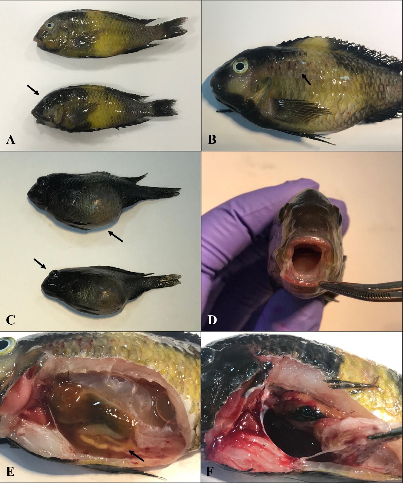
The affected Tropheus showed erratic behavior, swimming near the water surface, darkened skin, inappetence, abdominal distention, exophthalmus, petechial hemorrhages on the skin, and hemorrhages on the bases of the fins, around the anus and mouth, especially in the upper and lower jaws (Figure 1). At necropsy, enteritis, pale liver, and gronulomas were observed in the kidneys and spleen of infected individuals.

Figure 1
Figure 1.A: darkened area in the head, B: hemorrhages on the surface of the skin, C: darkened skin, exophthalmos, and ascites, D: hemorrhages in the upper and lower jaw, E: enteritis and presence of ascitic fluid in the peritoneal cavity, F: granulomas in the spleen.

Round, smooth, and opaque colonies were detected in TSA and the isolates were found to be motile Gram-negative rods. The API 20E profile of the bacteria is presented in Table 1. β-galactosidase, arginine dihydrolase, lysine decarboxylase, ornithine decarboxylase, citrate, hydrogen sulfide, gelatin, glucose, mannose, sorbitol, rhamnose, sucrose, melibiose, amygdalin and arabinose were detected to be positive, while urease, tryptophan deaminase, indole, Voges-Proskauer, and inositol were found to be negative in all isolates (Table 1).

Table 1.The morphological and biochemical properties of isolated Citrobacter freundii
Properties C.freundii Properties C.freundii
Gram stain - IND -
Shape Rod VP -
Motility + GEL +
Catalase + GLU +
Oxidase - MAN +
ONPG + INO -
ADH + SOR +
LDC + RHA +
ODC + SAC +
CIT + MEL +
H2S + AMY +
Urease - ARA +
TDA -

ONPG: β-galactosidase, ADH: Arginine dihydrolase, LDC: Lysine decarboxylase, ODC: Ornithine decarboxylase, CIT: Citrate, H2S: hytdrogen sulfide, TDA: Tryptophan deaminase, VP: Voges-Proskauer, GEL: Gelatin, GLU; Glucose, MAN: Mannose, INO: Inositol, SOR: Sorbitol, RHA: Rhamnose, SAC: Sucrose, MEL: Melibiose, AMY: Amygdalin, ARA: Arabinose.

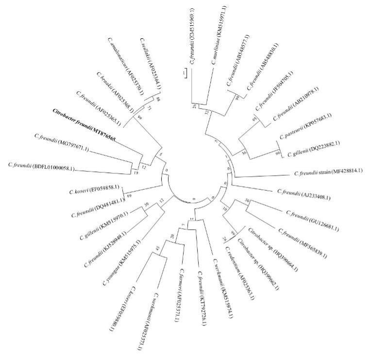
A 99 % sequence similarity with C. freundii was identified from the results of the 16 s rRNA sequence of the isolated bacterial pathogen in the BLASTN 2.6.1 database. The strain was registered in the NCBI GenBank with accession number (MT876565) and the phylogenetic tree of the isolated strain and the homogeneous sequences are presented in Figure 2.

Figure 2
Figure 2.Phylogenetic tree of Citrobacter freundii (MT876565) (in bold) and related Citrobacter sequences obtained from GenBank (NCBI). The Maximum Composite Likelihood Method was performed with Mega 7 software with 1000 bootstrap replicates.

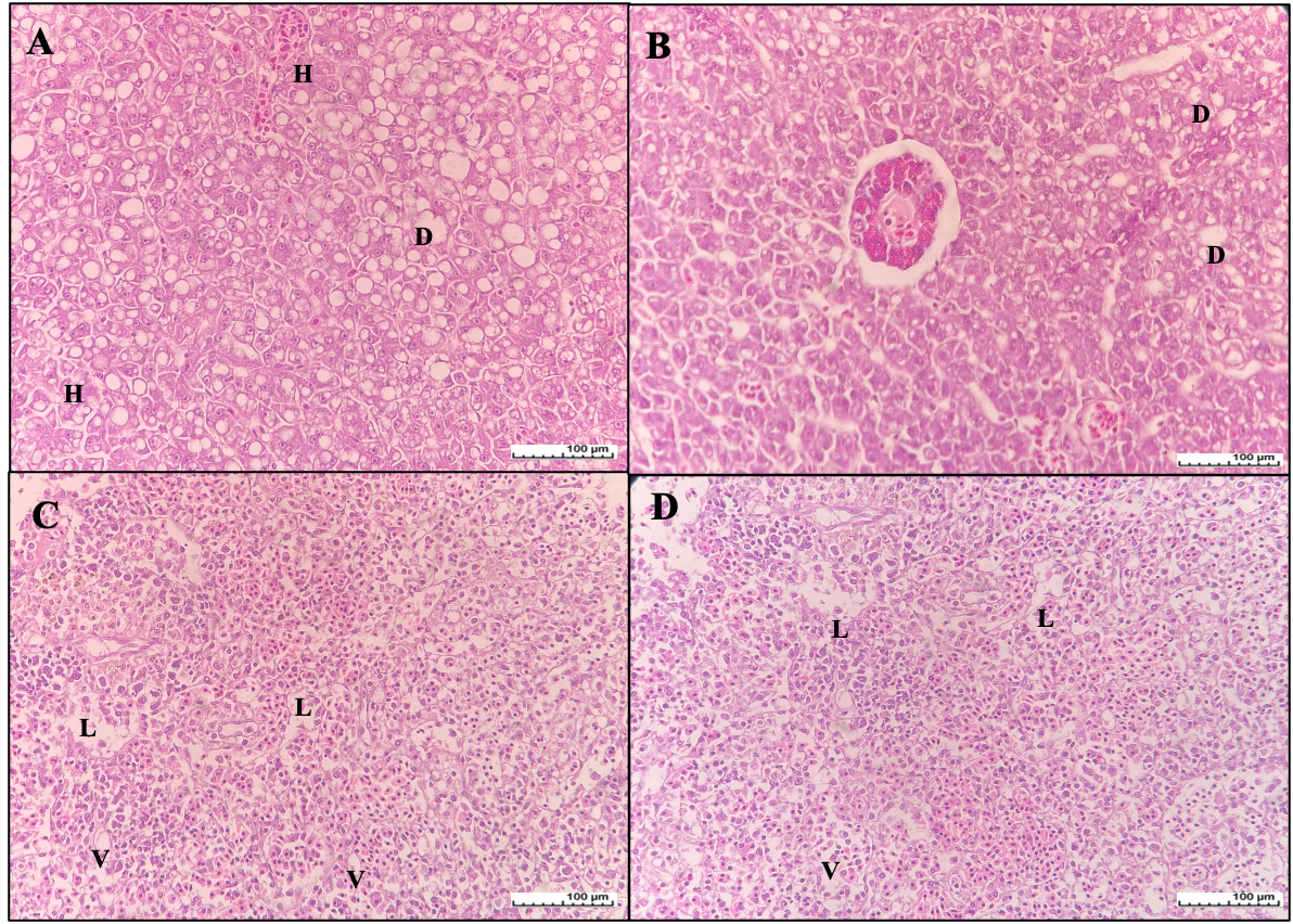
Histological changes of liver and spleen are presented in Figure 3. Loss of tissue structure, vacuolar degeneration and hepatocytes with nuclear loss were detected in the infected Tropheus spp.. Cellular degeneration was identified in the spleen tissue samples (Figure 3).

Figure 3
Figure 3.A-B: liver tissue of infected Tropheus spp. D: vacuolar degeneration; H: hepatocytes with nuclear loss- C-D: spleen tissue of infected Tropheus spp. L: depletion of lymphoid tissues; V: vacuole formation

Discussion

C. freundii has been found in several different natural sources, including soil, food, water, different animal species, and humans (Sedlák 1973; Schmidt et al. 1993; Nawaz et al. 2008; Baeck et al. 2009). In humans, Citrobacter infection affects the central nervous system and causes 75% of meningitis with brain abscess (Graham and Band 1981; Plakkal, Soraisham, and Amin 2013); it is also present in the gastrointestinal system and causes urinary tract infections, septicemia, and diarrhoea due to its opportunistic character (Schmidt et al. 1993; Metri, Jyothi, and Peerapur 2013; Liu et al. 2018). The pathogen is known for its ability to cause predominantly hemorrhages and hemorrhagic enteritis in fish, while an increase in the concentration of inorganic and organic matter in water, environmental pollution, and stress are reported as key parameters in the appearance of infection (Pandey 2013; de Pádua et al. 2014; Ture and Kutlu 2018).

Different mortality rates have been reported from C. freundii infections. Austin, Stobie, and Robertson (1992) detected low-level mortality in rainbow trout (20-30 mortalities/tank/day); however, in the infection experiment, disease signs and mortality were reported to be 40% within 72 h of injection. An experiment by Jeremić, Jakić-Dimić, and Veljović (2003) regarding infection in rainbow trout reported 60% mortality by the fifth day after injection, while Ture and Kutlu (2018) announced 10% cumulative mortality in rainbow trout. Sun et al. (2018) observed cumulative mortality rates of 60-100% in freshwater cultured stingray (Potamotrygon motoro) within 30 days. The current study found similar mortality rates of up to 100% in both juvenile and adult fish groups during the period of infection.

The clinical and pathological findings of this study show the general symptoms of C. freundii infection, such as erratic swimming, darkened skin, hemorrhages, enteritis, and pale liver. Especially, the abdominal distension was observed extensively with darkened skin. Austin, Stobie, and Robertson (1992) observed melanosis, hemorrhages in the eyes and internal organs, ascitic fluid in the peritoneal cavity, pale spleen, and gastroenteritis in rainbow trout individuals. Baeck et al. (2009) reported lethargy, abnormal swimming behavior, hemorrhages on the skin, fins, ventral part of the body, gills, and internal organs with reddish fluid in the abdominal cavity of doctor fish (Garra rufa obtusa). Thanigaivel et al. (2015) detected extensive skin hemorrhage, tail ulceration, and lesions in naturally infected tilapia fingerlings. Sun et al. (2018) found similar clinical findings in P. motoro, such as loss of appetite, lethargy, hemorrhages, and ascitic fluid in the abdomen, hemorrhages in the wall of the abdominal cavity, and bleaching of the liver. Lethargy, skin darkening, pale and friable liver, gallbladder distension, and splenomegaly were detected in silver catfish (Rhamdia quelen) by Junior et al. (2018).

The biochemical characteristics of the isolated C. freundii strains were found to be similar to those in previous reports (Sato, Yamane, and Kawamura 1982; Jeremić, Jakić-Dimić, and Veljović 2003; Baeck et al. 2009; de Pádua et al. 2014; Junior et al. 2018; Yang et al. 2021). Variable β-galactosidase, arginine dihydrolase, citrate, hydrogen sulfide, and amygdalin reactions have been reported in different studies; Sato, Yamane, and Kawamura (1982) reported negative detection of citrate and amygdalinwhile Padua et al. (2014). and this current study found positive detection, however, the biochemical profile from the API 20E test kit confirmed the identification (Table 2). The 16s rRNA gene sequence results also validated the diagnosis with a 99% sequence similarity to C. freundii. A number of researchers have recently provided various information about the 16s rRNA gene sequences of different C. freundii strains (Honein et al. 2016; Cao et al. 2016; Gallani et al. 2016; Ture and Kutlu 2018). Comparison of the 16s rRNA analyses showed that there were similarities to related Citrobacter strains (Figure 2). Comparing all of these data, the biochemical characteristics and 16s rRNA gene sequence results indicated that the isolated pathogen was C. freundii.

Table 2.Morphological and biochemical properties of Citrobacter freundii isolated from Tropheus spp.
Test or Substrate Current Study Soto et al. (1982) Jeremic et al. (2003) de Padua et al. (2014) Cao et al. (2016) Dong and Cai (2001)
ONPG + + +(3/3) +(2/2) - +
ADH + - -(3/3) +(2/2) - -
LDC - - -(3/3) -(2/2) - -
ODC - - -(2/3) -(2/2) - -
CIT + - -(3/3) +(2/2) + +
H2S + + +(3/3) -(2/2) + +
URE - - -(3/3) -(2/2) - -
TDA - - -(3/3) -(2/2) - -
IND - - -(3/3) -(2/2) - -
VP - - -(3/3) -(2/2) - -
GEL - - -(3/3) -(1/2) - -
GLU + + +(3/3) +(2/2) + +
MAN + + +(3/3) +(2/2) + +
INO - - -(3/3) -(2/2) - -
SOR + + +(3/3) +(2/2) + +
RHA + + +(3/3) +(2/2) + +
SAC + + +(2/3) +(2/2) + +
MEL + + -(2/3) +(2/2) + +
AMY + - +(3/3) +(1/2) - -
ARA + + +(3/3) +(2/2) + +

ONPG: β-galactosidase, ADH: Arginine dihydrolase, LDC: Lysine decarboxylase, ODC: Ornithine decarboxylase, CIT: Citrate, H2S: Hytdrogen sulfide, URE: Urease, TDA: Tryptophan deaminase, VP: Voges-Proskauer, GEL: Gelatin, GLU; Glucose, MAN: Mannose, INO: Inositol, SOR: Sorbitol, RHA: Rhamnose, SAC: Sucrose, MEL: Melibiose, AMY: Amygdalin, ARA: Arabinose.

The histopathological results showed vacuolar degeneration, hepatocytes with nuclear loss and tissue structure loss. In the spleen tissue, cellular degeneration was observed. The histopathological studies showed similar results with other Citrobacter infection cases : de Pádua et al. (2014) isolated C. freundii from infected native Brazilian catfish (Pseudoplatystoma) with histopathological symptoms of centrilobular necrosis in the liver. Junior et al. (2018) reported C. freundii infection in silver catfish (Rhamdia quelen) with tissue structure loss, hepatocytes with nuclear loss, and degeneration in liver tissue. Likewise, Jung et al. (2021) isolated C. tructae from diseased rainbow trout (Oncorhynchus mykiss) and reported hepatic necrosis from the liver and coagulative necrosis from the spleen samples in their histopathological studies.

The hobby of ornamental fishkeeping is the basis of one of the most popular industries around the world; it is a multimillion dollar sector and represents a major part of the pet market (Noga 2010; Gallani et al. 2016). However, aquarium fish are susceptible to several diseases that may occur due to temperature changes, poor water and food quality, etc. (Pandey 2013). C. freundii has been reported from several cases of disease in aquarium fish species, including giant gourami (Osphronemus goramy) (Honein et al. 2016) and freshwater angelfish (Pterophyllum scalare) (Gallani et al. 2016). However, there is limited information on the effects of this pathogenic bacteria in ornamental fish species. It is recommended that the disease agents in ornamental fish species be further clarified in order to implement prevention and control strategies.

Conclusion

In conclusion, an outbreak was observed in Tropheus spp. and the causative agent of the disease was identified as C. freundii by biochemical and molecular analyses. The diseased fish showed typical clinical symptoms and the mortality rate reached 100%. To our knowledge, this is the first report of C. freundii infection in Tropheus spp., which is an economically important ornamental cichlid species.


Funding

The authors received no financial support for the research.

Competing Interests

The authors have no relevant financial or non-financial interests to disclose.

Author Contributions

All persons who meet authorship criteria are listed as authors, and all authors certify that they have participated sufficiently in the work to take public responsibility for the content, including participation in the concept, design, analysis, writing, or revision of the manuscript equally.

Data availability statement

The authors confirm that the data supporting the findings of this study are available within the article.

Ethics approval

Ethics approval was not required for this study.

Submitted: March 20, 2023 CEST

Accepted: August 05, 2024 CEST

References

Austin, B., and D.A. Austin. 2007. Bacterial Fish Pathogens: Disease of Farmed and Wild Fish. 4th ed. Chichester: Springer.
Google Scholar
Austin, B., M. Stobie, and P. A. W. Robertson. 1992. “Citrobacter Freundii: The Cause of Gastro-Enteritis Leading to Progressive Low Level Mortalities in Farmed Rainbow Trout, Oncorhynchus Mykiss Walbaum, in Scotland.” Bulletin of the European Association of Fish Pathologists 12:166–67.
Google Scholar
Baeck, G. W., J. H. Kim, C. Choresca Jr., D. K. Gomez, S. P. Shin, J. E. Han, and S. C. Park. 2009. “Mass Mortality of Doctor Fish (Garra Rufa Obtusa) Caused by Citrobacter Freundii Infection.” Journal of Veterinary Clinics 26:150–54.
Google Scholar
Cao, H., X. Long, L. Lu, X. Yang, and B. Chen. 2016. “Citrobacter Freundii: A Causative Agent for Tail Rot Disease in Freshwater Cultured Japanese Eel Anguilla Japonica.” Israeli Journal of Aquaculture- Bamidgeh 68:1–7.
Google Scholar
Cardoso, P. H. M., A. M. Moreno, L. Z. Moreno, C. H. D. Oliveira, F. D. A. Baroni, S. R. D. L. Maganha, R. L. M. D. Sousa, and S. D. C. Balian. 2019. “Infectious Diseases in Aquarium Ornamental Pet Fish: Prevention and Control Measures.” Brazilian Journal of Veterinary Research and Animal Science 56:1–16.
Google Scholar
Collins, R. A., K. F. Armstrong, R. Meier, Y. Yi, S. D. J. Brown, R. H. Cruickshank, S. Keeling, and C. Johnston. 2012. “Barcoding and Border Biosecurity: Identifying Cyprinid Fishes in the Aquarium Trade.” PloS One 7:e28381. https:/​/​doi.org/​10.1371/​journal.pone.0028381.
Google ScholarPubMed CentralPubMed
Domínguez, L. M., and Á. S. Botella. 2014. “An Overview of Marine Ornamental Fish Breeding as a Potential Support to the Aquarium Trade and to the Conservation of Natural Fish Populations.” International Journal of Sustainable Development and Planning 9:608–32. https:/​/​doi.org/​10.2495/​SDP-V9-N4-608-632.
Google Scholar
Gallani, S. U., F. D. A. Sebastiao, G. M. Valladão, A. Z. Boaratti, and F. Pilarski. 2016. “Pathogenesis of Mixed Infection by Spironucleus Sp. and Citrobacter Freundii in Freshwater Angelfish Pterophyllum Scalare.” Microbial Pathogenesis 100:119–23.
Google Scholar
Graham, D. R., and J. D. Band. 1981. “Citrobacter Diversus Brain Abscess and Meningitis in Neonates.” JAMA: The Journal of the American Medical Association 245:1923–25.
Google Scholar
Helfman, G. 2007. Fish Conservation: A Guide to Understanding and Restoring Global Aquatic Biodiversity and Fisheries Resources. Washington DC: Island Press.
Google Scholar
Honein, K., S. S. S. D. S. Jagoda, A. Arulkanthan, H. Ushio, and S. Asakawa. 2016. “Draft Genome Sequences of Citrobacter Freundii Strains CF04 and A41 Isolated from Moribund, Septicemic Giant Gourami (Osphronemus Goramy) in Sri Lanka.” Genome Announcements 4:e00820-16. https:/​/​doi.org/​10.1128/​genomeA.00820-16.
Google ScholarPubMed CentralPubMed
Hulme, P. E. 2009. “Trade, Transport and Trouble: Managing Invasive Species Pathways in an Era of Globalization.” Journal of Applied Ecololgy 46:10–18. https:/​/​doi.org/​10.1111/​j.1365-2664.2008.01600.x.
Google Scholar
Jeremić, S., D. Jakić-Dimić, and L.J. Veljović. 2003. “Citrobacter Freundii as a Cause of Disease in Fish.” Acta Veterinaria 53:399–410.
Google Scholar
Jung, W. J., H. J. Kim, S. S. Giri, S. G. Kim, S. W. Kim, J. W. Kang, J. Kwon, et al. 2021. “Citrobacter Tructae Sp. Nov. Isolated from Kidney of Diseased Rainbow Trout (Oncorhynchus Mykiss).” Microorganisms 9:275.
Google Scholar
Junior, G. B., A. C. dos Santos, C. de Freitas Souza, M. D. Baldissera, K. L. dos Santos Moreira, M. L. da Veiga, M. I. U. M. de Rocha, A. C. C. de Vargas, A. M. Cunha, and B. Baldisserotto. 2018. “Citrobacter Freundii Infection in Silver Catfish (Rhamdia Quelen): Hematological and Histological Alterations.” Microbial Pathogenesis 125:276–80. https:/​/​doi.org/​10.1016/​j.micpath.2018.09.038.
Google Scholar
King, T. A. 2019. “Wild Caught Ornamental Fish: A Perspective from the UK Ornamental Aquatic Industry on the Sustainability of Aquatic Organisms and Livelihoods.” Journal of Fish Biology 94:925–36. https:/​/​doi.org/​10.1111/​jfb.13900.
Google Scholar
Lewbart, G. A. 2001. “Bacteria and Ornamental Fish.” Seminars in Avian and Exototic Pet Medicine 10:48–56. https:/​/​doi.org/​10.1053/​saep.2001.19543.
Google Scholar
Liu, L.H., N.Y. Wang, A.Y.J. Wu, C.C. Lin, C.M. Lee, and C.P. Liu. 2018. “Citrobacter Freundii Bacteremia: Risk Factors of Mortality and Prevalence of Resistance Genes.” Journal of Microbiology, Immunology and Infection 51:565–72. https:/​/​doi.org/​10.1016/​j.jmii.2016.08.016.
Google Scholar
Maceda-Veiga, A., O. Domínguez-Domínguez, J. Escribano-Alacid, and J. Lyons. 2016. “The Aquarium Hobby: Can Sinners Become Saints in Freshwater Fish Conservation?” Fish and Fisheries 17:860–74. https:/​/​doi.org/​10.1111/​faf.12097.
Google Scholar
Metri, B. C., P. Jyothi, and B. V. Peerapur. 2013. “Antibiotic Resistance in Citrobacter Spp. Isolated from Urinary Tract Infection.” Urology Annals 5:312–13.
Google Scholar
Nawaz, M., A.A. Khan, S. Khan, K. Sung, and R. Steele. 2008. “Isolation and Characterization of Tetracycline-Resistant Citrobacter Spp. from Catfish.” Food Microbiology 25:85–91. https:/​/​doi.org/​10.1016/​j.fm.2007.07.008.
Google Scholar
Noga, E.J. 2010. Fish Disease: Diagnosis and Treatment. 2nd ed. USA: Willey-Blackwell.
Google Scholar
Pádua, S. B. de, D. P. Marques, F. A. Sebastiao, F. Pilarski, M. L. Martins, and M. M. Ishikawa. 2014. “Isolation, Characterization and Pathology of Citrobacter Freundii Infection in Native Brazilian Catfish Pseudoplatystoma.” Brazilian Journal of Veterinary Pathology 7:151–57.
Google Scholar
Pandey, G. 2013. “Treatment and Control of Ornamental Fish Infection.” Universal Journal of Pharmacy Take Research to New Heights 2:52–56.
Google Scholar
Plakkal, N., A.S. Soraisham, and H. Amin. 2013. “Citrobacter Freundii Brain Abscess in a Preterm Infant: A Case Report and Literature Review.” Pediatrics and Neonatology 54:137–40. https:/​/​doi.org/​10.1016/​j.pedneo.2012.10.004.
Google Scholar
Raja, K., P. Aanand, S. Padmavathy, and J.S. Sampathkumar. 2019. “Present and Future Market Trends of Indian Ornamental Fish Sector.” International Journal of Fisheries and Aquatic Studies 7:6–15.
Google Scholar
Sato, N., N. Yamane, and T. Kawamura. 1982. “Systemic Citrobacter Freundii Infection among Sunfish Mola Mola in Matsushima Aquarium.” Bulletin of the Japanese Society of Scientific Fisheries 48:1551–57. https:/​/​doi.org/​10.2331/​suisan.48.1551.
Google Scholar
Schmidt, H., M. Montag, J. Bockemühl, J. Heesemann, and H. Karch. 1993. “Shiga-like Toxin II-Related Cytotoxins in Citrobacter Freundii Strains from Humans and Beef Samples.” Infection and Immunity 61:534–43. https:/​/​doi.org/​10.1128/​iai.61.2.534-543.1993.
Google ScholarPubMed CentralPubMed
Sedlák, J. 1973. “Present Knowledge and Aspects of Citrobacter.” In Current Topics in Microbiology and Immunology/Ergebnisse Der Mikrobiologie Und Immunitätsforschung, edited by R. Ahmed, S. Akira, A. Casadevall, J. E. Galan, A. Garcia-Sastre, B. Malissen, and R. Rappuoli, 41–59. Berlin · Heidelberg: Springer-Verlag. https:/​/​doi.org/​10.1007/​978-3-642-65772-6_2.
Google Scholar
Sun, H. Y., X. H. Cao, Y. F. Jiang, L. Y. Ni, Z. Q. Mo, Q. W. Qin, Y. W. Li, and X. M. Dan. 2018. “Outbreak of a Novel Disease Associated with Citrobacter Freundii Infection in Freshwater Cultured Stingray, Potamotrygon Motoro.” Aquaculture 492:35–39. https:/​/​doi.org/​10.1016/​j.aquaculture.2018.03.058.
Google Scholar
Thanigaivel, S., S. Vijayakumar, S. Gopinath, A. Mukherjee, N. Chandrasekaran, and J. Thomas. 2015. “In Vivo and in Vitro Antimicrobial Activity of Azadirachta Indica (Lin) against Citrobacter Freundii Isolated from Naturally Infected Tilapia (Oreochromis Mossambicus).” Aquaculture 437:252–55. https:/​/​doi.org/​10.1016/​j.aquaculture.2014.12.008.
Google Scholar
Toranzo, A.E., J.M. Cutrin, B.S. Roberson, S. Nune, J.M. Abell, F.M. Hetrick, and A.M. Baya. 1994. “Comparison of the Taxonomy, Serology, Drug Resistance Transfer, and Virulence of Citrobacter Freundii Strains from Mammals and Poikilothermic Hosts.” Applied and Environmental Microbiology 60:1789–97. https:/​/​doi.org/​10.1128/​aem.60.6.1789-1797.1994.
Google ScholarPubMed CentralPubMed
Ture, M., and I. Kutlu. 2018. “Isolation of Citrobacter Freundii from Rainbow Trout (Oncorhynchus Mykiss) in Freshwater Cage.” Journal of Limnology and Freshwater Fisheries Research 4:85–89. https:/​/​doi.org/​10.17216/​limnofish.396496.
Google Scholar
Yang, J., T. Tian, K. Xiao, Q. Zeng, C. Tan, and H. Du. 2021. “Pathogenic Infection and Immune-Related Gene Expression of Chinese Sturgeon (Acipenser Sinensis) Challenged by Citrobacter Freundii.” Developmental and Comparative Immunology 114:103872. https:/​/​doi.org/​10.1016/​j.dci.2020.103872.
Google Scholar

This website uses cookies

We use cookies to enhance your experience and support COUNTER Metrics for transparent reporting of readership statistics. Cookie data is not sold to third parties or used for marketing purposes.

Powered by Scholastica, the modern academic journal management system