

Introduction
Bosnia and Herzegovina (B&H) has long history of aquaculture, dating back to first salmonid farm established in 1894, followed by the first cyprinid farm in 1902 (Hamzić 2003). Aquaculture production is dominated by freshwater fish farming (95 percent). The main products are rainbow trout and common carp. Mariculture production consists of gilthead seabream, European seabass, oyster and mussel. The extent of fish farming in Bosnia and Herzegovina involves 91,026 square meters allocated for trout fish farms and 2,278 hectares dedicated to carp fisheries. The freshwater aquaculture production was 3 465 tonnes in 2018 (FAO 2023).
Consequently, aquaculture holds significant economic importance, playing a important role in the country’s culture and overall economy. The occurrence of carp diseases poses a substantial risk to aquaculture production, making prompt disease diagnosis essential for effective containment and prevention. This is particularly important when dealing with emerging diseases that can lead to significant mortality and are not currently regulated by state legislation.
It is worth noting that important viral diseases of carp, such as spring viremia of carp (SVC), koi herpesvirus disease (KHVD) and carp edema virus disease (CEVD) have not been diagnosed so far in Bosnia and Herzegovina.
CEVD or koi sleepy disease is caused by the dsDNA Carp edema virus (CEV), which belongs to the subfamily Chordopoxvirinae within the family Poxviridae. Initially diagnosed in 1974 in Japan, koi sleepy disease was primarily observed in ornamental koi (Cyprinus carpio koi) (Murakami et al. 1976). However, following the confirmation of CEV in carp (Cyprinus carpio carpio), the disease is referred to as carp edema virus disease. The disease typically manifests within a water temperature range of 19°C to 24°C; however, cases of the disease have been documented across a wide spectrum of water temperatures, spanning from 6°C to 24°C (Divya et al. 2019). In general, the disease follows an acute course when water temperatures range from 15°C to 25°C, while at lower temperatures of 6°C to 10°C, a chronic course with lower mortality rates is observed (Lewisch et al. 2015; Way et al. 2017; Toffan et al. 2020).
The primary feature of the disease is the extreme lethargy observed in affected fish, giving the “sleeping” appearance. Consequently, the fish succumb to anoxia. The clinical presentation of CEVD shares similarities with KHVD, wherein the main clinical signs include lethargy, pronounced enophthalmos (sunken eyes), increased production of skin and gill mucus, necrotic changes on the gills, and ulcerative lesions around the mouth and base of the fins (Antychowicz et al. 2005; Way et al. 2017).
Presently, there is no effective treatment available for CEVD. Some studies suggest that mortality can be reduced by salt baths using a 0.5% NaCl solution (Seno et al. 2003; Miyazaki, Isshiki, and Katsuyuki 2005).
The transmission of CEV to other species, including common bream (Alburnus alburnus), crucian carp (Carassius carassius), Prussian carp (Carassius gibelio), roach (Rutilus rutilus), tench (Tinca tinca), and perch (Perca fluviatilis), has been demonstrated experimentaly (Matras et al. 2019). Those species, despite testing positive for CEV, do not show clinical signs of the disease, nor does mortality occur. The presence of CEV was identified in wild carp populations in both Europe and the USA (Tolo et al. 2021; Lovy et al. 2018; Marsella et al. 2021). Way et al. (2017) and Matras et al. (2019) suggested that infected carp, acting as virus carriers, could have a crucial role in spreading viral infections not only to carp, but also to potential vector species. This mode of virus transmission poses a significant challenge in preventing and eradicating the disease.
CEV has been previously identified in the Balkan region, with detection reported in the Republic of Serbia in 2017 (Radosavljevic et al. 2018) and in the Republic of Croatia in 2018 (Zrnčić et al. 2020). Here, we describe the first detection of CEVD caused by carp edema virus in wild carp in Bosnia and Herzegovina.
Materials and Methods
Disease History and Clinical Examination
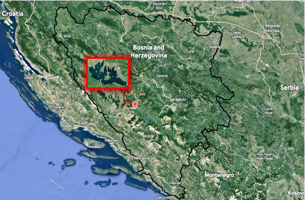
In early April 2023, with warmer temperatures, a sudden onset of daily carp mortalities occurred in Rama Lake (Figure 1).

Notably, specimens ranging from 40 to 50 cm in length and weighing between 2.5 and 3.5 kg were affected by this mortality. Among the various fish species present in the lake, such as a strugac (Squalius svallize), chub ( Squalius cephalus), rainbow trout (Oncorhynchus mykiss), brown trout (Salmo trutta), goldfish (Carassius auratus), perch (Perca fluviatilis), common carp (Cyprinus carpio), pumpkinseed (Lepomis gibbosus), silver crucian carp (Carassius auratus langsdorfii), and Prussian carp (Carassius gibelio), only carp suffered, while the other species remained unaffected with no clinical signs of disease. During the mortality event the water temperature was 11°C.
The affected carp exhibited clinical signs of the disease, gathering near the inflows of fresh water that entered the lake. Additionally, in the shallow parts of the lake, the carp congregated, displaying lethargic “sleeping” behaviour, gliding on the water surface. The lethargic carp was easily lifted out of the water by hand. When fishing boats approached, the “sleeping” carp would briefly “awaken” and swim away from the boat, only to calm down and resume their surface position.
The sample of 10 moribund fish was caught in a landing net and transported on ice to the Laboratory for Aquaculture of the Veterinary Institute Sarajevo. Necropsy was performed on all sampled fish.
PCR
For PCR detection, five pooled samples (gills, anterior kidney and brain or two fish per pool) from clinically affected fish was subjected to further analysis to detect the presence of CEV, KHV and SVCV. DNA was extracted directly from the homogenates using the QIAamp Cador Pathogen Mini Kit (Qiagen, Hilden, Germany), following the manufacturer instructions. For the amplification of the viral genome, a qRT real-time PCR was used. The master mix used included 10 μl of Luna Universal Probe qRT-PCR Master Mix (New England BioLabs, USA), 1 μl of RT Enzyme Mix (20x), 0.8 μl of each 10mM primer, and 0.4 μl of 10mM probe. Primers used were previously specified by Matras et al. (2017), Gilad et al. (2004), and Yue et al. (2008), respectively (Table 1).
Virus isolation
For virus isolation, five pooled samples (gills, anterior kidney and brain or two fish per pool) were homogenized using mortar and pestle with sterile quartz sand in minimum essential medium (MEM) with a 10% fetal bovine serum (FBS) and 1% antibiotic-antimycotic solution and centrifuged at 2500 g for 20 min. Supernatants were filtered through a 0.45 μL pore-size filter membrane and inoculated onto 24h old monolayers of the EPC and BF-2 cell lines. Fish cell lines, namely epithelioma papulosum cyprini (EPC, Fijan et al. 1983), and bluegill fry (BF2, Wolf, Gravell, and Malsberger 1966), were used for cell culture examination. Cell cultures were propagated in an incubator at 22.5°C, using ROTI Cell Eagle’s MEM with Earle’s salts (Carl Roth, Germany), without glutamine, supplemented with 10% FBS (Merck, Germany) and 1% antibiotic-antimycotic solution (containing 10,000 IU penicillin/mL, 10 mg streptomycin/mL, and 25 µg amphotericin B/mL). After the formation of a cell monolayer, the samples were inoculated in replicates into 96-well culture plates and incubated at 15°C. Daily observation for cytopathogenic effect (CPE) was conducted for 21 days, with subcultivation performed every 7 days.
Parasitology
For parasitological examination, a skin scraping, and pieces of gill tissue were examined for presence of parasites by light microscopy.
Results
A clinical examination of moribund carp common carp revealed a large amount of mucus covering the entire body of the fish. The gills exhibited oedema and a notable accumulation of thick mucus (Figure 2b). The eyes displayed a prominent haemorrhage line, enophthalmos (sunken appearance), and opacity (Figure 2a). Additionally, petechial haemorrhages were found on the body, abdomen, and at the bases of the fins (Figure 2c). A necropsy revealed severe fibrinous peritonitis (Figure 3). However, no significant changes were observed in other organs.


All tested samples of diseased carp from Rama Lake were positive for carp edema virus (Ct values 22 to 27).
When samples from clinically affected carp were analysed for the presence of KHV and SVCV, real-time PCR tests showed negative results.
No cytopathic effect was observed in inoculated BF-2 and EPC cells inoculated after three blind passages.
No parasites were observed from the microscopic examination of gills and skin scrapes. A minor incidence of fish parasites, namely Bothriocephalus acheilognathi and Khawia japonensis, was observed within the intestinal content of a limited number of specimens.
Discussion
Recently, carp oedema virus (CEV) has emerged as a notable pathogen, impacting common carp in several European countries, and has been designated as an emerging disease by the World Organisation for Animal Health (WOAH 2023). In the present case, characteristic disease manifestations of CEVD, such as lethargy and unresponsiveness accompanied by swollen gills covered in mucus (Way et al. 2017), were observed. Internally, no pathologies were present, except fibrinous peritonitis, as previously noted (Radosavljevic et al. 2021). Previous attempts have typically been unsuccessful in isolating CEV on cell lines (Jung-Schroers et al. 2015; Swaminathan et al. 2016). In this study, no cytopathic effect (CPE) was observed during three consecutive passages on EPC and BF2 cell cultures.
The presence of CEV was identified in wild carp populations in both Europe and the USA (Tolo et al. 2021; Lovy et al. 2018; Marsella et al. 2021), as well as in fish hosts that displayed no signs of disease (Matras et al. 2019). This can considerably enhance the potential for further spread of the disease. Unfortunately, in the case of Rama Lake, other native species were not captured and tested, thereby making it impossible to rule out the presence of CEV in those species within the lake.
In our case, origin of the infection remains unclear. However, considering the practice of regular stocking of carp in Rama Lake from aquaculture facilities, it could potentially serve as a source of the pathogen. Also, it is possible that it is the transfer of the pathogen by a species that carries it asymptomatically, especially considering the potentially large number of species which may play a role as animal vectors of CEV infection. Within European carp aquaculture, fish from diverse species within the local fish fauna exhibit a tendency to migrate into production or hibernation ponds, or are cultivated in polyculture alongside carp (Adamek et al. 2022). Unfortunately, in the case of Rama Lake, other native species were not captured and tested, thereby making it impossible to rule out the presence of CEV in those species within the lake.
The presence of CEV in native fish populations and the potential vector role of these species in spreading and distributing the virus become critical areas of investigation for future research. In order to determine the presence of CEV or other carp viruses in native fish species, it is crucial to perform testing during each carp mass mortality event. Adopting such an approach would yield valuable information regarding the transmission and distribution of CEV. Naturally, there is always the potential for the pathogen transfer through alternative carriers, such as parasitic invertebrates, piscivorous birds, and aquatic mammals (Matras et al. 2019).
Having in mind that CEV has to be regarded as a serious infectious agent rather than an opportunistic pathogen affecting weakened specimens (Adamek et al. 2022), it is essential to implement efficient biosecurity measures to prevent further spread of CEV. Additionally, it is crucial to restrict the movement of carp from ponds where the virus has been detected. These actions aim to minimize the risk of transmission and contain the spread of CEV, thereby protecting the carp population and preventing the further dissemination of the disease.
CEV infection is widespread in carp aquaculture, causing various mortality rates, depending on various factors (Radosavljevic et al. 2021). In the absence of a surveillance program, there exists insufficient data regarding the dissemination of CEV in B&H, raising the possibility that the true scope of CEV might be underestimated. Although CEVD is not currently included in the list of diseases of concern in B&H, it is crucial to implement mandatory control measures for CEV, in addition to SVC and KHV, to safeguard cyprinid production.
Conclusions
Here, we report the first outbreak of CEVD in B&H. This case highlights the importance of testing for CEV in cases of carp mortality and may indicate that CEV is emerging as an important cause of wild carp mortalities in the country. Having in mind the high importance of common carp in B&H, a surveillance and control program for CEV is warranted. Uncertainty regarding potential presence of CEV in asymptomatic fish complicates the efforts to contain disease and stop further spreading. Effective biosecurity is crucial for avoiding the emergence and dissemination of fish diseases. In B&H, there is an annual surveillance program in place with the goal of identifying the presence of SVC and KHV. Nevertheless, an enhancement to this program could involve the incorporation of CEV to more efficiently oversee and manage this disease within the country.
Acknowledgments
The authors would like to express their sincere gratitude to the Institute of Veterinary Medicine of Serbia for their invaluable assistance and support throughout the study. Additionally, special thanks are extended to the Sports Fishing and Tourist Association “Ramske vode” for their dedication to preserving Rama Lake and its diverse fauna, including the well-being of carp. The study was funded by the Serbian Ministry of Science, Technological Development and Innovation (Contract No. 451-03-47/2023-01/200030).