Loading [Contrib]/a11y/accessibility-menu.js
Skip to main content
null
Bulletin of the EAFP
  • Menu
  • Articles
    • Case study
    • Expert Opinion
    • Method
    • Note
    • Research article
    • Review
    • Workshop Report
    • All
  • For Authors
  • Editorial Board
  • About
  • Issues
  • search

RSS Feed

Enter the URL below into your favorite RSS reader.

http://localhost:39917/feed
Case study
December 21, 2024 CEST

Streptococcosis in Nile tilapia Farming: A Severe yet Overlooked Health Challenge in Mexico, a Study Case in Campeche, Mexico

Amelia Isabel Paredes-Trujillo, Manuel Mendoza-Carranza,
Aquaculturebacteriamortalityaquaculture health
Copyright Logoccby-4.0 • https://doi.org/10.48045/001c.127774
This article has a retraction notice: https://doi.org/10.48045/001c.131920
Photo by CDC on Unsplash
Bulletin of the EAFP
Paredes-Trujillo, Amelia Isabel, and Manuel Mendoza-Carranza. 2024. “Streptococcosis in Nile Tilapia Farming: A Severe yet Overlooked Health Challenge in Mexico, a Study Case in Campeche, Mexico.” Bulletin of the European Association of Fish Pathologists, December. https:/​/​doi.org/​10.48045/​001c.127774.
Save article as...▾
Download all (7)
  • Figure 1. Clinical signs lesions caused by Streptococcus agalactiae.
    Download
  • Figure 2. Clinical signs lesions caused by Streptococcus agalactiae.
    Download
  • Figure 3. Histopathology damage of liver, kidney and spleen affected Streptococcus agalactiae.
    Download
  • Figure 4. Histopathology damage of muscle, brain and intestine affected Streptococcus agalactiae.
    Download
  • Figure 5. a) Production (left axis) and losses by Streptococcus agalactiae (right axis) in a Nile tilapia farm in Campeche, losses; b) Prevalence in total harvest.
    Download
  • Figure 6. Total annual gross economic loss based on Monte Carlo simulation.
    Download
  • Figure 7. Management practices in an ­­­intensive farm located in Campeche.
    Download

Sorry, something went wrong. Please try again.

If this problem reoccurs, please contact Scholastica Support

Error message:

undefined

View more stats

Abstract

Streptococcosis is a bacterial disease that causes significant economic losses in Nile tilapia farming worldwide. An Outbreaks of Streptococcus agalactiae was detected in grow-out ponds on a farm in Campeche, Mexico. This study is the first to describe and analyze the histopathological damages, economic losses, and clinical manifestations of S. agalactiae in a commercial farm in Campeche, Mexico. A total of 30 Nile tilapia exhibiting clinical signs of the disease were collected in May 2023. Necropsies of the affected fish revealed characteristic signs of streptococcosis, including bilateral exophthalmia, nodules in the spleen, hemorrhagic brain, hepatomegaly, spleen congestion and accumulation of fluid in the abdominal cavity. Nile tilapia organs (brain, liver, kidney, muscle, spleen, stomach and intestine) were removed and fixed in 10 % formalin for histopathological examination. Histopathological alterations in tissues showed significant internal organ damage, characterized by a granulomatous reaction, melanomacrophage pigment, vacuolar degeneration, and granular eosinophilic cells containing evident bacterial bodies. Alterations in organs tissues were graded by severity scores. We analyzed information on biomass losses caused by S. agalactiae from January to December 2022. The Monte Carlo simulation show economic losses of streptococcosis at 30,740 kg of Nile tilapia per year, translating to a financial impact of averaged $1,537,671 ± 34,2587 Mexican pesos (76464.53 ± 17036 USD). Additionally, the disease reached an 80% prevalence rate, resulting in high mortality and morbidity rates among the fish. The prognosis for streptococcosis in Mexico is concerning, given that it is recognized as one of the most significant diseases affecting cultivated Nile tilapia. Despite the severe impacts of streptococcosis outbreaks on the environment, public health, and socio-economics, there remains a notable data gap regarding the disease’s epidemiology in Mexico. It is crucial to generate more scientific information on diagnosis, prevalence, pathogen distribution, natural historical variation, newly identified serotypes, and potential zoonotic cases.

Introduction

Tilapia farming is the second most significant freshwater fish industry globally, following carp (FAO 2020). Currently, worldwide Nile tilapia production grew from 700,000 in 2010 to 850,000 tons in 2020 (FAO 2020). Principal Nile tilapia producers are China, Indonesia, Egypt, and the Philippines, with Brazil and Mexico emerging as important producers in the Americas (FAO 2020). In 2017, Mexico reported a total of 4,634 Nile tilapia farms operating throughout the country, collectively generating an annual production of 117,806 t. The states with the highest production are Chiapas, contributing 30,912.11 t, following by Jalisco (8,178.08 t), and Nayarit with (7,618 t) (INAPESCA 2018).

Nevertheless, the elevated stocking densities required to achieve current Nile tilapia production levels in Mexico have given rise to various parasitic agents (bacteria, viruses, and other parasites). Streptococcosis has emerged as a prevalent bacterial disease in Nile tilapia production world systems, as documented by Pretto-Giordano et al. (2010), Delannoy, Samai, and Labrie (2021), Zhang et al. (2021), Delphino, Leal, Gardner, et al. (2019), and Delphino, Joshi, and Alvarez (2022). Since its initial record in Nile tilapia in 2009, streptococcosis has become the predominant pathogen, accounting for over 90% of detected and isolated pathogens in aquaculture. This has resulted in mortality rates ranging from 30 to 90% on a global scale (Sheehan, Lee, Wong, et al. 2009; Liu, Ye, Liu, et al. 2018). In Nile tilapia farming, streptococcosis outbreaks, are mainly associated with Streptococcus agalactiae group B and S. iniae. Economic losses can reach 23 billion US dollars in countries like the United States, Israel, Japan, Kuwait, Thailand, Mexico, Honduras, and Brazil (Evans, Klesius, Gilbert, et al. 2002; Evans et al. 2006; Suanyuk et al. 2008; Mian et al. 2009; Delannoy, Zadoks, Lainson, et al. 2012; Delannoy, Samai, and Labrie 2021; Li et al. 2016; Zheng, Wu, Hu, et al. 2018).

In Malaysia and Mexico, mortality rates exceeding 50% have been recorded during acute infections, typically occurring within 3 to 7 days post infection. Alternatively, the rates can be lower but still result in a sustained daily mortalities during chronic infections (Zamri-Saad et al. 2014; Ortega, García, Irgang, et al. 2018). In Malaysia, Nile tilapia weighing between 300 to 400 grams experienced mortalities of up to 60% due to S. agalactiae (Zamri-Saad et al. 2014). Floating cages in Brazil reported mortality rates ranging from 25 % to 35 % (Leal et al. 2019), while in China, rates ranged from 20 to 30% (Ye, Li, Lu, et al. 2011). In Northern Africa, cultivation ponds reported mortality rates ranging from 6 to 14% (Delannoy, Samai, and Labrie 2021). Regarding S. iniae, mortality rates from 30 to 50% have been observed in Iowa (Rahmatullah, Ariff, Kahieshesfandiari, et al. 2017), and in Mexico, mortality rates of 68 and 80% were documented in Oreochromis aureus production (Ortega, García, Irgang, et al. 2018).

The overall scenario appears discouraging, as Streptococcus spp. has already become well-established and widely distributed across production systems in nearly 10 states of the Mexican Republic (Table 1). Despite the significance of Nile tilapia culture in Mexico, the scarcity of reports regarding the status of this disease is notable.

Given the substantial potential for losses associated with streptococcosis, it is imperative to enhance our understanding of this disease in Mexico. This will support the proposal of sanitary control strategies for aquaculture facilities and the development of biosecurity programs, thereby reducing disease incidence and mitigating economic losses and health risks. In Mexico, comprehensive information on various pathological aspects of this disease is lacking, including data about its prevalence, geographical distribution, and the associated economic losses resulting from its growing presence. Therefore, this study aims to describe and analyze the histopathological damage, economic losses, and clinical manifestations of streptococcosis in a commercial farm in Campeche, Mexico.

Table 1.Presence of Streptococcus spp. in some locations of Mexico (table sorted by year)
State Locality Species Prevalence (%) Year Host species Culture type Reference
Campeche Chulbac Streptococcus agalactiae na 2023 Tilapia Recirculation system Not published data EPOMEX, 2023
Chiapas Presa Malpaso Streptococcus iniae 1.4 2023 Oreochromis niloticus Floating cages Soto-Rodríguez, Lopez, and Rendon 2023
Guerrero _ S. agalactiae 59 2023 O. niloticus Floating cages Soto-Rodríguez, Lopez, and Rendon 2023
Chiapas Presa Malpaso/Comitán, Palenque, Tzimol, Tecpatán, Reforma, Trinitaria y Chiapa de Corzo S. agalactiae 45.16- 6.45 2023 O. niloticus Floating cages and Recirculation system Pineda-Hernández et al. 2023
Guerrero Cd. Altamirano S. iniae na 2023 Tilapia Recirculation system Pineda-Hernández et al. 2023
Mexico Mexico S. iniae na 2022 Tilapia Recirculation system Alcántara-Jauregui, Valladares-Carranza, and Ortega 2022
Campeche Cd. Carmen S. agalactiae 100 2021 Tilapia Recirculation system CESAICAM 2021
Campeche Cd. Carmen S. iniae 100 2021 Tilapia Recirculation system CESAICAM 2021
Jalisco _ S. agalactiae na 2021 O. niloticus Recirculation system CESAJ 2021
Chiapas Malpaso Dam Streptococcus spp. na 2020 Tilapia Floating cages CESACH 2020
Chiapas Comitán S. agalactiae 15 2020 O. niloticus Recirculation system CESACH 2020
Chiapas Comitán S. iniae 15 2020 O. niloticus Recirculation system CESACH 2020
Chiapas Mezcalapa S. agalactiae 34 2020 O. niloticus Recirculation system CESACH 2020
Chiapas Mezcalapa S. iniae 17 2020 O. niloticus Recirculation system CESACH 2020
Chiapas Tecpatán S. iniae 17 2020 O. niloticus Recirculation system CESACH 2020
Chiapas Ostuacán S. agalactiae 17 2020 O. niloticus Recirculation system CESACH 2020
Guerrero El gallo S. agalactiae, S. iniae na 2020 O. niloticus Floating cages Castro-Ortíz 2020
Nayarit Ixtlan del Rio S. iniae, S. agalactiae na 2010-2019 Tilapia Recirculation system CESANAY,2010-2019
Santiago Ixcuintla, Estación Nanchi S. agalactiae na 2010-2019 Tilapia Recirculation system CESANAY 2010-2019
Ahuacatlan, Heriberto Jara S. iniae na 2010-2019 Tilapia Recirculation system CESANAY 2010-2019
Xalisco, Trigomil S. agalactiae na 2010-2019 Tilapia Recirculation system CESANAY 2010-2019
San Luis Potosí _ S. iniae 80 2018 O. aureus Recirculation system Ortega, García, Irgang, et al. 2018
Chiapas Malpaso Dam Streptococcus spp. na 2012 Tilapia Floating cages M. J. Hernández-Hernández et al. 2012
Querétaro _ S. iniae 60 2018 O. aureus Recirculation system Ortega, García, Irgang, et al. 2018
Rosario Streptococcus sp. na 2018 O. niloticus Recirculation system Valenzuela-Armenta, Díaz-Camacho, Cabanillas-Ramos, et al. 2018
Veracruz Alvarado Streptococcus sp. na 2017 O. niloticus Rustic ponds Huicab-Pech, Castaneda-Chavez, and Lango-Reynoso 2017
Sinaloa Adolfo López, Mateos, Sanalona y Dique IV Streptococcus spp. na 2009 O. aureus Recirculation system/Cages Soto 2009

Materials and Methods

Field and laboratory work

A follow-up study designed to elucidate Streptococcosis infection was carried out in an intensive farm located in Chulbac, Chiná, in the municipality of Campeche during May 2023. A total of 30 Nile tilapia (Oreochromis niloticus) with sign such as eroded fins, injured eyes, exophthalmia, ulcers, ascites, external lesions and desquamation were collected using a 2 m diameter cast net with a 2.5 cm mesh from a production tank. The fish collected were kept in tanks with artificial aeration and transported to laboratory. Fish were euthanized with benzocaine (100 mg/l) until opercular movements ceased (Fabiani et al. 2013). Each specimen was measured to obtain the total length (TL, cm), and total weight (W, g). The fish had an average weight of 280 g, ranging from 210 to 340 g, and an average total length of 17.50 cm, ranging from 15.34 to 19.53 cm. The genomic DNA of Streptococcus agalactiae was validated for one-step or real-time PCR (qPCR) in concurrent research conducted by Castillo-Avila et al. (2024); however, the serotype was not determined.

Histopathology procedures

Nile tilapia organs (brain, liver, kidney, muscle, spleen, stomach and intestine) were removed and fixed in 10 % neutral-buffered formalin during 72 h prior to processing using standard protocols for histology. Organ sections were dehydrated in 70, 80 and 96% ethanol baths, cleared with xylene, and embedded in paraffin wax, and sectioned at 5 µm on a Leica™ microtome (Mumford et al. 2007; Igeh and Avenant-Oldewage 2020). Slides were left overnight in an oven at 40°C to ensure adhesion. Subsequently, sections were stained with haematoxylin and eosin (H&E); some sections were stained with Ziehl-Neelsen and Fite–Faraco stains for acid-fast bacteria and examined under a light microscope (Igeh and Avenant-Oldewage 2020). Digital images were obtained using a Leica Digital Camera MC 120 HD and capture software.

Health condition by evaluation of histopathological index

To evaluate the health condition of Nile tilapia, the organ tissues alterations of each fish were analysed using the histopathological index proposed by Schwaiger et al. (2004). Additionally, the prevalence of each lesion per fish was calculated and expressed as percentage. According to the semi-quantitative method described by Schwaiger et al. (2004), histological alterations in tissue organs received damage severity scores. Following scores were used: 1) mild alterations or focal process in gills, 2) moderate alterations or multifocal, and 3) severe alterations or diffuse process. Moreover, the mean value of histological alteration was established for each fish into three categories mild (0.1–1.0), moderate (1.1–2.0), and severe (2.1–3.0) (Steckert et al. 2018). The histological alteration was analysed using the nonparametric Kruskal–Wallis statistical test (p<0.05).

Economic losses analysis

A database containing information about monthly biomass losses caused by S. agalactiae from January to December 2022 provided by the fish farm owner was analyzed graphically. Prevalence, defined as the percentage of infected hosts among those tested, was assessed to measure the impact on production (Bush et al. 1997). The gross economic impact was estimated based on 10,000 interactions using a Monte Carlo simulation based on the following simple parameters (Bezerra et al. 2022): total losses of tilapia by S. agalactiae in one year (30,740 kg), minimum and maximum selling price by tilapia kg (80 – 120 Mexican pesos), and minimum and maximum production cost of 1 tilapia kg (45 – 55 Mexican pesos) (Personal communication, Dr. Fernando V. Iriarte-Rodriguez, Juárez Autonomous University of Tabasco – Aquaculture Program).

Results

Clinical manifestations of collected Nile tilapia

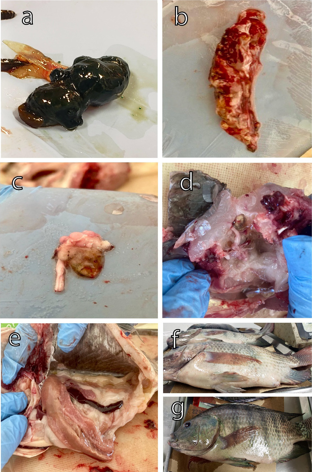
Outbreaks of S. agalactiae were detected in grow-out ponds in a Nile tilapia farm, reaching an 80% of prevalence, which resulted in high mortality and morbidity rates. Necropsies on the fish revealed classic signs of streptococcosis, including bilateral exophthalmia, fish exhibited protruding or swollen eyes (Figure 1a). Spleen with nodules typically showed focal areas of inflammation and bacterial proliferation within the spleen tissue (Figure 1b). The hemorrhagic brain was characterized by internal bleeding within the brain tissue (Figure 1c). Nervous cord with hyperplasia is characterized by the growth of nerve tissue, particularly in the spinal cord region, which can lead to neurological dysfunction (Figure 1d). Hepatomegaly and splenomegaly were observed post-mortem examination of affected fish (Figure 1e). Ascites accumulation of fluid was observed in the abdominal cavity, resulting in swelling or bloating of the abdomen in affected fish (Figure 1 f,g).

Figure 1
Figure 1.Clinical signs lesions caused by Streptococcus agalactiae.

a) Bilateral exophthalmia; b) Spleen with nodules; c) Hemorrhagic brain; d) Nervous cord with hyperplasia; e) Hepatomegaly and spleen congestor; f, g) Ascites.

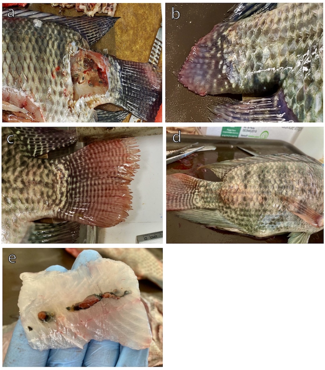
It is important to note that certain batches of fish did not exhibit any obvious disease signs before experiencing significant mortality. However, a high occurrence of secondary lesions (myositis and caudal fin deformities, spinal cord curvature, necrosis in and on the muscle) bleeding at the base of the fins, and tail fraying was observed during the necropsies of affected organisms, indicating a chronic pattern (Figure 2a-e). Additionally, some fish exhibited significant hemorrhaging on the lips and mouth, severe congestion, and congested kidneys. Upon dissection of the fish, we observed the presence of bloody, hemorrhagic ascitic fluid in the abdominal cavity, which appeared dark in color (Figure 2d).

Figure 2
Figure 2.Clinical signs lesions caused by Streptococcus agalactiae.

a) Muscle necrosis; b) Fraying of the tail; c) Slight deformation of the caudal fin; d) Hemorrhagic caudal fin; e) Muscle abscess.

Histopathological examination

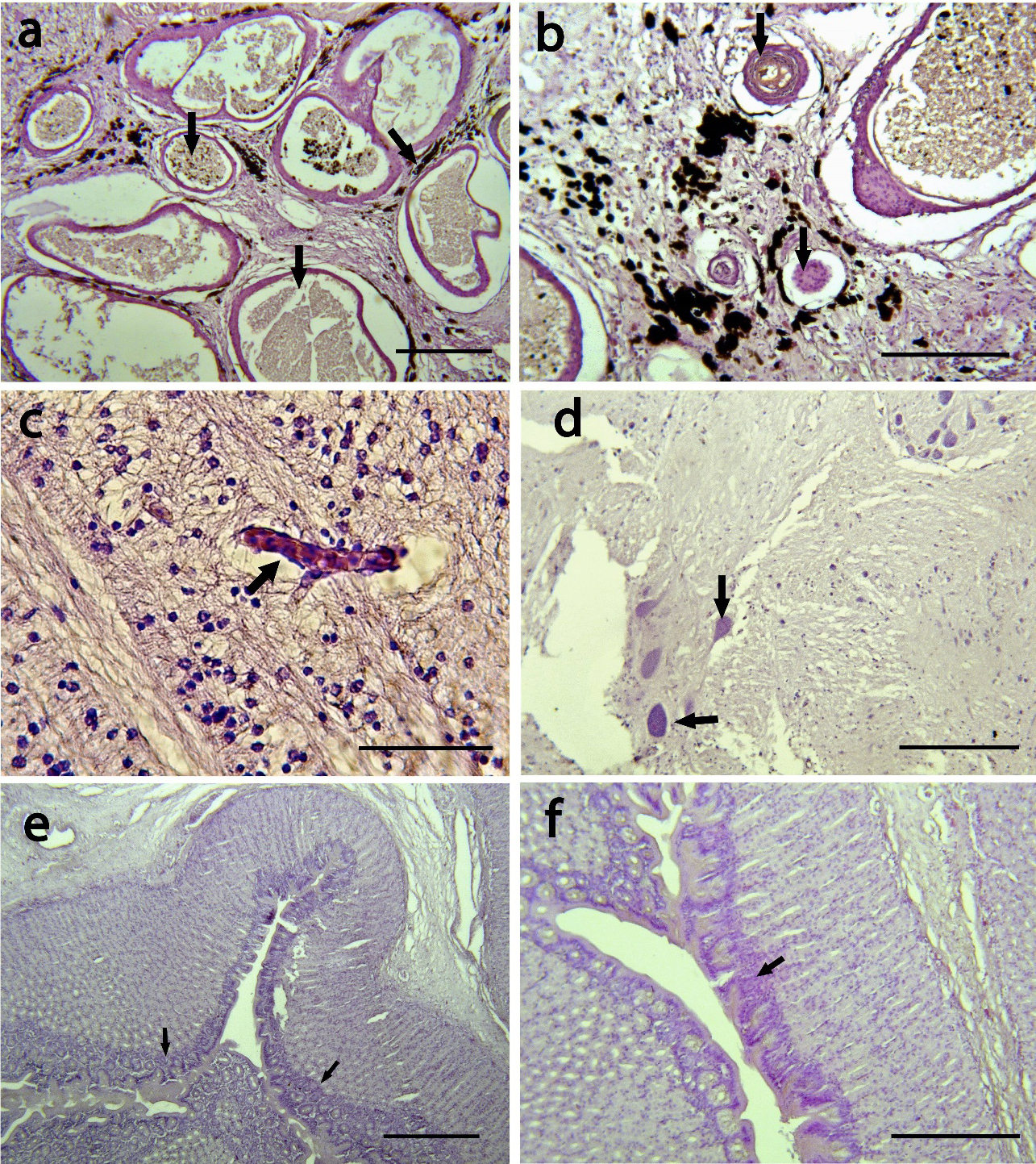
The brain, liver, kidney, muscle, spleen, stomach, and intestine of Nile tilapia infected with S. agalactiae displayed several degrees of histological alterations (Table 2). The histopathological examination of the liver tissue revealed the presence of melanomacrophage pigment (indicated by an arrow in Figure 3a), hemorrhage, and substantial eosinophilic infiltration surrounding the blood vessels. Additionally, there was evidence of edematous perivasculitis that progressed to congestion, thrombosis, and necrosis in the portal blood vessels (Figure 3a). Furthermore, vacuolation in hepatocytes was observed, along with lipid accumulation (indicated by an arrow in Figure 3b).

In the kidney, lesions were observed in both the excretory and hematopoietic tissues, accompanied by melanomacrophage activation (indicated by an arrow in Figure 3c). Additionally, there was evidence of lymphocyte depletion, as well as hemorrhage and necrosis (indicated by an arrow in Figure 3c, d).

In the spleen of the Nile tilapia, vascular congestion surrounded by inflammatory cell infiltration was observed (Figure 3e). Additionally, thrombosis of the splenic blood vessels was evident, along with an increase in the melanomacrophage center in the splenic parenchyma (Figure 3f).

Figure 3
Figure 3.Histopathology damage of liver, kidney and spleen affected Streptococcus agalactiae.

a) Liver hemorrhage (arrow) with blood vessels with large number of eosinophil infiltration around liver (arrow); b) Liver with vacuolation of hepatocytes (arrow); c,d) Kidney showing activation of melanomacrophages (arrow) with lymphocytic depletion (arrow); e,f) Spleen presented vascular congestion surrounded by inflammatory cell infiltration.

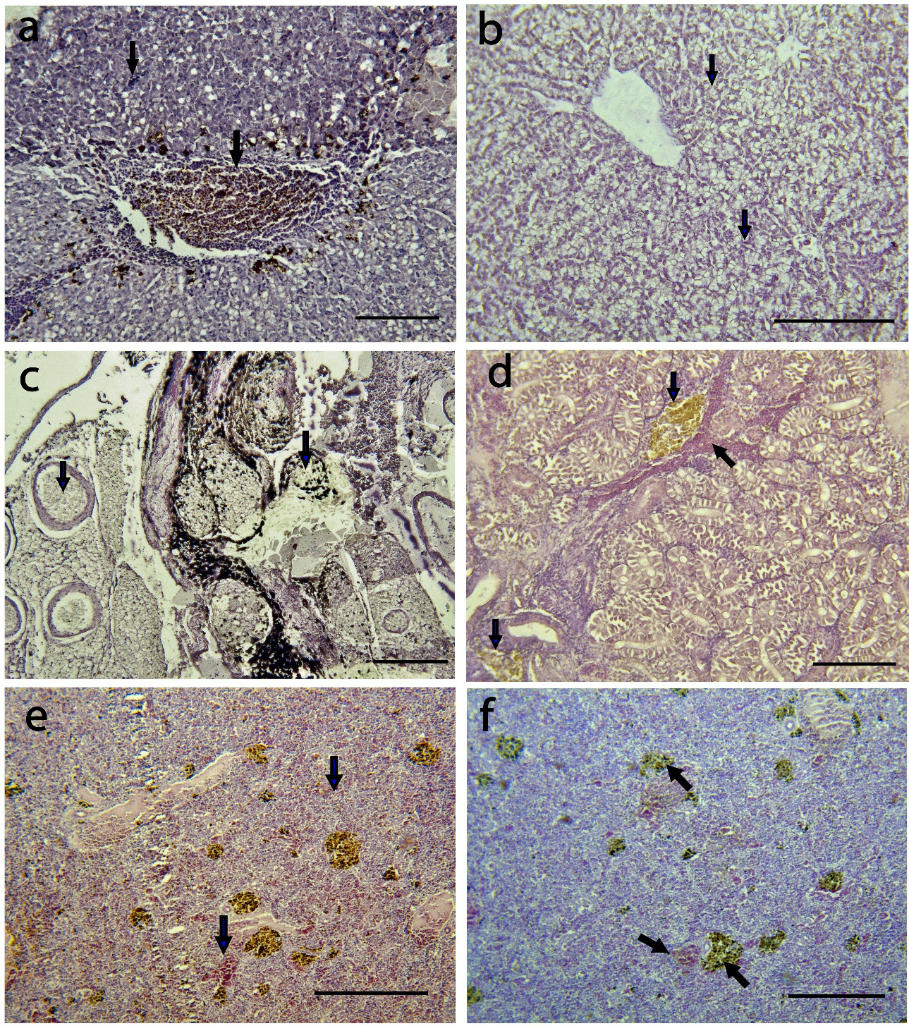
The muscle displays pigmentation attributed to the presence of melanomacrophages and the formation of granulomas (indicated by an arrow in Figure 4a, b). The brain exhibits signs of encephalitis along with vascular congestion (indicated by an arrow in Figure 4c, d). In the intestine mucosa, showed moderate eosinophilic infiltrates, necrosis (indicated by an arrow in Figure 4e), and inflammatory cell aggregation (indicated by an arrow in Figure 4f).

Figure 4
Figure 4.Histopathology damage of muscle, brain and intestine affected Streptococcus agalactiae.

a,b) muscle associated with the presence of melanomacrophages (arrow) and granuloma formation (arrow); c,d) brain encephalitis with vascular congestion (arrow); e,f) Intestine mucosa showed epithelia necrosis, and inflammatory cell aggregation.

Histopathological examination of the organ tissues revealed morphological changes in the tissue of all fish. The organs with most severe damage were liver, kidney and spleen (Table 2). The most prevalent histopathological damage reported in the fish was melanomacrophage pigment, granulomas, vacuolar degeneration, hemorrhage (Table 2). Melanomacrophage pigment and granulomas had severe damage with an alteration mean values of 2.81±1.90 and 2.3±1.85 respectively. While lymphocyte infiltration showed mild damage with an AMV of 0.24±0.19 (Table 2).

Table 2.Prevalence and alteration mean values (AMV) of damage histopathologic with Streptococcus agalactiae in Nile tilapia collected in an intensive farm from Campeche, Mexico
Liver Kidney Spleen Muscle Brain Intestine MVA (± SD)
Histopathological lesions
(n=30 fish)
Melanomacrophage pigment 70 67 85 78 2.81±1.90
Hemorrhage 50 65 45 40 40 35 1.80±1.02
Eedema 25 5 10 0.62±0.90
Eosinophil infiltration 8 10 15 5 8 3 0.75±097
Congestion 25 30 15 10 20 10 1.12±0.91
Focal necrosis 35 40 25 45 1.32±1.42
Vacuolar degeneration 75 45 30 1.23±0.95
Thrombosis 10 5 10 0.72±0.90
Granulomas 40 55 45 70 2.3±1.85
Vascular congestion 5 2 10 20 1±0.87
Inflamammation 45 30 35 25 37 40 1.43±1.05
Neutrophyl 3 5 5 0.50±0.30
Lymphocyte infiltration 10 5 5 0.24±0.19

Economic losses analysis by Streptococcus agalactiae

In the first three months, tilapia harvests display an increasing trend, reaching maximum values in February at 80 tons and in March at 79 tons. Following these months, production declines by approximately 50%, reaching its lowest levels in November and December, with 16 tons each month (Figure 5a). The quantity of fish loss due to muscular necrosis exhibits a similar trend at the beginning of the year, with a maximum loss of 5,076 kg recorded in January and 4,302 kg in October (Figure 5a). Prevalence remains stable from January to June, reaching its highest rates in August and October, at 88% and 89%, respectively (Figure 5b).

Figure 5
Figure 5.a) Production (left axis) and losses by Streptococcus agalactiae (right axis) in a Nile tilapia farm in Campeche, losses; b) Prevalence in total harvest.

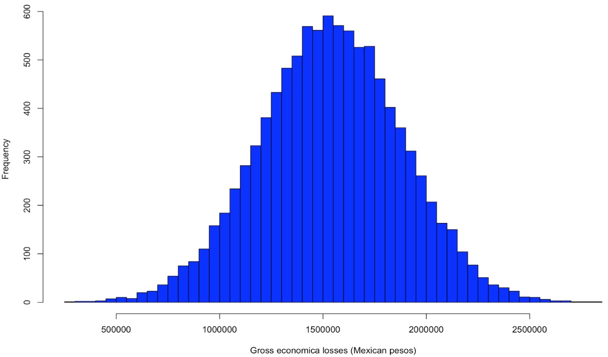
Based on the Monte Carlo simulation the total annual gross economic loss due to muscle necrosis often referred to by producers as “fish with lumps,” averaged $1,537,671 ± 34,2587 Mexican pesos (76464.53 ± 17036 USD). The Monte Carlo model estimated the minimum and maximum losses ranging from 299,302 to 2,824,909 Mexican pesos (14,883 – 140,475 USD) (Figure 6).

Figure 6
Figure 6.Total annual gross economic loss based on Monte Carlo simulation.

Discussion

This study represents the first report detailing the economic losses and describing the lesions and clinical changes caused by S. agalactiae infection in an intensive farm in Campeche. The aquaculture health committee of the state of Campeche (CESAICAM) reported the presence of S. agalactiae for the first time in 2001, specifically in the northeast of Ciudad del Carmen. Additionally, the current paper represents the first record of S. agalactiae in recirculation systems in the central region of Campeche state.

In Mexico, there is currently no available data to ascertain when or how Streptococcus spp. was introduced. The situation appears concerning, as these bacteria have become well-established and are widely distributed across nearly ten states in the Mexican Republic (Table 1). The economic impact of streptococcosis at the national level could be significant, as evidenced by the mortality rates, product loss, and high treatment costs observed on the studied farm.

The most prevalent clinical signs during the acute phase of the infection in production systems include swimming abnormalities, anorexia, corneal opacity, unilateral or bilateral exophthalmia, skin ulcers, abscesses, and hemorrhages on the skin, jaw, and at the base of the caudal peduncle (Anshary et al. 2014; Iregui et al. 2014; Sun, Fang, Ke, et al. 2016; Yi, Wang, Li, et al. 2019; Zhang 2021). Additionally, abdominal distension, spinal cord curvature, hemorrhagic ascites, and poor body condition are observed, all of which are attributed to bacteria of the Streptococcus genus (Zhang 2021). Hemorrhagic and inflammatory reactions are commonly observed in affected organs such as the eye, liver, spleen, kidney, and brain (Rahmatullah, Ariff, Kahieshesfandiari, et al. 2017; Ortega, García, Irgang, et al. 2018; Alazab, Sadat, and Younis 2022). Similarly, Hernández, Figueroa, and Iregui (2009) demonstrated that S. agalactiae exhibits a preference for infecting specific organs, as the brain, eyes, and heart (with 71, 44, and 37 %, of frequency respectively). M. Hernández-Hernández et al. (2023) observed lesions that included exophthalmia, ascites, and granulomas at the base of the tail with pus accumulations.

However, subclinical, and chronic infections in adult Nile tilapia have been consistently detected. For instance, yellow or dark red nodules in the musculature near the vertebrae, myositis, and deformities of the caudal fin are evident (Sun, Fang, Ke, et al. 2016; Zhang et al. 2021). These subclinical findings likely indicate an adaptation by the bacterium to environmental factors characteristic of production systems, such as osmotic pressure, acidity, elevated levels of nitrogenous waste, and indiscriminate antibiotic use (Evans et al. 2009).

The histopathological findings were similar as reported in recent studies that indicated that infected fish with streptococcal disease exhibited multiple pathological conditions of tissues internal organ mainly the presence de granulomatous reaction, melanomacrophage pigment, vacuolar degeneration, granular eosinophilic cells with evident bacterial bodies (Alsaid, Hassan, Mohd, et al. 2014; He, Huang, Wang, et al. 2017; Laith, Ambak, Hassan, et al. 2017). Melanomacrophage were the most apparent damage histologic affected in organs in livers and spleens of fish infected with bacteria. The melanomacrophages have the function of eliminating, detoxifying, or recycling waste materials from erythrocytic and cellular metabolic processes. Additionally, melanomacrophages play a crucial role in the response to foreign entities, such as infectious agents or immune conditions (Franco-Belussi and de Oliveira 2016).

Several risk factors have been associated with the occurrence of streptococcosis outbreaks (Zhang 2021). High temperatures can enhance the pathogenicity of S. agalactiae in Nile tilapia. Zhao, Zou, Han, et al. (2023) reported higher mortality rates (80 %) at temperatures of 31°C compared to those exposed to 22°C (10 %) in fish infected with S. agalactiae. M. J. Hernández-Hernández et al. (2012) reported an 80% prevalence of Streptococcus spp. in 10 production units at Malpaso Dam, Chiapas. Additionally, a mortality rate of 50%, along with clinical signs in 60% of the cases. Also, an association was identified between low oxygen levels ranging from 5 to 6.5 mg/l and temperatures exceeding 31°C with the incidence of Streptococcus spp. Poor sanitary management was observed on the farm where the fish were collected. This included elevated temperatures, high stocking densities, wastewater channels situated close to the farm, dead fish, insufficient disinfection of fishing equipment and ponds, and inadequate water changes (Figure 7a-d).

Figure 7
Figure 7.Management practices in an ­­­intensive farm located in Campeche.

a) High fish densities; b) Dead fish in the production ponds; c, d) Water channel with untreated biological waste.

In Mexican aquaculture, suboptimal management practices within production systems are common and may contribute to the increased presence and spread of pathogens. It has been consistently observed that the movement of fish for commercial purposes without the implementation of proper sanitary measures stands as the primary risk factor for the dissemination and introduction of both S. iniae and S. agalactiae (Amal et al. 2013; Paredes-Trujillo and Mendoza-carranza 2022). Notably, in Mexico, it is common practice to sell fingerlings without the issuance of sanitary certificates, coupled with the inadequate sanitary measures employed by many production farms. This combination promotes recurrent reinfections of the disease. It is imperative to identify and understand the routes and dynamics through which Streptococcus spp. enters in Nile tilapia farms.

Horizontal transmission appears to be the prevailing mechanism for the dissemination of Streptococcus spp. (Kim et al. 2007; Amal et al. 2013). It is well-established that certain elements, such as contaminated water, infected fingerlings, direct contact with deceased fish, high fish stocking densities, and fluctuations in environmental variables (elevated salinity, low dissolved oxygen levels, and high nitrogenous waste concentrations), can heighten the susceptibility of Nile tilapia to infection (Amal et al. 2013; Zamri-Saad et al. 2014). Additionally, infections can result from the excretions of diseased fish and cannibalism (Kim et al. 2007). Furthermore, there is substantial evidence supporting the possibility of vertical transmission as a route for this bacterial disease to infect the offspring (Pradeep, Suebsing, Sirthammajak, et al. 2016). Nevertheless, it remains essential to continually assess the specific risk factors to each production unit to confirm these introduction pathways, with a particular focus on the context of Mexico.

Mexico is situated in the subtropical and tropical zones, where temperature plays a crucial role in infection dynamics. It is widely recognized that elevated temperatures between 30 and 35 °C are closely linked to the occurrence of S. agalactiae outbreaks in Nile tilapia (Evans et al. 2009; Mian et al. 2009; Amal et al. 2013; Paredes-Trujillo and Mendoza-carranza 2022). Poor management practices, such as low water replacement and uncontrolled food supplies, can lead to increased nitrogenous waste, particularly ammonia. Elevated ammonia levels (0.24 mg/l, >10 mg/l, 0.3 mg/l) have been linked to Streptococcus agalactiae, which may, in turn, be associated with weakened host defenses due to stress (Evans et al. 2006). Water quality plays a pivotal role in maintaining the well-being of cultured fish populations, enhancing biomass production, and supporting their natural resistance to diseases (Amal et al. 2013). Variations in physicochemical parameters and contamination resulting from suboptimal management practices and inadequate water control can create conditions that favor pathogen survival, consequently affecting the immune response of fish (Amal et al. 2013).

The sanitary management of Nile tilapia is fundamental to the production process and must be conducted according to standardized procedures throughout all phases, beginning with the management of fingerlings and breeders (including genetic improvement and reproduction), as well as ensuring water quality and sanitary practices. Designing an effective biosecurity program requires a thorough risk analysis of fish diseases. This process includes identifying potential sources of pathogens, understanding transmission routes, and evaluating the susceptibility of the cultured species. The concept of biosecurity measures in Nile tilapia culture in Mexico has not yet been fully implemented. An ongoing concern in Mexican aquaculture systems is the excessive use of antibiotics, which are often administered by producers as the first line of treatment for Streptococcus spp. infections, without prior and accurate laboratory diagnosis. Precise identification of the bacterium is essential for determining the correct dosage and treatment. Improperly administered antibiotics not only pose significant risks of antibiotic residues but also contribute to the development of antibiotic resistance in the bacteria (Amal et al. 2013).

Conclusions

Streptococcosis in Nile tilapia represents a significant challenge in Mexico, marked by a lack of comprehensive historical data and an incomplete understanding of its current dynamics. There is limited documentation regarding the presence of Streptococcus spp. in major Nile tilapia production regions.

The economic losses linked to streptococcosis on Nile tilapia farms in Campeche raise serious concerns for farmers. Histopathological damage, chronic infections, and targeted mortalities are notable characteristics of the disease, further alarming the overall health of the fish population.

Given this context, it is imperative to intensify scientific research to gain precise insights into the strains present and their transmission dynamics, as well as to propose effective prevention and eradication measures. Additionally, urgent educational initiatives must be undertaken to inform producers about disease prevention, control, and identification. It is also essential to establish early intervention protocols involving producers and collaboration with national and state aquaculture health organizations.


Acknowledgments

This research was supported by the Consejo Nacional de Ciencia y Tecnología (CONAHCYT) under posdoctoral grant [253392]. Thanks are extended to the Laboratorio de Sanidad Acuícola, Instituto de Ecología, Pesquerías y Oceanografía (EPOMEX) de la Universidad Autónoma de Campeche. Thanks to Rodolfo Rodríguez del Río for facilitating the acquisition of the biomass data, Ana Delia Cu Escamilla, Ricardo Ávila Castillo. Thanks to owner of the Nile tilapia farm in Campeche for providing biomass loss data, as well as for sample collection.

Funding statement

This research was funded by Universidad Autónoma de Campeche (www.uacam.mx), with internal funds. The funder had no role in study design, data collection and analysis, decision to publish, or preparation of the manuscript.

Conflicts of interest

The authors have no conflict of interest to declare regarding this publication.

Ethical statement

We confirm that the animals in this study do not suffer any animal abuse. Approved by the research ethics committee of ECOSUR (Comité de Ética para la Investigación de El Colegio de la Frontera Sur) official letter number CEI-05-10-2023 that followed the International Guiding Principles for Biomedical Research Involving Animals.

Author contributions

Conceptualization: A Paredes-Trujillo.
Data curation: A, Paredes-Trujillo, M, Mendoza-Carranza.
Formal analysis: A, Paredes-Trujillo, M, Mendoza-Carranza.
Funding acquisition: A, Paredes-Trujillo.
Investigation: A, Paredes-Trujillo, M, Mendoza-Carranza.
Methodology: A, Paredes-Trujillo, M, Mendoza-Carranza.
Resources: A, Paredes-Trujillo, M, Mendoza-Carranza.
Supervision: A, Paredes-Trujillo, M, Mendoza-Carranza.
Writing – original draft: A, Paredes-Trujillo.
Writing – review and editing: M, Mendoza-Carranza.

Submitted: October 21, 2024 CEST

Accepted: December 21, 2024 CEST

References

Alazab, A., A. Sadat, and G. Younis. 2022. “Prevalence, Antimicrobial Susceptibility, and Genotyping of Streptococcus Agalactiae in Nile Tilapia Fish (Oreochromis ⁠niloticus).” Journal of Advanced Veterinary and Animal Research 9 (1): 95. https:/​/​doi.org/​10.5455/​javar.2022.i573.
Google Scholar
Alcántara-Jauregui, F. M., B. Valladares-Carranza, and C. Ortega. 2022. “Enfermedades bacterianas y sus agentes etiológicos identificados en peces de México - Una Revisión.” Revista MVZ Cordoba 27 (2): e2387. https:/​/​doi.org/​10.21897/​rmvz.2387.
Google Scholar
Alsaid, M., H. J. Hassan, D. Mohd, et al. 2014. “Pathologic Findings of Experimental Streptococcus Agalactiae Infection in Red Hyprid Nile Tilapia (Oreochromis ⁠sp.).” International Journal of Advances in Chemical Engineering 1 (1): 93–96. https:/​/​doi.org/​10.15242/​IJACEBS.C1213075.
Google Scholar
Amal, M. N. A., M. Zamri-Saad, A. Siti-Zahrah, and A. R. Zulkafli. 2013. “Transmission of Streptococcus Agalactiae from a Hatchery into a Newly Established Red Hybrid Nile Tilapia, Oreochromis ⁠niloticus (L.) × Oreochromis ⁠mossambicus (Peters), Farm.” Journal of Fish Diseases 36 (8): 735–39. https:/​/​doi.org/​10.1111/​jfd.12056.
Google Scholar
Anshary, H., R. A. Kurniawan, S. Sriwulan, R. Ramli, and D. V. Baxa. 2014. “Isolation and Molecular Identification of the Etiological Agents of Streptococcosis in Nile Nile Tilapia (Oreochromis ⁠niloticus) Cultured in Net Cages in Lake Sentani, Papua, Indonesia.” SpringerPlus 3 (1): 627. https:/​/​doi.org/​10.1186/​2193-1801-3-627.
Google Scholar
Avila-Castillo, R. 2024. “Identificación y Distribución de bacterias de los géneros: Francisella y Streptoccocus en Nile tilapias Oreochromis niloticus cultivadas en granjas intensivas y semi-intensivas en el estado de Campeche.” Universidad Autonoma de Campeche.
Bezerra, G. A., D. C. Pires, A. L. Watanabe, C. C. B. Neto, A. R. P. Simões, and H. Hisano. 2022. “Economic Feasibility and Risk Analysis of Nile Tilapia Juveniles Reared in a Biofloc Technology System.” Research Square. https:/​/​doi.org/​10.21203/​rs.3.rs-1922931/​v1.
Google Scholar
Bush, A. O., K. D. Lafferty, J. M. Lotz, A. W. Shostak, et al. 1997. “Parasitology Meets Ecology on Its Own Terms: Margolis et al. Revisited.” Journal of Parasitology 83 (4): 575. https:/​/​doi.org/​10.2307/​3284227.
Google Scholar
Castro-Ortíz, A. 2020. “Identificación y caracterización de patógenos bacterianos aislados de Nile tilapias del Nilo (Oreochromis niloticus) cultivadas en la presa de El Gallo, Guerrero, Mexico.” Universidad Michoacana de San Nicolas de Hidalgo.
CESACH. 2020. “Programa de Trabajo Del Proyecto 2020 Peces Del Incentivo Prevención y Control de Enfermedades Acuícolas En El Estado de Chiapas, Del Programa de Sanidad e Inocuidad Agroalimentaria Ejercicio Fiscal 2020 Con Recursos de Origen Federal.” Comité de Sanidad Acuicola del Estado de Chiapas.
CESAICAM. 2021. “Programa de Trabajo Del Proyecto Peces Del Subcomponente Prevención y Control de Enfermedades En Organismos Acuícolas En El Estado de Campeche Del Programa de Sanidad e Inocuidad. Agroaumentaria Ejercicio Fiscal 2021 Con Recursos de Origel Federal.”
CESAJ. 2021. “Buenas Prácticas Acuícolas.” Comité Estatal de Sanidad e Inocuidad Acuícola de Jalisco CESAJ A.C.
Delannoy, C. M. J., H. Samai, and L. Labrie. 2021. “Streptococcus Agalactiae Serotype IV in Farmed Nile Tilapia.” Aquaculture 544:737033. https:/​/​doi.org/​10.1016/​j.aquaculture.2021.737033.
Google Scholar
Delannoy, C. M. J., R. N. Zadoks, F. A. Lainson, et al. 2012. “Draft Genome Sequence of a Nonhemolytic Fish-Pathogenic Streptococcus Agalactiae Strain.” Journal of Bacteriology 194 (22): 6341–42. https:/​/​doi.org/​10.1128/​JB.01552-12.
Google Scholar
Delphino, M., R. Joshi, and A. T. Alvarez. 2022. “Economic Appraisal of Using Genetics to Control Streptococcus Agalactiae in Nile Nile Tilapia under Cage and Pond Farming System in Malaysia.” Scientific Reports 12 (1): 8754. https:/​/​doi.org/​10.1038/​s41598-022-12649-9.
Google Scholar
Delphino, M., C. A. G. Leal, I. A. Gardner, et al. 2019. “Seasonal Dynamics of Bacterial Pathogens of Nile Nile Tilapia Farmed in a Brazilian Reservoir.” Aquaculture 498:100–108. https:/​/​doi.org/​10.1016/​j.aquaculture.2018.08.023.
Google Scholar
Evans, J. J., P. H. Klesius, P. M. Gilbert, et al. 2002. “Characterization of Beta-Haemolytic Group B Streptococcus ⁠agalactiae in Cultured Seabream, Sparus Auratus L., and Wild Mullet, Liza Klunzingeri (Day), in Kuwait.” Journal of Fish Diseases 25 (9): 505–13. https:/​/​doi.org/​10.1046/​j.1365-2761.2002.00392.x.
Google Scholar
Evans, J. J., P. H. Klesius, D. J. Pasnik, and J. F. Bohnsack. 2009. “Human Streptococcus Agalactiae Isolate in Nile Nile Tilapia (Oreochromis ⁠niloticus).” Emerging Infectious Diseases 15 (5): 774–76. https:/​/​doi.org/​10.3201/​eid1505.080222.
Google Scholar
Evans, J. J., D. J. Pasnik, G. C. Brill, and P. H. Klesius. 2006. “Un-Ionized Ammonia Exposure in Nile Nile Tilapia: Toxicity, Stress Response, and Susceptibility to Streptococcus ⁠agalactiae.” North American Journal of Aquaculture 68 (1): 23–33. https:/​/​doi.org/​10.1577/​A05-032.1.
Google Scholar
Fabiani, B. M., W. R. Boscolo, A. Feiden, O. Diemer, F. Bittencourt, and D. H. Neu. 2013. “Benzocaine and Eugenol as Anesthetics for Brycon Hilarii.” Acta Scientiarum Animal Science 35 (2). https:/​/​doi.org/​10.4025/​actascianimsci.v35i2.16644.
Google Scholar
FAO. 2020. El estado mundial de la pesca y la acuacultura 2020. Roma. https:/​/​sustainablefisheries-uw.org/​el-estado-mundial-de-la-pesca-y-la-acuicultura-2020.
Google Scholar
Franco-Belussi, L., and C. de Oliveira. 2016. “The Spleen of Physalaemus Nattereri (Amphibia: Anura): Morphology, Melanomacrophage Pigment Compounds and Responses to α-Melanocyte Stimulating Hormone.” Italian Journal Zoology 83 (3): 298–305. https:/​/​doi.org/​10.1080/​11250003.2016.1194488.
Google Scholar
He, Y., J. L. Huang, K. Y. Wang, et al. 2017. “Pathogenicity of Streptococcus ⁠agalactiae in Oreochromis ⁠niloticus.” Oncotarget 9 (1): 401–13.
Google Scholar
Hernández, E., J. Figueroa, and C. Iregui. 2009. “Streptococcosis on a Red Nile Tilapia, Oreochromis Sp., Farm: A Case Study.” Journal of Fish Diseases 32 (3): 247–52. https:/​/​doi.org/​10.1111/​j.1365-2761.2008.00981.x.
Google Scholar
Hernández-Hernández, M., L. García-Márquez, J. Gutiérrez-Jiménez, J. Feliciano-Guzmán, R. López-Santiz, and G. Bautista-Trujillo. 2023. “Oreochromis niloticus exhibe una prevalencia mayor de Streptococcus beta hemolítico cuando se mantienen en jaulas en comparación con estanques.” Abanico Veterinario 13:1–16. https:/​/​doi.org/​10.21929/​abavet2023.19.
Google Scholar
Hernández-Hernández, M. J., B. Gutiérrez-Jiménez, G. Ruiz-Sesma, and G. Bautista-Trujillo. 2012. “Oxígeno-temperatura en la incidencia de Streptococcus spp. en jaulas flotantes de Nile tilapia (Oreochromis niloticus) en Malpaso, Chiapas.” Revista Espacio I+D Innovación más Desarrollo 27:131–42.
Google Scholar
Huicab-Pech, Z. G., M. R. Castaneda-Chavez, and F. Lango-Reynoso. 2017. “Pathogenic Bacteria in Oreochromis ⁠Niloticus Var. Stirling Nile Tilapia Culture.” Journal of Fisheries and Aquatic Sciences 08 (02). https:/​/​doi.org/​10.4172/​2150-3508.1000197.
Google Scholar
Igeh, P. C., and A. Avenant-Oldewage. 2020. “Pathological Effects of Cichlidogyrus Philander Douëllou, 1993 (Monogenea, Ancyrocephalidae) on the Gills of Pseudocrenilabrus Philander (Weber, 1897) (Cichlidae).” Journal of Fish Diseases 43 (2): 177–84. https:/​/​doi.org/​10.1111/​jfd.13121.
Google Scholar
Instituto Nacional de Pesca. 2018. “Acuacultura Tilapia.” 2018. https:/​/​www.gob.mx/​inapesca/​acciones-y-programas/​acuacultura-tilapia.
Iregui, C., P. Barato, A. Rey, G. Vasquez, and N. Verjan. 2014. “Epidemiology of Streptococcus Agalactiae and Streptococcosis in Nile Tilapia Fish (Oreochromis Sp.).” Epidemiology I: Theory, Research and Practice, 251–68.
Google Scholar
Kim, J. H., D. K. Gomez, C. H. Choresca, and S. C. Park. 2007. “Detection of Major Bacterial and Viral Pathogens in Trash Fish Used to Feed Cultured Flounder in Korea.” Aquaculture 272 (1–4): 105–10. https:/​/​doi.org/​10.1016/​j.aquaculture.2007.09.008.
Google Scholar
Laith, A. A., M. A. Ambak, M. Hassan, et al. 2017. “Molecular Identification and Histopathological Study of Natural Streptococcus Agalactiae Infection in Hybrid Nile Tilapia (Oreochromis ⁠niloticus).” Veterinary World 10 (1): 101. https:/​/​doi.org/​10.14202/​vetworld.2017.101-111.
Google ScholarPubMed CentralPubMed
Leal, C. A. G., G. A. Queiroz, F. L. Pereira, G. C. Tavares, and H. C. P. Figueiredo. 2019. “Streptococcus Agalactiae Sequence Type 283 in Farmed Fish, Brazil.” Emerging Infectious Diseases 25 (4): 776–79. https:/​/​doi.org/​10.3201/​eid2504.180543.
Google Scholar
Li, K., L. Liu, J. H. Clausen, M. Lu, and A. Dalsgaard. 2016. “Management Measures to Control Diseases Reported by Nile Tilapia (Oreochromis Spp.) and Whiteleg Shrimp (Litopenaeus Vannamei) Farmers in Guangdong, China.” Aquaculture 457:91–99. https:/​/​doi.org/​10.1016/​j.aquaculture.2016.02.008.
Google Scholar
Liu, G., Z. Ye, D. Liu, et al. 2018. “Influence of Stocking Density on Growth, Digestive Enzyme Activities, Immune Responses, Antioxidant of Oreochromis ⁠niloticus Fingerlings in Biofloc Systems.” Fish & Shellfish Immunology 81:416–22. https:/​/​doi.org/​10.1016/​j.fsi.2018.07.047.
Google Scholar
Mian, G. F., D. T. Godoy, C. A. G. Leal, T. Y. Yuhara, G. M. Costa, and H. C. P. Figueiredo. 2009. “Aspects of the Natural History and Virulence of S. Agalactiae Infection in Nile Nile Tilapia.” Veterinary Microbiology 36 (1–2): 180–83. https:/​/​doi.org/​10.1016/​j.vetmic.2008.10.016.
Google Scholar
Mumford, S., J. Heidel, C. Smith, J. Morrison, B. MacConnell, and V. Blazer. 2007. Fish Histology and Histopathology. Amerika Serikat: US Fish and Wildlife National Conservation Training Center.
Google Scholar
Ortega, C., I. García, R. Irgang, et al. 2018. “First Identification and Characterization of Streptococcus Iniae Obtained from Nile Tilapia (Oreochromis Aureus) Farmed in Mexico.” Journal of Fish Diseases 41 (5): 773–82. https:/​/​doi.org/​10.1111/​jfd.12775.
Google Scholar
Paredes-Trujillo, A., and M. Mendoza-carranza. 2022. “A Systematic Review and Meta-Analysis of the Relationship between Farm Management, Water Quality and Pathogen Outbreaks in Nile Tilapia Culture.” Journal of Fish Diseases 45 (10): 1529–48. https:/​/​doi.org/​10.1111/​jfd.13679.
Google Scholar
Pineda-Hernández, I., L. Escobar Sarabia, F. Zavala Hernández, P. Carachure Olmos, G. Álvarez Díaz, and C. A. Rodríguez Torres. 2023. “La temperatura como un factor de crecimiento en juveniles de Nile tilapia roja en las condiciones climáticas de CD. Altamirano, Guerrero.” Ciencia Latina Revista Científica Multidisciplinar 1:10321–32. https:/​/​doi.org/​10.37811/​cl_rcm.v7i1.5216.doi:10.31644/​IMASD.27.2021.a08.
Google Scholar
Pradeep, P. J., R. Suebsing, S. Sirthammajak, et al. 2016. “Evidence of Vertical Transmission and Tissue Tropism of Streptococcosis from Naturally Infected Red Nile Tilapia (Oreochromis Spp.).” Aquaculture Reports 3:58–66. https:/​/​doi.org/​10.1016/​j.aqrep.2015.12.002.
Google Scholar
Pretto-Giordano, L. G., E. E. Müller, J. C. Freitas de, and V. G. da Silva. 2010. “Evaluation on the Pathogenesis of Streptococcus ⁠⁠agalactiae in Nile Nile Tilapia (Oreochromis ⁠niloticus).” Brazilian Archives of Biology and Technology 53 (1): 87–92. https:/​/​doi.org/​10.1590/​S1516-89132010000100011.
Google Scholar
Rahmatullah, M., M. Ariff, M. Kahieshesfandiari, et al. 2017. “Isolation and Pathogenicity of Streptococcus ⁠iniae in Cultured Red Hybrid Nile Tilapia in Malaysia.” Journal of Aquatic Animal Health 29 (4): 208–13. https:/​/​doi.org/​10.1080/​08997659.2017.1360411.
Google Scholar
Schwaiger, J., H. Ferling, U. Mallow, H. Wintermayr, and R.D. Negele. 2004. “Toxic Effects of the Non-Steroidal Anti-Inflammatory Drug Diclofenac.” Aquatic Toxicology 68 (2): 141–50. https:/​/​doi.org/​10.1016/​j.aquatox.2004.03.014.
Google Scholar
Sheehan, B., Y. S. Lee, E. S. Wong, et al. 2009. “Aquavac Strep Sa: Una novedosa vacuna para el control de las infecciones causadas por Streptococcus agalactiae Biotipo 2 en Nile tilapia de granja.” In En memorias: Manejo de Streptococcus en peces de aguas cálidas., 21–26.
Google Scholar
Soto, R. S. 2009. Investigación de Presas En Sinaloa. Calidad Del Agua y Bacterias Presentes En Nile tilapia Cultivada. Mazatlán.
Google Scholar
Soto-Rodríguez, S., S. A. Lopez, and K. G. A. Rendon. 2023. “Prevalence of Pathogenic Bacteria Detected by qPCR from Cultured Nile Nile Tilapia (Oreochromis ⁠niloticus Linnaeus, 1758) in Southwest Mexico.” BioRxiv, 6. https:/​/​doi.org/​10.1101/​2023.06.27.546743.
Google Scholar
Steckert, L. D., L. Cardoso, G. T. Jerônimo, S. B. Pádua, and M. L. Martins. 2018. “Investigation of Farmed Nile Nile Tilapia Health through Histopathology.” Aquac. 486:161–69. https:/​/​doi.org/​10.1016/​j.aquaculture.2017.12.021.
Google Scholar
Suanyuk, N., F. Kong, D. Ko, G. L. Gilbert, and K. Supamattaya. 2008. “Occurrence of Rare Genotypes of Streptococcus Agalactiae in Cultured Red Nile Tilapia Oreochromis Sp. and Nile Nile Tilapia O. Niloticus in Thailand—Relationship to Human Isolates?” Aquac. 284 (1–4): 35–40. https:/​/​doi.org/​10.1016/​j.aquaculture.2008.07.034.
Google Scholar
Sun, J., W. Fang, B. Ke, et al. 2016. “Inapparent Streptococcus Agalactiae Infection in Adult/Commercial Nile Tilapia.” Sci. Rep. 6 (1): 26319. https:/​/​doi.org/​10.1038/​srep26319.
Google Scholar
Valenzuela-Armenta, J. A., S. P. Díaz-Camacho, J. A. Cabanillas-Ramos, et al. 2018. “Microbiological Analysis of Nile Tilapia and Water in Aquaculture Farms from Sinaloa.” Biotecnia 20 (1): 20–26. https:/​/​doi.org/​10.18633/​biotecnia.v20i1.525.
Google Scholar
Ye, X., J. Li, M. Lu, et al. 2011. “Identification and Molecular Typing of Streptococcus Agalactiae Isolated from Pond-Cultured Nile Tilapia in China.” Fisheries Science 77 (4): 623–32. https:/​/​doi.org/​10.1007/​s12562-011-0365-4.
Google Scholar
Yi, M., M. Wang, Z. Li, et al. 2019. “An Investigation into the Effects of Streptococcus ⁠agalactiae on the 5-HT System and the Behavior of GIFT Nile Tilapia (Oreochromis ⁠niloticus).” Aquac. Rep. 15:100232. https:/​/​doi.org/​10.1016/​j.aqrep.2019.100232.
Google Scholar
Zamri-Saad, M., M. N. A. Amal, A. Siti-Zahrah, and A. R. Zulkafli. 2014. “Control and Prevention of Streptococcosis in Cultured Nile Tilapia in Malaysia: A Review.” Pertanika J. Trop. Agric. Sci. 37 (4): 20.
Google Scholar
Zhang, Z. 2021. “Research Advances on Nile Tilapia Streptococcosis.” Pathogens 10 (5): 558. https:/​/​doi.org/​10.3390/​pathogens10050558.
Google Scholar
Zhang, Z., F. Zhang, J. Du, D. Chen, and W. Zhang. 2021. “Impacts of Land Use at Multiple Buffer Scales on Seasonal Water Quality in a Reticular River Network Area.” PLOS ONE 16 (1). https:/​/​doi.org/​10.1371/​journal.pone.0244606.
Google Scholar
Zhao, Z., Q. Zou, S. Han, et al. 2023. “Omics Analysis Revealed the Possible Mechanism of Streptococcus Disease Outbreak in Nile Tilapia under High Temperature.” Fish Shellfish Immunol. 134:108639. https:/​/​doi.org/​10.1016/​j.fsi.2023.108639.
Google Scholar
Zheng, Y., W. Wu, G. Hu, et al. 2018. “Hepatic Transcriptome Analysis of Juvenile GIFT Nile Tilapia (Oreochromis ⁠niloticus), Fed Diets Supplemented with Different Concentrations of Resveratrol.” Ecotoxicol. Environ. Saf. 147:447–54. https:/​/​doi.org/​10.1016/​j.ecoenv.2017.08.006.
Google Scholar

This website uses cookies

We use cookies to enhance your experience and support COUNTER Metrics for transparent reporting of readership statistics. Cookie data is not sold to third parties or used for marketing purposes.

Powered by Scholastica, the modern academic journal management system