
Introduction
A neoplasm is an abnormal mass of tissue that is not coordinated with the growth of normal tissue and exceeds the growth of the normal tissues; it may be benign or malignant. The benign one is a confined development of well-structured, non-invasive tissue; its growth occurs through expansion, and it can exert pressure on neighboring healthy tissues. Generally, benign tumors expand at a slow rate and are typically not life-threatening unless they disrupt essential functions of the body (Gomes 2022). Contrarily, the malignant neoplasm (cancer) is life-threatening and recently defined as unregulated growth of altered cells influenced by evolution through natural selection and has a character of metastasis or distance spread to different parts of the body other than its original site (Brown et al. 2023). Localized invasion of surrounding tissues, anaplasia, dysplasia, cellular polymorphism, and atypical mitosis, together with other criteria of malignancy, are the additional behavioral and cellular characteristics of the malignant neoplasms (Baba and Catoi 2007).
Fish tumors received attention, especially those with a relationship to marine pollution (Sindermann 1976, 1979, 1990; Stich et al. 1977; Overstreet 1988; Fournie and Vogelbein 1994). However, fish are good bioindicators for environmental pollution because of their relatively long lifespans, and they are especially vulnerable to water contamination from the environment (Morado, Araújo, and Gomes 2017).
Jubail Province is a heavily industrialized region on the Arabian Gulf in the eastern region of Saudi Arabia, with the potential of environmental pollution (Jawad and Ibrahim 2021). Many industrial contaminants are known to be teratogenic as well as mutagens and carcinogens (Sindermann 1990). Several classes of chemical carcinogens have been shown to induce neoplasia in laboratory fishes with polycyclic aromatic hydrocarbons (PHAs) (Oishi, Yamazaki, and Harada 1976; Watermann, Dethlefsen, and Happenheit 1982). Different fish species could be affected by different types of neoplasms originating from different tissues, either epithelial or mesenchymal, where spindle cell tumors are common spontaneous cutaneous neoplasms observed in fish, and it was noticed in many fish species (Groff 2004). Fibromas are benign tumors composed of bundles of spindle-shaped cells having a fibrocytic or fibroblastic appearance arranged in a whirling pattern along with abundant collagen (Handharyani et al. 2008). In different animal species other than teleosts, the neoplastic overgrowth may be originate from more than one tissue of origin: fibrous and bony tissues, with the end result of fibro-osteomas (Soltero-Rivera et al. 2015; Nottle et al. 2021; Razmyar et al. 2008). In contrast to fibromas in different animal species, including fishes, the available literature about fibro-osteomas in fishes is scarce. On the other side, fibromas were recorded in many ornamental fishes, such as goldfish (Shokrpoor et al. 2016; Kamalesh, Chandy, and Bedre 2021); angelfish (Grosset, ME, and Groff 2016); white skirt tetra (Shalizar-Jalali et al. 2020); parrotfish (Grizzle and Williams 1983); and anciently in some edible fishes, such as trout (kreyberg 1937); major carp (Pillai and Thampy 1990); and recently in largescale mullet (Planiliza macrolepis) (Mariasingrayan, Danaraj, Dinh-Hung, et al. 2024).
To the best of our knowledge, there were few previous studies on inconsiderable details, especially on fibromas and fibro-osteomas found in the commercial fish species inhabiting the water of the Arabian Gulf in the Jubail region. During the course of investigation for fish health at El- Jubial fish markets, remarkable growths were found on the external surfaces of some edible fishes (four fish species) at different sites on their bodies, so the present study was undertaken to describe the gross morphology and associated histopathological lesions of these neoplastic masses, their distribution (positioning) on the body of the affected fishes, and to discuss the potential impact of the anthropogenic activities and pollution on the occurrence of fish neoplasms at Jubial Province.
Materials and Methods
Fish samples
The present study was carried out for two years during the veterinary investigation of fish health conditions at El-Jubial Fish market, the main fish landing center station at the marine fisheries port from the coastal waters of the Jubial province (27’°0’28’‘N, 49° °39’36’'E). Fishes showing the neoplastic lesions were identified according to Carpenter et al. (1997). The total length and weight of each fish were measured to the nearest cm and gm or kg. The body surface, fins and head of each fish were inspected both visually and by palpation by hand, together with internal post-mortem examination for any neoplastic masses in the internal organs. A remarkable single abnormal growth was observed on each of the four examined commercial fish species. In 250 examined Bartail flathead, two fish showed a rounded, well-circumscribed neoplastic overgrowth; one of them was on the head region, and it was firmly attached to the lower jaw (the fish was 35 cm in length and 1.5 kg in weight); in the second affected fish of this species, the neoplasm was hard and attached to the dorsal fin. In the spangled emperor, one out of 300 examined fish showed a tumor mass at the posterior end of the left side of the operculum; the affected fish sample was 55 cm in length and 2.250 kg in weight. A neoplastic overgrowth in the areolated grouper was observed as a swelling underneath the muscular tissue in the ventral region of the fish (one out of 200 examined fish), near the caudal peduncle (the fish was 30 cm in length and 2.75 kg in weight). In Javan flounder, 150 fish were examined, and the neoplasm was found in one case at the ventral aspect of the fish near to the dorsal fin, and the fish was 20 cm in length and 280 g in weight.
For histopathological examination
A part of the abnormal growths was fixed in Bouin’s solution for 48 hr. and processed for routine paraffin histology. Sections were cut at 5 µm and stained with Harris, hematoxylin, and eosin (Luna 1968). Tumors were examined under Olympus BX50 and its associated software. Special histochemical Masson’s trichrome staining (MT stain) was performed to demonstrate collagen fiber in the neoplastic tissue and to confirm bony tissue proliferation (differentiate newly formed and old bones) (Lim, Lee, Yun, et al. 2013).
Results
Gross lesions
Tumor occurrence
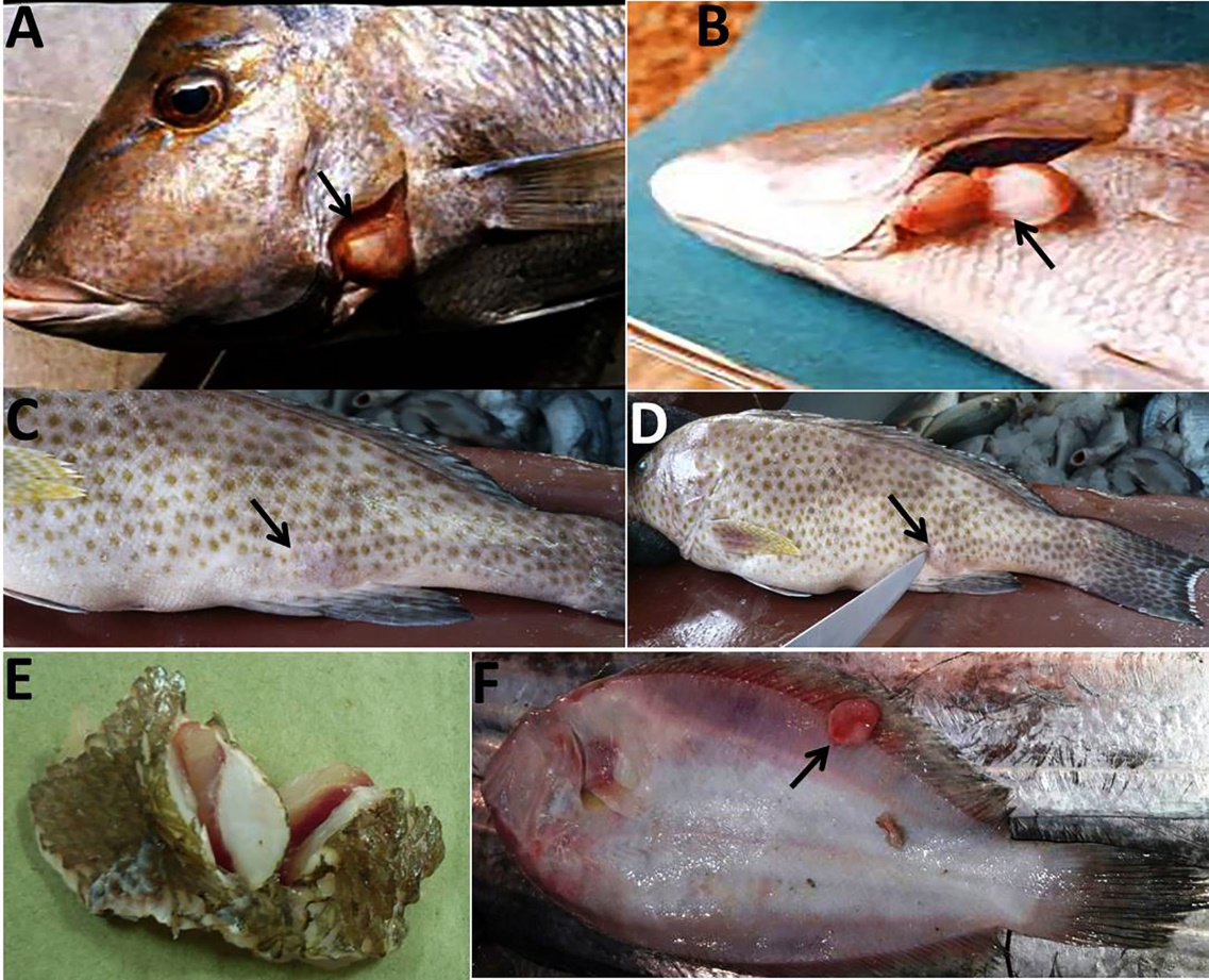
A single neoplastic tissue mass was observed in each of the following four fish species: the neoplastic overgrowth of one fish, Bartail flathead observed externally on the head region (Fig. 1A), where the tumor mass appeared well circumscribed and brownish, related to the skin coloration of this fish species. It was about 2.5 cm in diameter and attached to the anterior aspect of the lower jaw of the mouth opening (Fig. 1B). The tumor mass was large, round and forming a constriction ring at the site of its attachment to the skin (Fig. 1C). In the cut section, the tumor was fleshy in consistency, white in appearance, and covered with intact skin (Fig. 1D). In another fish, the Bartail flathead, the tumor was observed as a focal pedunculated, round, very hard swelling on the dorsal fin of the examined fish. (Fig. 1E); the neoplastic mass measured 0.5 cm and extended bilaterally on the dorsal fin (Fig. 1F).
_observed_externally_on_the_head_region_of_bartail.jpeg)
In Spangled emperor, the tumor mass was observed at the posterior end of the left side of the operculum (Fig. 2A), and it was well circumscribed in its shape (20 mm in diameter) and firm in consistency, and in the cut section , the color appeared white and avascular (Fig. 2B).
In Areolated grouper, the neoplasm was observed as a fleshy, nodular, and swollen mass deep in the lateral aspect of the muscular tissue near the anal fin (Fig. 2C&D). In the cut section, it appeared pale and well circumscribed, and it was about 1.5 cm in diameter (Fig. 2E). In Javan flounder, the neoplasm was round, fleshy, soft in consistency, and white in cut section (Fig. 2F).
In the necropsy examination, there were no gross lesions for any neoplastic overgrowth, where all the internal organs were apparently normal.
_observed_in_spangled_emperor_showing_**(a)_**_the.jpeg)
Histopathological findings
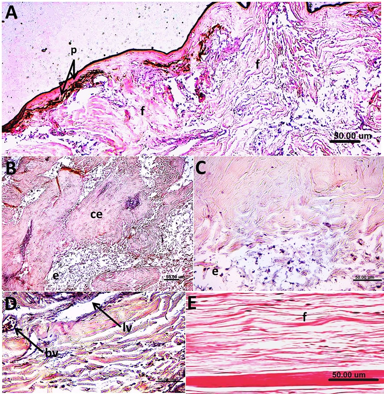
The fibromas were diagnosed in Bartail flathead, and nearly the same histopathological picture was observed in Spangled emperor, Areolated grouper, and Javan flounder. In Bartail flathead, the outermost part of the neoplasm was covered with a pigmented skin layer (Fig. 3A), and the core of the neoplastic tissue underlying the skin was formed of fibrous connective tissue proliferation with the characteristic spindle shape and deposition of collagen fibers, indicating mature fibroblasts and a tumor, fibroma (Fig. 3B and C). The collagen fibers were separated by edema (Fig. 3C). Dilated lymph and blood vessels (Fig. 3D) were not uncommon findings. The fibro-osteoma was diagnosed in another fish, the Bartail flathead, where the histopathological examination in such a case showed the typical histopathological evidence of fibro-osteoma.
_**_in_bartail_flathead_and_*.jpeg)
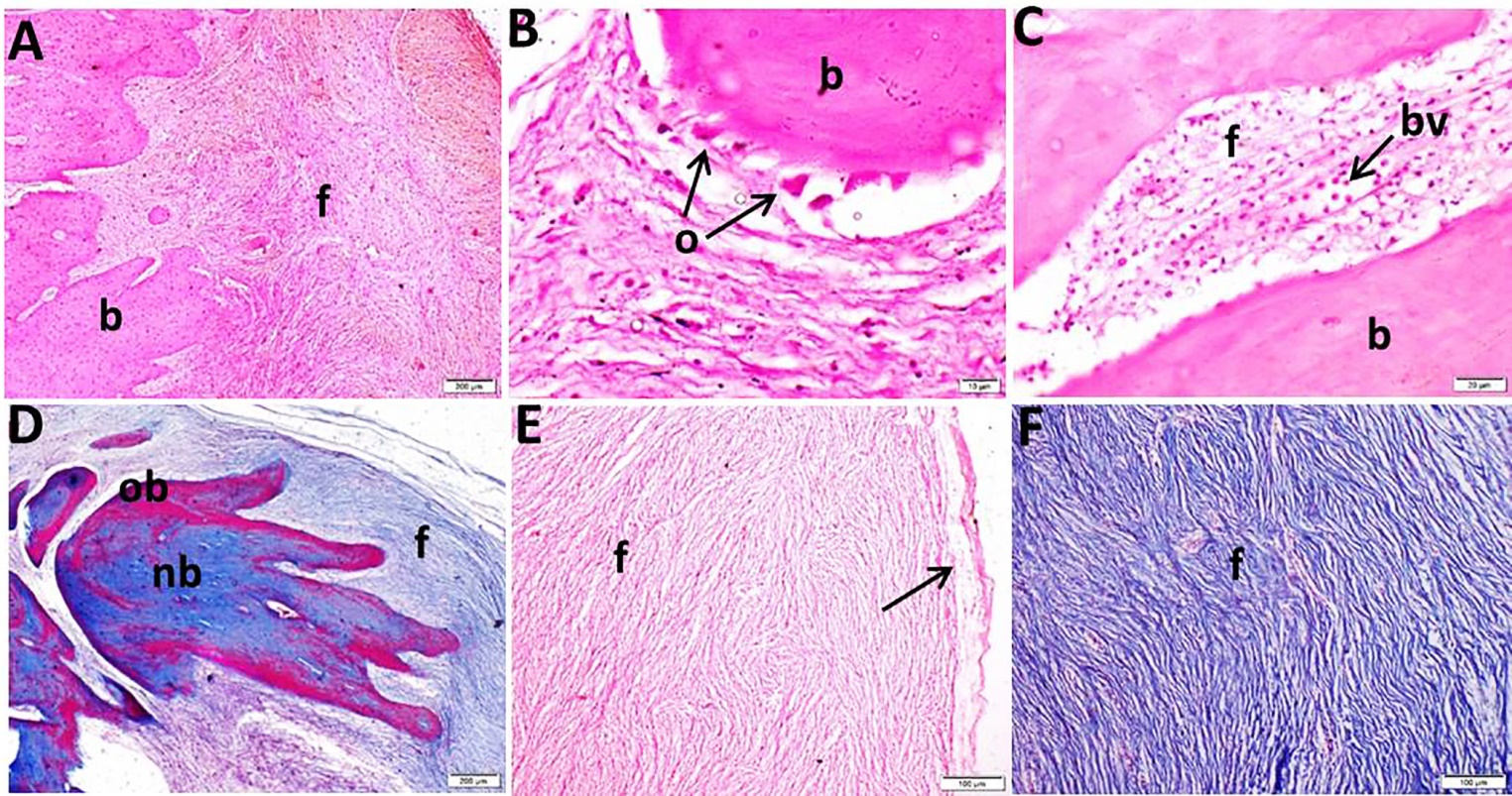
The neoplastic tissues formed of mixed tissue proliferation of both bony trabeculae and prominent fibroblasts (Fig. 4A). The bony trabeculae are surrounded with osteoblasts and osteocytes (Fig. 4B). The interlacing fibrous tissue between the bony trabeculae showed dilated blood vessels (Fig. 4C). Using special histochemical staining of the neoplastic tissue (MT stain), it was clear that the neoplastic tissue wasS formed of both old bones, which stained red, and new bone stained blue. Also, the formed trabeculae were intermingled with prominent proliferation of fibroblasts tissue with a positive reaction as collagen fibers (Fig. 4D). The proliferated fibroblasts appeared regularly arranged, edematous, with a whirling appearance and without criteria of malignancy (Fig. 4E). The characteristic positive blue color histochemical staining of the neoplastic fibrous tissue using MT stain for demonstration of fibrous connective tissues and collagen is demonstrated in Fig. 4F.
_**_mixed_.jpeg)
Discussion
Teleost’s marine fishes resemble other vertebrates in their basic susceptibility to developing neoplastic lesions. The present study revealed proliferative dermal lesions in four commercial marine fish species caught from the water of the Arabian Gulf. Our results were in harmony with the findings reported by Sindermann (1979), Overstreet (1988), Grosset, ME, and Groff (2016), Kamalesh, Chandy, and Bedre (2021), and Mariasingrayan, Danaraj, Dinh-Hung, et al. (2024), where the neoplastic overgrowth, fibromas, was recorded in many fish species at different sites in the affected fishes. The gross description of the neoplastic overgrowth in fish is varied and related to the stage of tumorigenesis and affected fish species (Anekhy and Mehana 2022). In fibroma, the cellular and collagen fibers’ contents are the most crucial factor for the texture of this tumor and indicate whether it is soft, with more cellular contents, or hard made up of numerous fibers and a small number of cells (John, Chirayath, and Paulson 2015). On the other side, dilated lymph and blood vessels associated with edema were recorded in solitary fibrous tumors (Davanzo et al. 2018).
In Bartail flathead, a more complicated neoplastic lesion was noticed on the dorsal fin, an osteo-fibromas. The tumor in this fish was rounded, hard in consistency, and firmly attached to the spines of the dorsal fin. In histopathological sections, the neoplastic mass was formed of both proliferated fibroblasts and osteoblasts forming interlacing bony trabeculae. The proliferated bone showed variable degrees of maturation when stained by MT stain. In this regard, the characteristic feature of the neoplastic cells is their rapid and new proliferation. MT stain is used to differentiate the old and newly formed bone (Lim, Lee, Yun, et al. 2013). However, the newly formed bone in the bony trabeculae could be an indication of active proliferation of the osteoblasts in the neoplastic tissue. On the other hand, it could be part of the dorsal fin bone that is undergoing remodeling in response to the presence of the tumor. The fact that there is a combination of both old and new bone does not necessarily indicate that the tumor is the source of the bone. To the best of our knowledge, the records of the mixed tumor, fibro-osteoma in marine fishes are scarce. In our study, it is worth mentioning that the observed neoplasms, fibromas and osteo-fibroma were observed in the tissues near the bony structure (lower jaw and fins). In this regard, the origin of fibroma was in question, and many ancient theories supposed the origin of fibroma, among which was the possibility of its origin from periosteum (Maurice and Milad 1981). The etiological agents of fibromas in teleosts are multifactorial, including congenital malformation, repeated physical trauma, radiation, and environmental pollution (Black et al. 1982; Annahita, Zahra, and Rahim 2017; Meyers et al. 2019), some extent some viruses, especially retroviruses (Francis-Floyd et al. 1993; Coffee, Casey, and Bowser 2013). However, the fibromas caused by viruses usually have a characteristic tissue reaction and appear as multifocal lesions at different sites of the fish body, associated with lymphocytes’ aggregation and vacuolated cytoplasm of the neoplastic cells (Anders, Hilger, and Moller 1991); thus, the possibility of viral etiology is considered unlikely.
In this study, the potential chemical contamination as well as manmade pollutants that might be discharged to the water of the Arabian Gulf at the El-Jubial region could be expected to be considered in the etiology of the neoplasms in these fish species, which is supported by Oishi, Yamazaki, and Harada 1976; Sindermann 1976; Watermann, Dethlefsen, and Happenheit 1982 and Anders, Hilger, and Moller 1991. Hashim, AL-Hussaini, and Al-Baz 1994, confirmed that some fish species caught from the water of the Arabian Gulf are affected by oil pollution in this region. Predisposing factors such as carcinogenic compounds, viruses, irritants, oncogenes, and parasites have all been reported in teleosts and should be considered as potential sources for tumor induction in the tropical fish (Stoskopf 1993). As the etiology and significance of fibroma in other commercial marine fish at El-Jubial are not well documented, further research is needed to evaluate the potential role of the environmental pollution in this location on the occurrence of the neoplasms in this fish species.
Acknowledgment
Our grateful appreciation and thanks to Mr. S.I. Al-Fayadh, Deputy Minister for Fisheries affairs, Ministry of Agriculture, Kingdom of Saudi Arabia, for facilitating the research work during this study. Great thanks to A. El-Zaharani, Ministry of Environment, Water, and Agriculture, Fish Welfare Branch, El-Jubail Province, Saudi Arabia, for his support and encouragement.
Author Contributions
MAM and MMI developed the research concept. This study was conducted in cooperation between all authors; MAM and MMI conceived and designed the study. MMI performed fish sampling and gross fish examination and identification of fish species. MAM, MMI, F.A.A., and A.A. shared in gross and histopathological examination of the fish neoplasms. All authors wrote, drafted, revised, and approved the final manuscript.
Compliance and ethical standards
All authors declare that the finding in this study was conducted on fish in markets. This article does not contain any studies with animals performed by any of the authors.
Conflicts of interest
The authors declare that they have no conflicts of interest to disclose.
Informed consent
Informed consent was obtained from all individual participants included in the study.
Funding
This research did not receive any specific grant from funding agencies in the public, commercial, or not-for-profit sectors.
Data availability statement
The data that support the findings of this study are available from the corresponding author upon reasonable request.