


INTRODUCTION
In spite of the relatively low production amount, among shellfish species produced in Türkiye carpet shell clam has the highest economic value (Serdar 2021). Unfortunately, due to overfishing and diseases, the production amount of this species, which forms natural populations in the world and in our country, has been decreasing from year to year (Serdar 2021).
Parasitic, bacterial and viral infections in Ruditapes decussatus and its genetically close species Ruditapes phillipinarum have substantially contributed to the pathologies and mortalities in both species. Ample research has been done recently regarding these pathogenic agents primarily focused on pathologies inflicted by Perkinsus olseni (Soudant, Chu, and Volety 2013; Nam et al. 2018; Sambade et al. 2025; Estȇvâo et al. 2023; Delisle et al. 2025). Another protozoan parasite Eomarteilia granula was also accused of mortalities in reared Manila clam from Korea (H. M. Lee et al. 2025). High metazoan parasitic burden consisting of trematodes Monorchis parvus and Curtuteria arguinae in different bivalve species such as cockle (Cerastoderma edule) has also been on focus and their negative impact on their hosts were thoroughly investigated by Jensen et al. (2025) and Stout et al. (2025). Vibrio species such as Vibrio splendidus and Vibrio toranzoniae were among the bacterial agents suspected of inflicting damage to Ruditapes decussatus (Papadopoulos et al. 2025) and Crassostrea gigas (Song et al. 2025). The last but not the least, unknown virus may have also contributed to the mortalities of Manila clam in natural beds from England (Bateman, White, and Longshaw 2012). Histopathology was extensively used in most mortality cases to reveal the real cause of pathological change and address aetiology as in the case of another bivalve mollusc species such as oysters (Crassostrea virginica) from North Carolina (Ben-Horin et al. 2024).
Although there are viral, bacterial and fungal agents which cause pathologies in Ruditapes decussatus and its relative species Ruditapes phillipinarum (FAO 2025; WOAH 2024), parasitic agents are the ones that share the largest part in their diseases and mortalities (EURL 2025; WOAH 2024). In the database of the World Organization for Animal Health (WOAH 2024), 5 out of 7 notifiable diseases in bivalve mollusc species are parasitic diseases.
Carpet shell clam (Ruditapes decussatus L.), considered to be one of the most economically important species among the bivalve molluscs produced in Türkiye, being one of the main exported aquatic products and finally and most importantly the fact that its natural population has been decreasing in recent years, makes the investigation of its parasite fauna urgent need. There are some research works from Türkiye regarding parasites in mussels (Özer and Güneydağ 2015; Yılmaz 2016). However, there is no comprehensive report of parasites and the pathology they inflicted in carpet shell clams dispersed in the Turkish seas. As the only natural bed, legally open for fisheries exploitation of carpet shell clam, İzmir bay was chosen as the only area of sampling (Anonymous 2022). Thus, the aim of the present study was to investigate and reveal the parasite species spectrum of carpet shell clam (Ruditapes decussatus L.) from İzmir Bay in order to serve for the long-term purpose of its natural conservation and its aquaculture production.
MATERIALS AND METHODS
The sampling material, which consisted of randomly selected 360 carpet shell clams (30 clams/month) and collected from İzmir Bay at the depth of 1.5 meter during the period between October 2023 and September 2024 (12 months), were freshly provided by local fishermen on a monthly basis and in a lump, brought at the collection day to the Parasitology Laboratory of İzmir/Bornova Veterinary Control Institute via uninterrupted cold chain.
The decision for the collection of 30 clams per month was based on the assumption of at least 10% parasite prevalence, at 95% confidence interval in detecting at least 1 parasite and population size of the host over 100,000 individuals (Ossiander and Wedemeyer 1973).
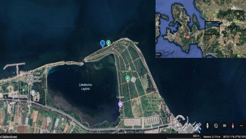
On the clam collection day, the parameters of sea water such as temperature and salinity at the sampling point Üçkuyular/İzmir, coordinates: 38º25’11’‘N 27º02’13’'E (figure 1) were measured in situ by temperature sensor SevenEasy (Mettler Toledo) and salinity refractometer (ATC).
Upon arrival of the samples to the laboratory, each clam was individually examined while the remaining clams were kept at +4 ºC. Before examination, each clam length and width were measured by a millimetric ruler and subsequently weighted with a scale (Shimadzu). Before proceeding to the cytological and histological processing of the clams, the whole body weight, shell weight and soft tissues were weighted individually in order to calculate condition index of each clam.
After dimension measurements, each clam was subsequently scrutinized by fresh examination, cytology and histology. For histology, the whole body was sliced and a piece of tissue 3-4 mm in thickness, which included all important organs (gills, mantle, heart, kidney, alimentary canal and digestive glands, gonads), was removed, placed in Davidson’s fixative and subsequently processed by routine histology techniques. The remaining tissues were used for the preparation of tissue smears, which were further fixed in 96% ethanol for 2 minutes, subsequently stained with Hemacolor®(Sigma-Aldrich) and mounted with Entellan^TM ®^(Merck). Gills, mantle, gonads, alimentary tract were smashed between two slides and examined fresh for alive parasites. Fixed ciliate protozoans were silver impregnated according the method described by Waggoner, Newman, and Tarbutton (2016).
The prevalence and mean intensity of the parasites were calculated according to Bush et al. (1997). Accordingly, the prevalence was calculated as the number of the infected(infested) host(s) divided by the total number of hosts investigated for that particular parasite and was given as a percentage. The concept of mean intensity was applied as the total number of individuals of the particular parasite species in a sample divided by the total number of the hosts infected with that parasite.
The prevalence and mean intensities of all found metazoan parasites were calculated accordingly to the formulas explained above. Although, the prevalence of Ancistrum spp. was also expressed as the number of the infected hosts by Ancistrum spp. divided by the total number of hosts investigated for this parasite, the mean intensity of that protozoan was expressed semi-quantitatively by method proposed in the research work of Xu, Song, and Warren (2015). Respectively, 1 - light infection (less than 20 protozoans per host) , 2 - moderate infection (20-100 protozoans per clam), 3 - heavy (more than 100 protozoans per host). The mean intensity of Ancistrum spp. was calculated based only on native observation of fresh smears, no histological data was used for this purpose.
The parasites were identified according to taxonomic keys provided in related scientific literature (table 1).
Statistical analyses were performed with the aid of Microsoft Excel upon application of statistical concepts revealed by Sümbülüoğlu and Sümbülüoğlu (2016). Obtained data was kept as Microsoft Excel files. All calculated Pearson correlation coefficients were tested by the student t-test, at 95% confidence interval for their statistical significance.
RESULTS
On the clam collection day, the parameters of sea water such as temperature and salinity at the sampling point Üçkuyular/İzmir were measured and are provided in figure 1 along with the monthly prevalences and mean intensities of the identified parasites.
_and_intensities_of_each_parasite_along_with_in_situ_measured_sea_w.png)
Examination of the clams by cytological and histological methods revealed rich parasite fauna consisting of one protozoan and five metazoans, respectively a protozoan Ancistrum spp. and metazoans Cercaria tapidis (Monorchis parvus Looss 1902), Urastoma cyprinae, symbiotic copepod, nematode larva and cestode larva.
The dispersion of each parasite in clams’ tissues varied, according to the intrinsic habitat preference of the parasite. Parasite locations for each parasite species and organ preferences are given in table 2.
The overall (annual) prevalences and mean intensities of the identified one protozoan and five metazoans parasites are shown in table 3.
Ancistrum spp.
Observed protozoa possessed following features; motile with laterally flattened bodies, armoured with numerous rows of cilia, anterior ends were narrower than posterior ends, conspicuous cytostome was present at the posterior part of the ciliates, existence of numerous food vacuoles, single macronucleus with numerous micronuclei, filter feeding mode, attachment to the gills by thigmotactic cilia or moving around them with affinity, straight adoral rows which started at the anterior end, somatic kineties were more numerous on the left side of the body than at the right, existence of naked area at the anterior used for attachment to the gill lamellae.
The average body length was measured as 109,13 µm, with its standard deviation of 17,78 µm. Although the prevalence and mean intensity were relatively high in all seasons (figure 2, table 3), pathological changes were not observed on the gill lamellae (figure 3-a). There was moderate positive correlation (r=0.32) between prevalence and mean intensity and moderate positive correlation (r=0.36) between sea water salinity and prevalence (figure 2). Ancistrum spp. preferred gills as the living habitat (table 2).
Cercaria tapidis (Monorchis parvus Looss 1902)
Only sporocysts of the trematodes, which utilize clams as intermediate hosts and the adults are obligate parasites in vertebrates, were found, metacercariae were not encountered. The gross morphology of the sporocysts (colourless, thickwalled) filled with cercariae was enough to classify them as Cercaria tapidis, which encompass the cercariae of many trematode species. However, after puncture of the sporocyst, the escaped cercariae were obtained and identified down to the species level by identification key provided by De Montaudouin et al. (2009).
Motile larval digeneans (cercariae) were found inside small bags (sporocysts) attached to the gills. The bags were also motile showing periodic contractions. Disrupted sporocysts revealed ovoid cercariae with small tail. The digenean larvae according to their morphology (diastome, pharynx present, no eyespots, long non-furcate tail) were identified as Monorchis parvus.
Although there were no observable histopathological changes such as hyperplasia, metaplasia, atrophy or necrosis, vacuolisation or most typical sign of pathology in bivalves such as hemocytic infiltration with development of granulocytoma, there were signs of displacement and compression of digestive glands and gonads. There were signs of parasitic castration in clams examined in January (figure 3-c).
There was strong positive correlation (r=0.65) between prevalence and mean intensity, moderate positive correlation (r=0.48) between sea water temperature and prevalence and weak positive correlation (r=0.18) between sea water temperature and mean intensity (figure 2). Cercaria tapidis preferred gonads as the living habitat (table 2). However, in heavy infestations the parasite was also found in the digestive glands and gills.
Urastoma cyprinae (Graff 1882)
Encountered parasites were bilaterally symmetrical, flattened and their external surfaces ciliated with two eye spots at the anterior part of the body, possessed mucous and frontal glands, actively swam on the clams gills’ surfaces. Possession of dispersed vitellaria and existence of terminally located genital (oral) pore, along with the close contact with bivalve mollusc (in this case clam) was enough to classify them as Urastoma cyprinae.
The parasites was detected only on fresh gill smears, actively swimming.
There was strong positive correlation (r=1.00) between prevalence and mean intensity, strong positive correlation (r=0.87) between sea water temperature and prevalence and strong positive correlation (r=0.87) between sea water temperature and mean intensity (figure 2). Urastoma cyprinae preferred gills as the living habitat (table 2).
Symbiotic copepod
Observed morphological features: cylindrical metameric animal plan with tagmatization (cephalothorax and abdomen) and existence of exoskeleton, aquatic, on the head region possession of two pairs of antennae, one pair of mandibles and two pairs of maxillae, where maxillipeds are specialised for feeding, abdomen free of appendages.
There was strong positive correlation (r=0.54) between prevalence and mean intensity, weak negative correlation (r = -0.28) between sea water temperature and prevalence and strong negative correlation (r = -0.54) between sea water temperature and mean intensity (figure 2). The symbiotic copepod preferred digestive tract as the living habitat (table 2). No pathology was observed (figure 3-h).
Nematode larva
The visible anatomical features of the encountered parasite were triploblastic architecture with vermiform bilaterally symmetrical body shape, possessing cuticle, epidermis and pseudocoelom.
There were typical signs of pathology in bivalves such as hemocytic infiltration with development of granulocytoma in the connective tissue, where hemocytes encapsulated the parasites (figure 3-f). Implicitly, there were signs of displacement and compression of digestive glands with hindrance of the haemolymph movement.
As a habitat, the parasite preferred mantle and digestive glands (table 2). There was strong seasonal variation in the parasite abundance as the parasite was encountered solely in spring months such as March and April (figure 2).
Cestode larva
The primordial scolex with suckers and a rostellum were discernible from one specimen in histological slide (figure 3-g). Proglottids or strobila were not visible as it is expected for larval cestodes (metacestodes).
Only one parasite was found in a microcyst in one clam foot. No pathological changes were observed.
**_histological_section_from_the_gills_(thick_arrow)_with_numerous_*ancistrum*_spp._tr.jpg)
DISCUSSION AND CONCLUSION
From the viewpoint of the ecological and disease dynamics, the antagonistic relation between the parasites and their bivalve molluscan hosts was extensively scrutinized by many researchers (Lauckner 1983; Bower 2006; Elston and Ford 2011). These authors reported the overall impact of the parasite on their hosts, respectively, along with the tissue destruction, lowering condition and fecundity and in some parasite species inflicting parasitic castration, energy disbalance and growth impairment, increasing probability of predation and respectively reduction of the survival, increasing the risk of contracting secondary bacterial and viral infections.
Specifically, parasitic diseases encountered in clams worldwide have been on focus due to the aforementioned disease causative associations. Infections and infestations with species of Perkinsus, Marteilia, Haplosporidium, Marteilioides, Mikrocytos, microsporidia, amoeba, ciliated protozoa, coccidians, gregarins, turbellarians and trematod cercaria and metacercaria in clams have been thoroughly investigated and reported by Bower (2022).
Among these parasite groups some were encountered in the present research.
Ancistrum species were frequently reported as usual inhabitants of the bivalve mollusc gills (Bower 2022; Xu, Song, and Warren 2015; Fenchel 1965). Investigation of their pathological effect revealed contradictory results. Berilli et al. (2000) did not report any evidence of pathogenicity associated with the presence of Ancistrum species on the gills of Chamelea gallina from the Adriatic sea. Similar observations were made by Figueras, Jardon, and Caldas (1991) in mussels. On the contrary, Pauley, Chew, and Sparks (1967) detected severe pathological changes such as necrosis, hemocytic infiltration and metaplastic change of epithelial cells (from columnar to cuboidal) associated with the presence of Thigmotrichid ciliates in the gills of experimentally debilitated oysters (Crassostrea gigas).
The present research work confirms the results of Berilli et al. (2000) and Figueras, Jardon, and Caldas (1991) in different bivalve species, in this case carpet shell clam (R. decussatus). No apparent seasonality of the occurrence of the parasite was found indicating that regardless of the season, clams are host of core thriving population of Ancistrum spp. which is not affected by the sea water temperature. Failure to calculate the buccal field/body length ratio (Xu, Song, and Warren 2015) from alive specimens let to inability to assign the parasite to any Ancistrum species. However, depending on its host preference, it can be speculated that the found parasite is Ancistrum crassum, closely associated with clam species such as Ruditapes philippinarum (Xu, Song, and Warren 2015).
Cercariae of different digenean species are reported to inflict serious damage to their bivalve hosts via parasitic castration. Hindrance of the gonadal development due to presence of digenean sporocysts and metacercariae were reported in the works of Carballal et al. (2001), Navas et al. (1992), Cremonte et al. (2001), Jensen et al. (2025) and Stout et al. (2025). Parasites according to some of these authors were responsible for the total inhibition of the gonadal development. Moreover, Jonsson and Andre (1992) pointed that Cercaria cerastodermae was responsible for a mass mortality event of Cerastoderma edule from the Swedish coast of Baltic sea. However, no damage to the bivalve hosts due to infection by digenean trematodes was attributed in the works of M. K. Lee et al. (2001) and Figueras, Jardon, and Caldas (1991). In this research, no evidence of pathological change associated with the sporocysts of Cercaria tapidis were observed, except displacement of the gonads by the sporocysts. In figure 3-c numerous sporocysts and no presence of oocytes are visible in the gonadal tissue of a clam dissected in January when the gonads are in the state of winter rest. In this case, lack of developing gametes cannot be directly associated with the phenomenon of parasitic castration. Instead, it is more plausible to explain the observed lack of gametes by the natural reproductive cycle of clams, otherwise ample number of developing gamets along with the presence of numerous sporocysts in figure 3-d at the peak of gonadal maturation of clams in June would not have been seen. It is reasonable that association of the parasitic castration with digenean trematodes should always be done in connection with the annual reproductive cycle of the bivalves (Serdar and Lök 2005) and should not depend solely on histopathological conclusions.
Turbellarians of bivalve molluscs are generally regarded as ectocommensals (Fleming 1986; Goggin and Cannon 1989), parasites (Gonzalez et al. 2005; Özer and Güneydağ 2015) or between ectocommensals and parasites (Bower 2022). In this research, the prevalence and intensity of U. cyprinae was so low (1.38/1.00) and there was lack of histological specimen, that no comments could be made upon its effect on the health status of the clams. However, the strong positive correlation of the prevalence and intensity with the sea water temperature (figure 2), distinctive pattern of occurrence (only in summer) perfectly matches with the results of Özer and Güneydağ (2015) and Flemming (1986) who detected U. cyprinae only in the warmer months of the year. Additionally, relatively low overall prevalence (1.38) and intensity (1.00) reflects the results of the other researchers who found only few parasites per host (Goggin and Cannon 1989; Özer and Güneydağ 2015).
There are multiple reports about pathological changes of the digestive tract of bivalves especially of mussels caused by the parasitic copepod Mytilicola orientalis (Steele and Mulcahy 2001; Borkens and Koppe 2022). As such, Carballal et al. (2001) found metaplastic changes of the epithelium (replacement of the columnar epithelium by cuboidal) around Mytilicola-like copepods in the intestinal lumen of Galician cockles and haemocytic response to the hook attachments of M. intestinalis to the intestinal epithelium of rafted mussels from Ria de Arosa Spain by Figueras, Jardon, and Caldas (1991).
In the present research no Mytilicola-like parasites were found in the examined clams but the detected copepods were similar in external morphology to the copepods found by Francisco, Hermida, and Santos (2010) in mussels from Aveiro Estuary Portugal which they identified as Bathylaophonte azorica. No observable pathological changes to the epithelial lining of the intestine led us to conclude that copepod which we found was symbiotic commensal living in the digestive tract of the clams and uses clams as living habitat mostly in cooler months of the year.
There are not many nematode species which utilize bivalve molluscs as the final host (Bower 2006, 2022). However, there are research works which stressed the importance of bivalve molluscs as an intermediate hosts for anisakid nematodes, particularly Sulcascaris sulcata which is a principal parasite of the sea turtle Caretta caretta (Marcer et al. 2020; Santoro et al. 2020; Santoro, Palomba, and Modica 2022). These researchers isolated Sulcascaris sulcata from purple dye murex, mussels and scallops. Due to the scarcity of the parasite material (only 1 alive nematode larva), we were not able to assign the parasite down to the genus or species level. However, it might be speculated that the parasite was actually Sulcascaris sulcata solely based on its gross appearance, its preference for bivalve mollusc host (in this case clam) and presence of sea turtle population in İzmir Bay (personal observation).
Among the isolated parasites in the present research, only the nematode was associated with discernible pathology in the histological slides. There was heavy hemocytic infiltration around the nematodes in the digestive glands. This is in line with the results of Santoro et al. (2020) who found S. sulcata larvae entrapped in hemocytic capsules.
Marked seasonality in occurrence of the nematode, respectively detected only in spring (see figure 2) coincided with the migration of the sea turtles into their feeding grounds in İzmir Bay (Bentivegna 2002).
There is not much available scientific literature regarding cestodes of bivalve molluscs. Generally, molluscs serve as intermediate hosts for some cestodes species living as adults in different vertebrate species (Bower 2006; Lauckner 1983). The found metacestode was not alive specimen and was discernible only in one histological slide. Thus was technically impossible to be assigned to any cestode taxonomic group. No evidence of pathology was obtained.
Parasites as indicators of biodiversity and ecosystem health were thoroughly investigated by many ecological parasitologists (Mouritsen and Poulin 2002; Hudson, Dobson, and Lafferty 2006). The richness of parasites indicates resilience of the ecosystem, in other words the more abundant are their hosts the richer is the parasite fauna of that ecosystem, hence more biodiversity. From ecological point of view it is unfortunate that the results of the present study indicate relatively low parasite abundances in the carpet shell clam population of İzmir Bay. For example trematod (Cercaria tapidis) overall prevalence was calculated as 3.89%, which is relatively lower compared to prevalence of trematodes found by other researchers, respectively 4.5% (Özer and Güneydağ 2015), 58% (Francisco, Hermida, and Santos 2010), 9.7% (M. K. Lee et al. 2001), 20% (Jonsson and Andre 1992). Moreover, the apicomplexan parasites such as Nematopsis spp. and Porospora spp., which utilize bivalve molluscs as intermediate host and are parasites of crustaceans were not encountered at all. For comparison, Canestri-Trotti et al. (2000) detected Nematopsis sp. in 39.7% and Porospora sp. in 86.7% of the examined bivalves from the Adriatic Sea. It might be concluded that at least fish and bird biodiversity for which the indicator is the trematode fauna and copepod biodiversity for which apicomplexan fauna is an indicator are seriously reduced in İzmir Bay, and the cause could be attributed to the heavy urbanization, overfishing, destruction of the feeding grounds and sea water pollution (Kacar 2011; Kacar and Omuzbuken 2017).
Generally, diseases of aquatic organisms are multifactorial in their origin and closely associated with a kind of pathological change (Roberts 2012). On the contrary, parasites found in the present research, except the nematode, were not found to inflict any visible pathology. However, the absence of an evidence is not an evidence of absence regarding the pathology and under certain debilitating conditions of the hosts, such as low nutrition, presence of other pathogens, reproductive stress, environmental pollution, some harmless commensals or parasites can become real enemies of their hosts and inflict serious pathology (Kinne 1983). In this respect, special attention deserves Ancistrum spp. It might seem to be harmless in the natural population of clams, however under hatchery conditions, these ciliates can become real treat to the bivalve spats (Tokşen and Çilli 2010).
In conclusion, investigation of the clams unveiled parasite fauna composed of one protozoan and five metazoans. The most severe pathological impact was observed in the tissues infected with the nematode larvae. Other parasites were apparently harmless except Cercaria tapidis, which was responsible for displacement of the gonads by the sporocysts. Correlations between prevalences and intensities were positive and varied from moderate to strong. There was discernible pattern in seasonal abundances, respectively more parasites encountered in warmer months except the symbiotic copepod which preferred clams as its host in cooler months.
In future, effort must be spent to identify down to the species level the nematode, cestode and copepod species encountered in the present research and molecular methods of identification to be applied to all parasites detected in the current work.
Acknowledgements
Authors are grateful to the laboratory support provided by İzmir Bornova Veterinary Control Institute Parasitology Laboratory and its staff and especially Dr. Öznur Yazıcıoğlu and Okan Çeven for providing technical support during histology work.
Author’s Contributions
E. Çilli: conceptualization, methodology, investigation, writing - original draft, funding acquisition. A. Lök: conceptualization, methodology, writing - review & editing.
Conflict Of Interest
The authors declared that there are no conflicts of interest regarding the publication of this article. Views and opinions expressed are however those of the authors only and do not necessarily reflect those of the Republic of Türkiye Ministry of Agriculture and Forestry. Neither the Republic of Türkiye Ministry of Agriculture and Forestry can be held responsible for them.
Ethical Approval
Experimental Animals Ethical Commitee of İzmir Bornova Veterinary Control Institute, upon application and evaluation of Esat Çilli’s PhD proposal “İzmir Körfezinde Akivadeslerde (Ruditapes decussatus (Linnaeus, 1758)) Parazit Faunasının Araştırılması/ Investigation of the Parasite fauna in Grooved Carpet Shell (Ruditapes decussatus (Linnaeus, 1758)) in Izmir Bay”, made the decision on 5th September 2023 that there is no need for ethical approval.
Funding
The study presented here was a part of the Esat Çilli`s PhD research project in Aquaculture, Graduate School of Natural and Applied Sciences, Ege University, Türkiye with supervisor Professor Dr. Aynur Lök. Funding was provided by the Republic of Türkiye Ministry of Agriculture and Forestry under TAGEM grant scheme for the year 2024 (Project number: TAGEM/HSGYAD/T1/23/A6/P3/6425-İzmir Körfezinde Akivadeslerde (Ruditapes decussatus (Linnaeus, 1758)) Parazit Faunasının Araştırılması).