Loading [Contrib]/a11y/accessibility-menu.js

This website uses cookies

We use cookies to enhance your experience and support COUNTER Metrics for transparent reporting of readership statistics. Cookie data is not sold to third parties or used for marketing purposes.

Skip to main content
null
Bulletin of the EAFP
  • Menu
  • Articles
    • Case study
    • Method
    • Note
    • Opinion
    • Research article
    • Review
    • Workshop Report
    • All
  • For Authors
  • Editorial Board
  • About
  • Issues
  • search
  • Facebook (opens in a new tab)
  • LinkedIn (opens in a new tab)
  • RSS feed (opens a modal with a link to feed)

RSS Feed

Enter the URL below into your favorite RSS reader.

https://eafpbulletin.scholasticahq.com/feed
P-ISSN 0108-0288
E-ISSN 3005-4648
Research article
Vol. 44, Issue 4, 2025October 14, 2025 CEST

A survey to detect viral pathogens in wild-caught ornamental fish from ornamental fish wholesale facilities in the Peruvian Amazon

Fernando Carlos Ramos-Espinoza, Karin Bances-Chávez De Moya, Francisco Ulloa-Stanojlovic, Mauro Estrella-Ortiz, José Rodriguez-Callan, Yerson Duran-Ramírez, Vanessa Quevedo-Alvarado, Ignacio De Blas Giral, Rodolfo Velazco-Peña, Muriel Gómez-Sánchez Orezzoli,
Ornamental fishMegalocytivirusRanavirusHistopathology
Copyright Logoccby-4.0 • https://doi.org/10.48045/001c.145929
Photo by Dave Hoefler on Unsplash
Bulletin of the EAFP
Ramos-Espinoza, Fernando Carlos, Karin Bances-Chávez De Moya, Francisco Ulloa-Stanojlovic, Mauro Estrella-Ortiz, José Rodriguez-Callan, Yerson Duran-Ramírez, Vanessa Quevedo-Alvarado, Ignacio De Blas Giral, Rodolfo Velazco-Peña, and Muriel Gómez-Sánchez Orezzoli. 2025. “A Survey to Detect Viral Pathogens in Wild-Caught Ornamental Fish from Ornamental Fish Wholesale Facilities in the Peruvian Amazon.” Bulletin of the European Association of Fish Pathologists 44 (4). https:/​/​doi.org/​10.48045/​001c.145929.
Download all (2)
  • Figure 1. Diagrammatic view of study design.
    Download
  • Figure 2. A monogenoid in the gills of Pterophyllum scalare
    Download

Sorry, something went wrong. Please try again.

If this problem reoccurs, please contact Scholastica Support

Error message:

undefined

View more stats

Abstract

Peruvian ornamental fish industry is based on the trade of wild-caught Amazonian species, which are exported to several countries. Throughout the ornamental fish value chain, fish are exposed to different stressors, which enhance their susceptibility to pathogens. Major emerging viral diseases in ornamental fish may result in trade restrictions. Therefore, it is important for authorities to monitor the health status of ornamental fish populations. The aim of this study was to detect the presence of the genera Megalocytivirus and Ranavirus in five ornamental fish species from the Peruvian Amazon. A total of 600 wild-caught ornamental fish of five species were collected from ornamental fish wholesale facilities in the city of Iquitos, Peru, between June and September 2022. The samples included the species Hyphessobrycon erythrostigma, Corydoras splendens, Carnegiella strigata, Pterophylum scalare and Ancistrus temminckii and were analyzed by quantitative PCR (qPCR) and histopathology. The qPCR results did not detect Megalocytivirus and Ranavirus. In addition, histopathology revealed the presence of monogenoids and Piscinoodinium sp. in the gills and metacercarial cysts in the liver. Furthermore, histopathological examination revealed an unusual finding of Ichthyophthirius sp. in the esophagus of A. temminckii. The results showed that Megalocytivirus and Ranavirus were not detected during the sampling periods in at least five wild-caught ornamental fish species from the Peruvian Amazon and there are no histopathological lesions related to Megalocytivirus-infected fish. Finally, we advise that additional monitoring is necessary to detect the occurrence of the World Organization for Animal Health (WOAH) listed diseases in ornamental fish and develop strategies that ensure surveillance plans to consider the wide variety of Amazonian fish species in the ornamental fish trade, as well as the presence of disease-susceptible ornamental fish species.

1. INTRODUCTION

The Amazon Basin is a significant source of wild-caught ornamental fish for the global aquarium industry (De Sousa et al. 2021). Colombia, Peru, and Brazil are the main suppliers of Amazonian ornamental fish with most traded species belonging to the families Potamotrygonidae, Osteoglossidae, Characidae, Loricariidae, and Callichthyidae (Ortiz and Iannacone 2008). The city of Iquitos, located in the Peruvian Amazon, is the most important region for ornamental fish exports. In 2024, Peruvian ornamental fish exports reached a Free on Board (FOB) value of US$ 4 850 000 and the main destination markets were China, the United States, Germany, Japan, Thailand, Singapore, United Kingdom and Taiwan (PromPerú 2025). Some of the main species of wild-caught ornamental fish from the Peruvian Amazon are “Bleeding-heart Tetra” Hyphessobrycon erythrostigma, “Emerald Catfish” Corydoras splendens, “Marbled Hatchetfish” Carnegiella strigata, “Angelfish” Pterophyllum scalare, and “Xenocara” Ancistrus temminckii, (PNIPA 2021).

Inappropriate management practices such as poor water quality, improper nutrition, and high stocking density as well as poor biosecurity measures in ornamental fish wholesale facilities can cause the emergence of infectious diseases (Cardoso et al. 2019). One of the largest threats to the ornamental fish industry are viruses from the Iridoviridae family, such as Megalocytivirus and Ranavirus, which can lead to high mortality rates and economic losses (Maganha et al. 2018; Sivasankar et al. 2017). According to the Committee on Taxonomy of Viruses (ICTV) Megalocytivirus pagrus 1 is a species within the genus Megalocytivirus (subfamily Alphairidovirinae, family Iridoviridae), which was previously called infectious spleen and kidney necrosis virus (ISKNV), and includes three genotypes: red seabream iridovirus (RSIV), reddish body iridovirus (TRBIV) and ISKNV (Fusianto et al. 2023). Recently, the WOAH Aquatic Animal Health Code has included the Megalocytivirus pagrus 1 in its list of fish diseases (WOAH 2024).

The presence of these viruses has been investigated in several studies involving wild-caught ornamental Amazonian fish. In Brazil, a study reported that 47% of 24 fish species collected from ornamental fish wholesale facilities (including Arapaima gigas, Hypostomus plecostomus, Pterophyllum scalare and Pygocentrus nattereri) tested positive by PCR for the genus Megalocytivirus (Maganha et al. 2018). Also, the genus Megalocytivirus has been detected in Amazonian species including Astronotus ocellatus, Symphysodon sp., Apistogramma cacatuoides and Paracheirodon innesi (Jeong et al. 2008; Nolan et al. 2015; Baoprasertkul and Kaenchan 2019). Furthermore, in Germany, PCR assays confirmed the presence of ISKNV in samples from angel fish Pterophyllum altum associated with mortality events (Jung-Schroers et al. 2016).

Besides, the genus Ranavirus has a wide range of fish hosts and is listed by WOAH (Leiva-Rebollo et al. 2024). Regarding Amazonian fish, a study on the importation of ornamental species into the Europe Union found no conclusive evidence of Ranavirus infection in certain Amazonian species (Vesely et al. 2011). To the best of our knowledge, no virus detection studies have been developed in ornamental fish from the Peruvian Amazon.

The great diversity of ornamental fish species, the scarcity of research on their diseases, and the lack of regulation in the ornamental fish trade industry in most countries may have contributed to the limited implementation of surveillance plans for ornamental fish by authorities. Australia is one of the few countries that has developed a surveillance plan for Megalocytivirus in ornamental fish production facilities (Hood 2021). In recent years, some health authorities are beginning to introduce new requirements for the certification of ornamental fish to prevent the spread of diseases, which makes the early detection and active surveillance of viral pathogens important to prevent and control diseases in ornamental fish (Girisha et al. 2021).

The present study is the first to investigate and survey the presence of the genus Megalocytivirus and Ranavirus in wild-caught Amazonian fish species located in ornamental fish wholesale facilities from Iquitos, Peru, according to the guidelines from the WOAH Aquatic Animal Health Code.

2. MATERIAL AND METHODS

2.1. Fish

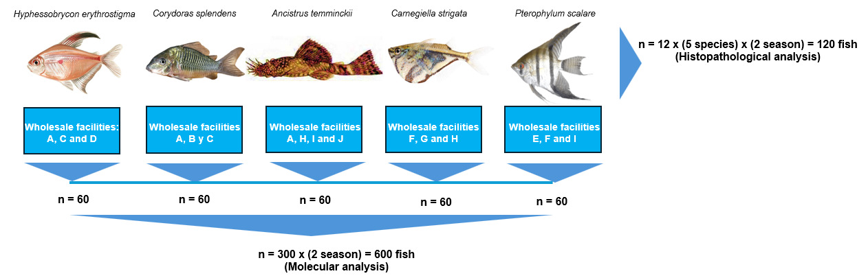
This study focused on five wild-caught Amazonian fish species traded as ornamental fish: “Bleeding-heart Tetra” Hyphessobrycon erythrostigma, “Emerald Catfish” Corydoras splendens, “Marbled Hatchetfish” Carnegiella strigata, “Angelfish” Pterophyllum scalare, and “Xenocara” Ancistrus temminckii. All the fish come from the main rivers of the Amazon Basin and are kept in wholesale facilities located in Iquitos, Peru (Table 1). These facilities are supplied with ground water, and the fish are kept in glass tanks equipped with handmade filters.

Table 1.Origin, total length and live stage of the ornamental fish kept in the wholesale facilities.
River Species Total length mean (cm) Standard deviation (SD) Live stage Wholesale facilities
(origin of the fish)
Rainy season
Itaya Corydoras splendens 4.83 0.25 Fry A
Napo Corydoras splendens 3.92 0.47 Fry B
Nanay Hyphessobrycon erythrostigma 2.75 0.26 Fry C
Nanay Hyphessobrycon erythrostigma 3.58 0.19 Fry D
Nanay Pterophylum scalare 3.29 0.26 Fry E
Nanay Pterophylum scalare 5.88 2.18 Fry F
Nanay Carnegiella strigata 3.02 0.06 Fry G
Tapiche Carnegiella strigata 3.33 0.49 Fry H
Blanco Ancistrus temminckii 6.91 0.97 Fry A
Momon Ancistrus temminckii 6.73 0.57 Fry H
Dry season
Napo Corydoras splendens 4.53 0.58 Fry A
Amazonas Corydoras splendens 4.45 0.47 Fry D
Nanay Hyphessobrycon erythrostigma 3.28 0.17 Fry A
Nanay Hyphessobrycon erythrostigma 3.25 0.29 Fry C
Nanay Pterophylum scalare 4.82 0.54 Fry E
Nanay Pterophylum scalare 5.72 0.79 Fry I
Blanco Carnegiella strigata 3.07 0.36 Fry F
Napo Carnegiella strigata 3.63 0.13 Fry H
Itaya Ancistrus temminckii 8.93 1.29 Fry I
Itaya Ancistrus temminckii 8.37 1.38 Fry J

2.2. Study design

In 2022, fish from 10 ornamental fish wholesale facilities were sampled in two different periods: one during the dry season (August to September) and the second during the rainy season (June to July). The sample size was determined to provide 95 % confidence in detecting Megalocytivirus and Ranavirus with a design prevalence (minimum expected prevalence) of 5%, according to the WOAH Aquatic Animal Health Code, which resulted in a sample size of 60. So, during the study, 60 fish of each of the five target species were collected from ornamental fish wholesale facilities in each season, resulting in a total of 600 fish for molecular analyses that were tested as pool of five fish. Additionally, during the same visits to ornamental fish wholesale facilities in both seasons, 24 fish from each target species were collected for histopathological analysis, totaling 120 fish. The study design is shown in Figure 1.

Figure 1
Figure 1.Diagrammatic view of study design.

2.3. Fish collection

Fish from the five target species showing nonspecific clinical signs (melanosis, fin erosion, hemorrhagic skin lesions) were collected at each wholesale facility after being held for a minimum period of one week. The selected fish was showing unspecific clinical signs as (fin erotion, melanosis, erythema, discoloration). They were placed in plastic bags containing 3 to 5 L of water, filled with oxygen, sealed with rubber bands, and transported to the National Authority for Health and Safety in Fisheries and Aquaculture (SANIPES) facilities, approximately a 30-45 min drive. Upon arrival at SANIPES, all fish were euthanized by overdose with a benzocaine solution (100 mg L1) (Sigma-Aldrich) and immediately examined for gross pathology. Kidneys, spleens, and livers from samples were then removed, pooled, and placed in tubes containing 95 % ethanol for molecular analysis and kept at -20°C until use. Tissues (esophagus, liver, spleen, kidney, gastrointestinal tract and gills) from each fish were immediately fixed in 10% neutral buffered formalin for histopathological analysis.

2.4. PCR detection of Megalocytivirus and Ranavirus

a) DNA Extraction

The ReliaPrep™ gDNA Tissue Miniprep System (Promega, Madison, WI, USA) was used to extract total DNA from 25 mg of pooled tissue (Kidney, spleen and liver), following the manufacturer’s instructions. The purified DNA was quantified using a Qubit 4 fluorometer (Thermo Scientific) before the PCR assays. DNA quality was assessed using an Epoch 2 microplate spectrophotometer (Biotek Instruments) and diluted to 25 ng μL–1. The final product was stored at -20 °C until use.

b) qPCR for Megalocytivirus and Ranavirus

The qPCR protocol involved a reaction mixture of 20 μL, consisting of 10 μL of TaqManTM Universal Master Mix II with UNG (Applied Biosystems), 0.4 μL of Probe (Applied Biosystems), 0.8 μL of each primer (0.4 μM), 2 μL of template DNA, and 6 μL of nuclease-free water. Table 2 shows the primer sets and probes and Table 3 shows the cycling conditions. The analysis was performed using QuantStudioTM Real-Time PCR Instrument and QuantStudioTM and Design and Analysis Software version 1.4 (Applied Biosystems). The synthetic sequence of 125 bp part of the MCP (major capsid protein) gene of the KagYT-96 isolate (GenBank: MK689686.1) inserted into plasmid pCR2.1-TOPO vector RSIV_RT Bam HI (GENSCRIPT-USA) was used as a positive PCR control for Megalocytivirus and the synthetic sequence of 316 bp part of the MCP (major capsid protein) gene of the strain LY-FV3-20161023 (GenBank: MG637360.1) inserted into the pCR2.1-TOPO vector MCP-1_RT_RANAVIRUS was used as a positive PCR control for Ranavirus. Molecular biology grade water was used as a negative PCR control.

Table 2.Primer/probe sets with sequences and amplicon sizes used for qPCR
Pathogen/Gene Primer/Probe 5’-3′ Sequences Amplicon size Reference
Megalocytivirus RSIV-RT F TGACCAGCGAGTTCCTTGACTT 125bp Mohr et al. (2015)
RSIV-RT R CATAGTCTGACCGTTGGTGATACC
RSIV Probe FAM-AACGCCTG CATGATGCCTGGC-TAMRA
Ranavirus MCP-RT F GTCCTTTAACACGGCATACCT 110bp Leung et al. (2017)
MCP-RT R ATCGCTGGTGTTGCCTATC
MCP Probe FAM-TTATAGTAGCCTRTGCGCTTGGCC-QSY7
Table 3.Thermal profile for qPCR
Pathogen Step Temperature (°C) Time
Megalocytivirus Pre-denaturation 95 10 min
Denaturation (45 cycles) 95 15 s
Annealing/extension 60 1 min
Ranavirus Pre-denaturation 95 10 min
Denaturation (50 cycles) 95 15 s
Annealing/extension 60 1 min

2.5. Histopathology

The tissue was fixed in 10% buffered formalin for 24 h. Then, the samples were dehydrated in increasing concentrations of ethanol, diaphanized in xylol and embedded in paraffin wax using HistoCore PEARL (Leyca Byosistems). Finally, samples were sectioned at a thickness of 3.5-4 µm, stained with haematoxylin and eosin (H&E) and examined using light microscopy to determine histopathological alterations. The identification of parasites in tissue sections was performed according to Bruno, Nowak, and Elliott (2006).

3. RESULTS

The assays detected no Megalocytivirus or Ranavirus DNA in any of the ornamental fish samples collected from the ten ornamental fish wholesale facilities in the city of Iquitos during both the rainy and dry seasons of 2022 (Table 4).

Table 4.Results of molecular analyses for the detection of Megalocytivirus and Ranavirus in five ornamental fish species collected in 2022.
Species Wholesale facilities Sample size qPCR positive/negative
Megalocytivirus
qPCR positive/negative
Ranavirus
Rainy season
Corydoras splendens A 30 Negative Negative
B 30 Negative Negative
Hyphessobrycon erythrostigma C 30 Negative Negative
D 30 Negative Negative
Pterophylum scalare E 30 Negative Negative
F 30 Negative Negative
Carnegiella strigata G 30 Negative Negative
H 30 Negative Negative
Ancistrus temminckii A 30 Negative Negative
H 30 Negative Negative
Dry season
Corydoras splendens A 30 Negative Negative
D 30 Negative Negative
Hyphessobrycon erythrostigma A 30 Negative Negative
C 30 Negative Negative
Pterophylum scalare E 30 Negative Negative
I 30 Negative Negative
Carnegiella strigata F 30 Negative Negative
H 30 Negative Negative
Ancistrus temminckii I 30 Negative Negative
J 30 Negative Negative
Figure 2
Figure 2.A monogenoid in the gills of Pterophyllum scalare

(A) Piscinoodinium sp. in the gills of Corydoras splendens (B) Ichthyophthirius sp. in the esophagus of Ancistrus temminckii (C) and digeneans in the gastrointestinal tract (D).

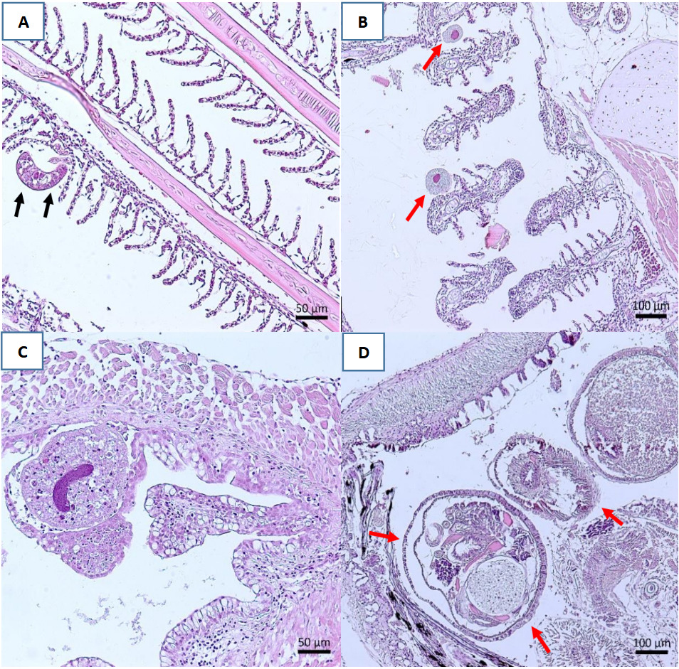
The histopathological examination did not reveal hypertrophied cells and inclusion bodies characteristic of Megalocytivirus-infected fish; however, some parasites were detected (Table 5), including the presence of monogenoids in the gills of C. splendens, P. scalare, C. strigata and A. temminckii. Additionally, Piscinoodinium sp., a dinoflagellate parasite, was found in the gills of C. splendens. Metacercarial cysts of digeneans were observed in the gastrointestinal tract of H. erythrostigma and A. temminckii. Finally, Ichthyophthirius sp. was detected beneath the mucosal epithelium of the esophagus of A. temminckii (Figure 2).

Table 5.Histopathological results of parasitic infections in five ornamental fish species collected in 2022.
Lesions Corydoras splendens Hyphessobrycon erythrostigma Pterophylum scalare Carnegiella strigata Ancistrus temminckii
Rainy season
Gills
Piscinoodinium sp. 1/12 0/12 0/12 0/12 0/12
Monogenoids 3/12 0/12 3/12 2/12 1/12
Esophagus
Ichthyophthirius sp. 0/12 0/12 0/12 0/12 2/12
Gastrointestinal tract
Digeneans 0/12 1/12 0/12 0/12 0/12
Dry season
Gills
Monogenoids 1/12 0/12 4/12 0/12 2/12
Gastrointestinal tract
Digeneans 0/12 0/12 0/12 0/12 4/12

4. DISCUSSION

The current study did not detect DNA of viruses within the genus Megalocytivirus in five species of wild-caught ornamental fish from the Peruvian Amazon. Studies on the presence of Megalocytivirus genus have been reported in Amazonian ornamental fish from Brazil, including P. nattereri (Cardoso et al. 2017), H. plecostomus and P. scalare (Maganha et al. 2018). In addition, this genus has been reported in two Amazonian fish species for consumption, Arapaima gigas and Pseudoplatystoma corruscans (Maganha et al. 2018; Fonseca et al. 2022). To better understand the circulation of this virus in the Peruvian Amazon, further surveys should include a wide range of wild-caught ornamental fish species, based on the fish diversity present in the region and the potential risk of undetected infections. Recently, the technical document “2022 Report of the WOAH ad hoc Group on susceptibility of fish species to infection with WOAH listed diseases” showed which species are susceptible to the three genotypes RSIV, TRBIV and ISKV, including the species P. scalare as susceptible to ISKNV genotype.

Regarding Ranavirus, the five species of wild-caught ornamental fish showed no positive results. A study on the importation of ornamental fish into the Europe Union found no conclusive evidence of Ranavirus genus presence in species such as Carnegiella marthae, C. strigata, Corydoras hastatus, Corydoras julii, H. erythrostigma, H. plecostomus, Monocirrhus polyacanthus, Potamotrygon hystrix and Pseudoplatystoma fasciatum (Vesely et al. 2011).

Pooled sampling was employed in this study in accordance with the WOAH Aquatic Manual, considering that pooling is an effective strategy for reducing analytical costs in surveillance plans (Muniesa et al. 2014). Nevertheless, diagnostic sensitivity may be reduced due to a dilution effect (Laurin et al. 2019) meaning that a larger number of samples is required to demonstrate the absence of Megalocytivirus and Ranavirus.

Although no viral DNA was detected in the current study, the potential risk of disease introduction cannot be ruled out, particularly through the importation of exotic ornamental fish into aquarium facilities (Koda et al. 2023). Supporting this concern, in Australia, ISKNV-like viruses, although the not been detected in wild fish populations, have been identified in imported ornamental fish and has been associated with high mortalities during quarantine (Mohr et al. 2015; Nolan et al. 2015).

The “2023 Report of the Meeting of WOAH Aquatic Animal Health Standards Commission” proposed a new chapter concerning the international movement of ornamental aquatic animals, in response to the potential risk of introducing and spreading fish pathogens. In this context, disease surveillance plans for ornamental fish are particularly important in countries with a significant ornamental fish industry.

Recent studies have employed diagnostic tests for disease surveillance in both wild and captive fish populations (Johnson et al. 2019; Koda et al. 2023). The Australian government has conducted surveillance plans for Megalocytivirus in ornamental fish facilities (Hood 2021) and has developed a risk-based surveillance system for megalocytiviruses, spring viraemia of carp virus and Aeromonas salmonicida in imported ornamental fish (Hood et al. 2019). In Peru, the official surveillance plan for diseases is conducted by SANIPES and focused on farmed aquatic species including whiteleg shrimp (Penaeus vannamei), rainbow trout (Oncorhynchus mykiss) and Tilapia (Oreochromis sp.) (SANIPES 2024). Nevertheless, no targeted surveillance activities are currently conducted for viral diseases in ornamental fish, despite Peru being one of the top exporters of Amazonian ornamental fish. The present study represents a first attempt to assess the presence of viral pathogens in wild-caught fish from the Peruvian Amazon. Although no viral DNA was detected, this baseline data highlights the need for further research focused on disease-susceptible ornamental fish species. Such data is essential to evaluate whether implementing a national surveillance program for ornamental fish is justified.

The study also includes histopathological examination, an important tool for detecting fish diseases. This method is considered as one of the 12-point checklists for designing and applying active disease surveillance in aquatic organisms (Bondad-Reantaso et al. 2021). The histopathological results revealed no lesions typically associated with Megalocytivirus-infected fish, such as enlarged cells and inclusion bodies, as described in previous studies (Jung-Schroers et al. 2016). Also, the histopathological results revealed the presence of monogenoids attached to the secondary gill lamellae of C. splendens, P. scalare and A. temminckii. These findings are consistent with previous histopathological studies on Amazonian ornamental fish, which have reported the presence of gill and skin monogenoids in fish species (Jerônimo et al. 2014; Ramos-Espinoza, Chuquipiondo, and Serrano-Martínez 2017; Dias, Ferreira, and Videira 2021). In heavy infections, monogeneans can damage fish by feeding on mucus and epithelial cells of the skin and gills (Chong 2022). However, the examination of the gills showed a low presence of monogenoids in the fish. According to Jerônimo et al. (2022), monogenoids infestations tend to increase under conditions of high fish density, low water flow, and elevated levels of organic matter.

In addition, the study identified Piscinoodinium sp. trophonts located between the gill filaments of C. splendens. These dinoflagellate parasites have previously been reported in Brazilian ornamental fish, including Corydoras spp. and C. splendens, and are known to cause hypertrophy, hyperplasia and edema in the gills (Ferraz and Sommerville 1998). Piscinoodinium pillulare* was also detected in eight species of ornamental fish farmed in Southern Brazil which it exhibited a higher prevalence compared to other protozoa parasites (Florindo et al. 2017).

This study revealed the presence of Ichthyophthirius sp. embedded beneath the mucosal epithelium of the esophagus of A. temminckii. An experimental study in the catfish Ictalurus punctatus also reported the presence of this protozoan in the peritoneal cavity (Maki, Brown, and Dickerson 2001). These locations are unusual since I. multifiliis is typically found on mucosal surfaces such as skin and gills (Matthews 2005). Among eight ornamental fish species from the Brazilian Amazon, C. strigata, C. martae and P. scalare showed the highest infection rates due to I. multifiliis (Tavares-Dias, Lemos, and Martins 2010).

The study found metacercariae and adult digeneans in the gastrointestinal tract of A. temminckii fish. Digeneans are parasites that require multiple hosts to complete their life cycle, involving mollusks as the first intermediate host, fish as the second intermediate host, and piscivorous birds as the definitive host (Hoshino, Hoshino, and Tavares-Dias 2018). As a result, most digeneans species cannot complete their life cycle within aquarium facilities due to the absence of intermediate hosts. Previous studies have reported the presence of several species of digeneans in Peruvian ornamental fish, including Potamotrygon motoro (Ramos-Espinoza, Chuquipiondo, and Serrano-Martínez 2017), Oxidoras niger (Pantoja et al. 2018) and Brochis multiradiatus (Cuadros et al. 2018). Migration of metacercariae can cause massive tissue erosion with inflammation and necrosis at the site of infection (Williams, Hernandez-Jover, and Shamsi 2023).

Overall, the Amazonian ornamental fish supply chain involves fishermen, intermediaries, and exporters. Throughout these stages fish are exposed to stressors such as handling, overcrowding, and poor water quality, which can lead to mortality and immunosuppression, increasing susceptibility to pathogens (Larcombe et al. 2025).

Therefore, it is necessary for ornamental fish wholesale facilities to implement biosecurity measures to prevent the introduction and spread of pathogens, especially in those facilities that export fish and are required to meet the aquatic animal heath requirements of importing countries.

In conclusion, this study showed that Megalocytivirus and Ranavirus were not detected in at least five wild-caught fish species from ornamental fish wholesale facilities. In addition, this study represents a preliminary survey of ornamental fish species from the Peruvian Amazon aimed at detecting viral pathogens known to cause diseases in ornamental fish worldwide. However, further research is needed to detect the occurrence of emerging, and WOAH-listed diseases in ornamental fish, as well as to develop strategies for designing surveillance plans in these species considering the great variety of Amazonian fish species.


Funding

This work was supported by the Programa Nacional de Innovación en Pesca y Acuicultura (PNIPA), Peru.

Competing interest

The authors declare that there are no conflicts of interest.

Submitted: June 16, 2025 CEST

Accepted: October 14, 2025 CEST

References

Baoprasertkul, P., and N. Kaenchan. 2019. “Distribution and Detection of Megalocytivirus in Ornamental Fish in Thailand.” Journal of Fisheries and Environment 43 (1): 11–23. https:/​/​li01.tci-thaijo.org/​index.php/​JFE/​article/​view/​149182.
Google Scholar
Bondad-Reantaso, M. G., N. Fejzic, B. MacKinnon, D. Huchzermeyer, S. Seric-Haracic, F. O. Mardones, C. Moham, et al. 2021. “A 12-Point Checklist for Surveillance of Diseases of Aquatic Organisms: A Novel Approach to Assist Multidisciplinary Teams in Developing Countries.” Reviews in Aquaculture 13 (3): 1469–87. https:/​/​doi.org/​10.1111/​raq.12530.
Google Scholar
Bruno, D. W., B. Nowak, and D. G. Elliott. 2006. “Guide to the Identification of Fish Protozoan and Metazoan Parasites in Stained Tissue Sections.” Disease of Aquatic Organisms 70 (1–2): 1–36. https:/​/​doi.org/​10.3354/​dao070001.
Google Scholar
Cardoso, P. H. M., S. R. Maganha, R. L. M. D. Sousa, and S. D. C. Balian. 2017. “First Report of Megalocytivirus in Red Piranhas (Pygocentrus Nattereri) by Molecular Diagnosis in Brazil.” International Journal of Avian & Wildlife Biology 2 (3): 82–84. https:/​/​doi.org/​10.15406/​ijawb.2017.02.00023.
Google Scholar
Cardoso, P. H. M., A. M. Moreno, L. Z. Moreno, C. H. D. Oliveira, F. D. A. Baroni, S. R. D. L. Maganha, R. L. Sousa, and S. D. C. Balian. 2019. “Infectious Diseases in Aquarium Ornamental Pet Fish: Prevention and Control Measures.” Brazilian Jounal of Veterinary Research and Animal Science 56 (2): 1–16. https:/​/​doi.org/​10.11606/​issn.1678-4456.bjvras.2019.151697.
Google Scholar
Chong, R. S. M. 2022. “Monogenean Infections.” In Aquaculture Pathophysiology, edited by F. S. B. Kibenge, B. Baldisserotto, and R. S. M. Chong, 517–26. Academic Press. https:/​/​doi.org/​10.1016/​B978-0-12-812211-2.00043-3.
Google Scholar
Cuadros, R. C., N. L. Rivadeneyra, J. C. Malta, E. Serrano-Martínez, and P. D. Mathews. 2018. “Morphology and Surface Ultrastructure of Dadaytrema Oxycephala (Digenea: Cladorchiidae) with a New Host Record from Peruvian Amazon Floodplain.” Biologia 73 (6): 569–75. https:/​/​doi.org/​10.2478/​s11756-018-0072-z.
Google Scholar
De Sousa, L. M., O. Lucanus, J. P. Arroyo-Mora, and M. Kalacska. 2021. “Conservation and Trade of the Endangered Hypancistrus Zebra (Siluriformes, Loricariidae), One of the Most Trafficked Brazilian Fish.” Global Ecology and Conservation 27:e01570. https:/​/​doi.org/​10.1016/​j.gecco.2021.e01570.
Google Scholar
Dias, M. T. D., G. V. Ferreira, and M. N. Videira. 2021. “Histopathological Alterations Caused by Monogenean Parasites of the Gills of Tambaqui Colossoma Macropomum (Serrasalmidae).” Semina: Ciências Agrárias 42 (3): 2057–64. https:/​/​doi.org/​10.5433/​1679-0359.2021v42n3Supl1p2057.
Google Scholar
Ferraz, E., and C. Sommerville. 1998. “Pathology of Piscinoodinium Sp. (Protozoa: Dinoflagellida), Parasites of the Ornamental Freshwater Catfishes Corydoras Spp. and Brochis Splendens (Pisces: Callichthyidae).” Disease of Aquatic Organisms 33 (1): 43–49. https:/​/​doi.org/​10.3354/​dao033043.
Google Scholar
Florindo, M. C., G. T. Jerônimo, L. D. Steckert, M. Acchile, E. L. T. Gonçalves, L. Cardoso, and M. L. Martins. 2017. “Protozoan Parasites of Freshwater Ornamental Fish.” Latin American Journal of Aquatic Research 45 (5): 948–56. https:/​/​doi.org/​10.3856/​vol45-issue5-fulltext-10.
Google Scholar
Fonseca, A. A., M. Laguardia-Nascimento, A. P. S. Ferreira, C. A. Pinto, T. R. P. Freitas, A. V. R. Júnior, V. S. T. Homem, and M. F. Camargos. 2022. “Detection of Megalocytivirus in Oreochromis Niloticus and Pseudoplatystoma Corruscans in Brazil.” Disease of Aquatic Organisms 149:25–32. https:/​/​doi.org/​10.3354/​dao03657.
Google Scholar
Fusianto, C. K., J. A. Becker, K. Subramaniam, R. J. Whittington, S. A. Koda, T. B. Waltzek, Murwantoko, and P. M. Hick. 2023. “Genotypic Characterization of Infectious Spleen and Kidney Necrosis Virus (ISKNV) in Southeast Asian Aquaculture.” Transboundary and Emerging Diseases 2023 (1): 6643006. https:/​/​doi.org/​10.1155/​2023/​6643006.
Google Scholar
Girisha, S. K., K. B. Kushala, M. S. Nithin, T. G. Puneeth, B. T. Naveen Kumar, T. N. Vinay, T. Suresh, S. K. Ajay, M. N. Venugopal, and K. S. Ramesh. 2021. “First Report of the Infectious Spleen and Kidney Necrosis Virus (ISKNV) Infection in Ornamental Fishes in India.” Transboundary and Emerging Diseases 68 (2): 964–72. https:/​/​doi.org/​10.1111/​tbed.13793.
Google Scholar
Hood, Y. 2021. “National Survey for Megalocytivirus in Ornamental Fish Production Facilities.” Animal Health Surveillance Quarterly Report 26 (4): 7–10.
Google Scholar
Hood, Y., J. Sadler, J. Poldy, C. S. Starkey, and A. P. Robinson. 2019. “Biosecurity System Reforms and the Development of a Risk-Based Surveillance and Pathway Analysis System for Ornamental Fish Imported into Australia.” Preventive Veterinary Medicine 167:159–68. https:/​/​doi.org/​10.1016/​j.prevetmed.2018.11.006.
Google Scholar
Hoshino, É. D. M., M. D. F. G. Hoshino, and M. Tavares-Dias. 2018. “Parasites of Ornamental Fish Commercialized in Macapá, Amapá State (Brazil).” Revista Brasileira de Parasitologia Veterinária 27:74–79. https:/​/​doi.org/​10.1590/​S1984-29612018002.
Google Scholar
Jeong, J. B., H. Y. Kim, L. J. Jun, J. H. Lyu, N. G. Park, J. K. Kim, and H. Do Jeong. 2008. “Outbreaks and Risks of Infectious Spleen and Kidney Necrosis Virus Disease in Freshwater Ornamental Fishes.” Disease of Aquatic Organisms 78 (3): 209–15. https:/​/​doi.org/​10.3354/​dao01879.
Google Scholar
Jerônimo, G. T., M. G. Cruz, E. D. A. Bertaglia, W. E. Furtado, and M. L. Martins. 2022. “Fish Parasites Can Reflect Environmental Quality in Fish Farms.” Reviews in Aquaculture 14 (3): 1558–71. https:/​/​doi.org/​10.1111/​raq.12662.
Google Scholar
Jerônimo, G. T., S. B. Pádua, D. Bampi, E. Gonçalves, P. Garcia, M. M. Ishikawa, and M. L. Martins. 2014. “Haematological and Histopathological Analysis in South American Fish Piaractus Mesopotamicus Parasitized by Monogenean (Dactylogyridae).” Brazilian Journal of Biology 74:1000–1006. https:/​/​doi.org/​10.1590/​1519-6984.09513.
Google Scholar
Johnson, S. J., P. M. Hick, A. P. Robinson, A. E. Rimmer, A. Tweedie, and J. A. Becker. 2019. “The Impact of Pooling Samples on Surveillance Sensitivity for the Megalocytivirus Infectious Spleen and Kidney Necrosis Virus.” Transboundary and Emerging Diseases 66 (6): 2318–28. https:/​/​doi.org/​10.1111/​tbed.13288.
Google Scholar
Jung-Schroers, V., M. Adamek, P. Wohlsein, J. Wolter, H. Wedekind, and D. Steinhagen. 2016. “First Outbreak of an Infection with Infectious Spleen and Kidney Necrosis Virus (ISKNV) in Ornamental Fish in Germany.” Disease of Aquatic Organisms 119 (3): 239–44. https:/​/​doi.org/​10.3354/​dao02995.
Google Scholar
Koda, S. A., K. Subramaniam, P. M. Hick, E. Hall, T. B. Waltzek, and J. A. Becker. 2023. “Partial Validation of a TaqMan Quantitative Polymerase Chain Reaction for the Detection of the Three Genotypes of Infectious Spleen and Kidney Necrosis Virus.” PLoS One 18 (2): e0281292. https:/​/​doi.org/​10.1371/​journal.pone.0281292.
Google Scholar
Larcombe, E., M. E. Alexander, D. Snellgrove, F. L. Henriquez, and K. A. Sloman. 2025. “Current Disease Treatments for the Ornamental Pet Fish Trade and Their Associated Problems.” Reviews in Aquaculture 17 (1): e12948. https:/​/​doi.org/​10.1111/​raq.12948.
Google Scholar
Laurin, E., K. Thakur, P. G. Mohr, P. Hick, M. S. J. Crane, I. A. Gardner, N. J. G. Moody, A. Colling, and I. Ernst. 2019. “To Pool or Not to Pool? Guidelines for Pooling Samples for Use in Surveillance Testing of Infectious Diseases in Aquatic Animals.” Journal of Fish Diseases 42 (11): 1471–91. https:/​/​doi.org/​10.1111/​jfd.13083.
Google Scholar
Leiva-Rebollo, R., A. M. Labella, J. Gémez-Mata, D. Castro, and J. J. Borrego. 2024. “Fish Iridoviridae: Infection, Vaccination and Immune Response.” Veterinary Research 55 (1): 88. https:/​/​doi.org/​10.1186/​s13567-024-01347-1.
Google Scholar
Leung, W. T., L. Thomas-Walters, T. W. Garner, F. Balloux, C. Durrant, and S. J. Price. 2017. “A Quantitative-PCR Based Method to Estimate Ranavirus Viral Load Following Normalisation by Reference to an Ultraconserved Vertebrate Target.” Journal of Virological Methods 249:147–55. https:/​/​doi.org/​10.1016/​j.jviromet.2017.08.016.
Google Scholar
Maganha, S. R., P. H. M. Cardoso, S. Carvalho, S. R. Almeida-Queiroz, A. M. Fernandes, and R. L. M. Sousa. 2018. “Molecular Detection and Phylogenetic Analysis of Megalocytivirus in Brazilian Ornamental Fish.” Archives of Virology 163:2225–31. https:/​/​doi.org/​10.1007/​s00705-018-3834-6.
Google Scholar
Maki, J. L., C. C. Brown, and H. W. Dickerson. 2001. “Occurrence of Ichthyophthirius Multifiliis within the Peritoneal Cavities of Infected Channel Catfish Ictalurus Punctatus.” Disease of Aquatic Organisms 44 (1): 41–45. https:/​/​doi.org/​10.3354/​dao044041.
Google Scholar
Matthews, R. A. 2005. “Ichthyophthirius Multifiliis Fouquet and Ichthyophthiriosis in Freshwater Teleosts.” The Journal of Advances in Parasitology 59:159–241. https:/​/​doi.org/​10.1016/​S0065-308X(05)59003-1.
Google Scholar
Mohr, P. G., N. J. Moody, L. M. Williams, J. Hoad, D. M. Cummins, K. R. Davies, and M. S. Crane. 2015. “Molecular Confirmation of Infectious Spleen and Kidney Necrosis Virus (ISKNV) in Farmed and Imported Ornamental Fish in Australia.” Disease of Aquatic Organisms 116 (2): 103–10. https:/​/​doi.org/​10.3354/​dao02896.
Google Scholar
Muniesa, A., C. Ferreira, H. Fuertes, N. Halaihel, and I. De Blas. 2014. “Estimation of the Relative Sensitivity of qPCR Analysis Using Pooled Samples.” PLoS One 9 (4): e93491. https:/​/​doi.org/​10.1371/​journal.pone.0093491.
Google Scholar
Nolan, D., F. Stephens, M. Crockford, J. B. Jones, and M. Snow. 2015. “Detection and Characterization of Viruses of the Genus Megalocytivirus in Ornamental Fish Imported into an Australian Border Quarantine Premises an Emerging Risk to National Biosecurity.” Journal of Fish Diseases 38:187–95. https:/​/​doi.org/​10.1111/​jfd.12222.
Google Scholar
Organismo Nacional de Sanidad Pesquera (SANIPES). 2024. “Reporte del Plan de Sanidad 2021-2023.”
Ortiz, N., and J. Iannacone. 2008. “Estado actual de los peces ornamentales amazónicos del Perú que presentan mayor demanda de exportación.” The Biologist 6 (1): 54–67. https:/​/​doi.org/​10.24039/​rtb200861526.
Google Scholar
Pantoja, C., T. Scholz, J. L. Luque, and A. Jones. 2018. “New Genera and Species of Paramphistomes (Digenea: Paramphistomoidea: Cladorchiidae) Parasitic in Fishes from the Amazon Basin in Peru.” Systematic Parasitology 95 (7): 611–24. https:/​/​doi.org/​10.1007/​s11230-018-9808-y.
Google Scholar
Programa Nacional de Innovación en Pesca y Acuicultura (PNIPA). 2021. “Cadena de valor de peces ornamentales. Estudio Prospectivo.”
PromPerú. 2025. “Exportaciones de peces ornamentales.” 2025. https:/​/​exportemos.pe/​descubre-oportunidades-de-exportacion/​producto/​0301110000.
Ramos-Espinoza, F. C., C. T. Chuquipiondo, and E. M. Serrano-Martínez. 2017. “Histopathological Study in Wild Freshwater Stingrays Potamotrygon Motoro in the Peruvian Amazon.” Comparative Clinical Pathology 26:525–29. https:/​/​doi.org/​10.1007/​s00580-017-2411-9.
Google Scholar
Sivasankar, P., K. R. John, M. R. George, P. Mageshkumar, M. M. Manzoor, and M. P. Jeyaseelan. 2017. “Characterization of a Virulent Ranavirus Isolated from Marine Ornamental Fish in India.” Virus Disease 28:373–82. https:/​/​doi.org/​10.1007/​s13337-017-0408-2.
Google Scholar
Tavares-Dias, M., J. R. G. Lemos, and M. L. Martins. 2010. “Parasitic Fauna of Eight Species of Ornamental Freshwater Fish Species from the Middle Negro River in the Brazilian Amazon Region.” Revista Brasileira de Parasitologia Veterinária 19:103–7. https:/​/​doi.org/​10.4322/​rbpv.01902007.
Google Scholar
Vesely, T., K. Cinkova, S. Reschova, F. Gobbo, E. Ariel, M. Vicenova, D. Pokorova, P. Kulich, and G. Bovo. 2011. “Investigation of Ornamental Fish Entering the EU for the Presence of Ranaviruses.” Journal of Fish Diseases 34 (2): 159–66. https:/​/​doi.org/​10.1111/​j.1365-2761.2010.01224.x.
Google Scholar
Williams, M., M. Hernandez-Jover, and S. Shamsi. 2023. “Parasites in Imported Edible Fish and a Systematic Review of the Pathophysiology of Infection and the Potential Threat to Australian Native Aquatic Species.” Diversity 15 (4): 470. https:/​/​doi.org/​10.3390/​d15040470.
Google Scholar
Word Organization for Animal Health (WOAH). 2024. “Chapter 1.4. Aquatic Animal Disease Surveillance.” In Aquatic Animal Health Code. Paris: WOAH.
Google Scholar

Powered by Scholastica, the modern academic journal management system