
Introduction
The African catfish (Clarias gariepinus) holds considerable economic importance worldwide, particularly in intensive and semi-intensive aquaculture systems. In Hungary, C. gariepinus and its hybrids represent one of the most common species cultured in intensive aquaculture systems, primarily due to their high adaptability, stress tolerance, and robust survival capacity (Popp, Váradi, et al. 2018; Van Weerd 1995).
In intensive aquaculture systems, a variety of environmental and management-related factors may contribute to fish health issues (Fruscella, Passantino, et al. 2026; Holan, Good, et al. 2020). Enteritis is among the most common health disorders and is frequently associated with improper feeding practices or deteriorating water quality, such as elevated ammonia and nitrite concentrations (Song, Hu, et al. 2017; Tomasso 1994). In addition, various pathogenic microorganisms, particularly bacteria such as Aeromonas spp. and Clostridium spp. (Cai, Chen, et al. 2025; Praveen, Debnath, et al. 2016; Song, Zhao, et al. 2014; Tran, Zhang, et al. 2018), as well as environmental toxins, may affect fry and fingerlings, especially when their immune systems are compromised. Overstocking and chronic stress further exacerbate the risk of disease outbreaks (van de Nieuwegiessen, Boerlage, et al. 2008). Effective prevention strategies rely on appropriate feed selection, continuous monitoring of water quality parameters, and strict adherence to hygiene and biosecurity measures (Wright, Li, et al. 2023).
The clinical presentation of bacterial enteritis in fish commonly includes anorexia or reduced feed intake, the passage of mucoid or whitish faeces, abdominal distension, and hyperaemia of the anal and caudal regions. Affected fish typically exhibit lethargy, increased shelter-seeking behaviour, and darkening of body pigmentation. Prolonged disease may impair growth performance, whereas severe cases can result in elevated mortality (Huang, Wei, et al. 2023).
The present study describes an outbreak of bacterial enteritis in African catfish hybrid (C. gariepinus× H. longifilis) fingerlings reared in an intensive aquaculture facility in Hungary, with a particular focus on the isolation and characterisation of bacterial isolates putatively associated with enteritis.
Materials and Methods
Source of samples
Following an increased mortality of a hybrid African catfish stock, diagnostic examination of affected fingerlings (5–6 g) was initiated. According to the farm records, mortality in the fingerling stock began two days after the fish had been transferred from the fry rearing system to the production unit at the age of 5 weeks. Affected fish showed reduced feed intake but continued to feed. Progressive paling of the caudal region was subsequently observed, followed by rapid death of affected individuals. Mortality increased markedly over the following two days. The fish were reared in an intensive flow-through aquaculture system supplied with a mixture of cold groundwater and geothermal well water. The farm has different rearing units (hatchery, fry rearing system, fingerling rearing system and market size system), but all units operate on the same water source. No changes in feed or feeding regime had been introduced, and the fish continued to feed throughout the course of the outbreak. The feed was a commercial 1.5 mm pellet with 52% protein and 20% fat. The daily feeding ratio was 5% of biomass. Water quality parameters remained within the recommended range, and the water temperature was maintained at 27.5± 0.57 °C, which is considered optimal for the species.
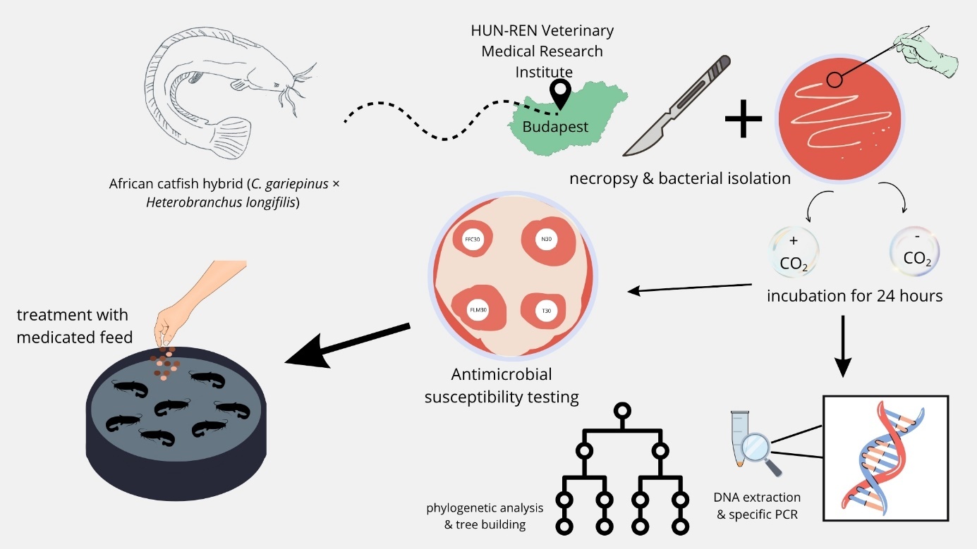
Following the detection of clinical signs, affected fish were selected by the farm owner and submitted alive for diagnostic examination by fish health professionals. Fish were selected to ensure that the characteristic pale discolouration of the caudal region was clearly visible and that they were in a condition suitable for live transport. Fish were transported alive in an aerated plastic bag to the animal facility of the HUN-REN Veterinary Medical Research Institute, Budapest, Hungary. For necropsy, fish were anaesthetised with 200 mg/L MS222 (Sigma, Merck, USA) solution and killed by a cervical cut. Sampling was performed in accordance with the relevant Hungarian legislation (Section 49 of Act No. XXVIII/1998 on the protection and preservation of animals, and Hungarian Government Decree No. 40/2013) and Directive 2010/63/EU on the protection of animals used for scientific purposes. The gills, skin, gut and internal organs of specimens were examined macroscopically and microscopically. A general parasitological examination was also performed by microscopic examination of skin scrapings and gill samples for the presence of common ectoparasites, including Ichthyophthirius multifiliis, other pathogenic protozoans, and skin and gill monogeneans.
Bacteriological examinations
Bacteriological samples were taken from the intestinal mucosa of five specimens exhibiting clinical signs of infection. Samples were inoculated onto Columbia blood agar and incubated at 25 °C for 24 h under both aerobic and anaerobic conditions. Anaerobic conditions were generated using an Oxoid AnaeroGen 2.5L Atmosphere Generation System (Thermo Fisher Scientific, USA). Colonies exhibiting distinct phenotypic characteristics on the primary culture plates were individually subcultured to obtain pure cultures, which were subsequently used for Gram staining, oxidase test, biochemical profiling and molecular identification as well as antimicrobial susceptibility testing. For biochemical identification, API 20 NE (bioMérieux, France), a standardised system for the identification of non-fastidious, non-enteric Gram-negative rods was used following the manufacturer’s guidelines.
Disk diffusion antimicrobial susceptibility testing (AST) was performed on Mueller-Hinton agar using ten antibiotics according to the guidelines of the CLSI VET03 standard. The plates were incubated at 28°C in triplicate with E. coli ATCC 25922 as quality control (QC) under aerobic conditions for 24 hours. To the best of our knowledge, validated clinical breakpoint values are not available for the bacterial strains examined in the present study, therefore breakpoint values for Aeromonas spp. were used based on the standards CLSI VET04 Ed3, CLSI VET09 Ed2, CLSI M45 Ed3 and EUCAST.
Molecular methods
Molecular identification was carried out using isolates from aerobic cultures. DNA was extracted using Quick-DNA Fungal/Bacterial Miniprep Kit (Zymo, USA), and DNA concentration and quality was measured with NanoDrop 2000c (Thermo Fisher Scientific, USA) spectrophotometer. Over 100 ng/µL DNA concentration was measured for all samples and the average A260/A280 ratio was 1.84. The approximately 900-bp fragment of the RNA polymerase sigma factor 70 subunit (rpoD) gene was amplified with rpoD70F (5′ACG ACT GAC CCG GTA CGC ATG TAY ATG MGN GAR ATG GNA CNG T 3′) and rpoD70R (5′ ATA GAA ATA ACC AGA CGT AAG TTN GCY TCN ACC ATY TCY TTY TT 3′) primers using the applied touchdown PCR protocol described by Yamamoto, Kasai, et al. (2000). As for the 16S ribosomal RNA (16S rRNA) gene, an approximately 1500-bp fragment was amplified using 16SF (5′ AGA GTT TGA TCA TGG CTC AG 3′) and 16SR (5′ GGT TAC CTT GTT ACG ACT T 3′) described by Borrell et al. (1997). The PCR products were purified using ExoSAP-IT PCR Product Cleanup Reagent (Thermo Fisher Scientific, USA) and subjected to Sanger sequencing using BigDye Terminator v3.1 Cycle Sequencing Kit (Thermo Fisher Scientific, USA). For DNA sequencing, the amplification primers of the 16S rRNA PCR product and the following rpoD sequencing primers were used: rpoD70Fs (5′ACG ACT GAC CCG GTA CGC ATG TA 3′) and rpoD70Rs (5′ ATA GAA ATA ACC AGA CGT AAG TT 3′). The resulting sequences were identified using BLASTn Megablast analysis tools of NCBI GenBank. In parallel, the 16S rRNA sequences were identified by similarity search in the EzBioCloud database. The 16S rRNA and rpoD genes were sequenced for three isolates representing the dominant colony morphotype and one isolate from each of the other two colony morphotypes. The consensus DNA sequences of Aeromonas dhakensis strain FGY5A were submitted to GenBank under the accession numbers: PZ725175 (1390-bp partial 16S rRNA) and PZ729943 (771-bp partial rpoD).
Phylogenetic analyses
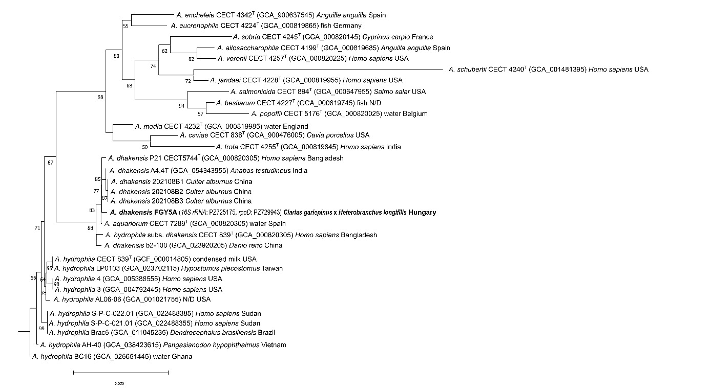
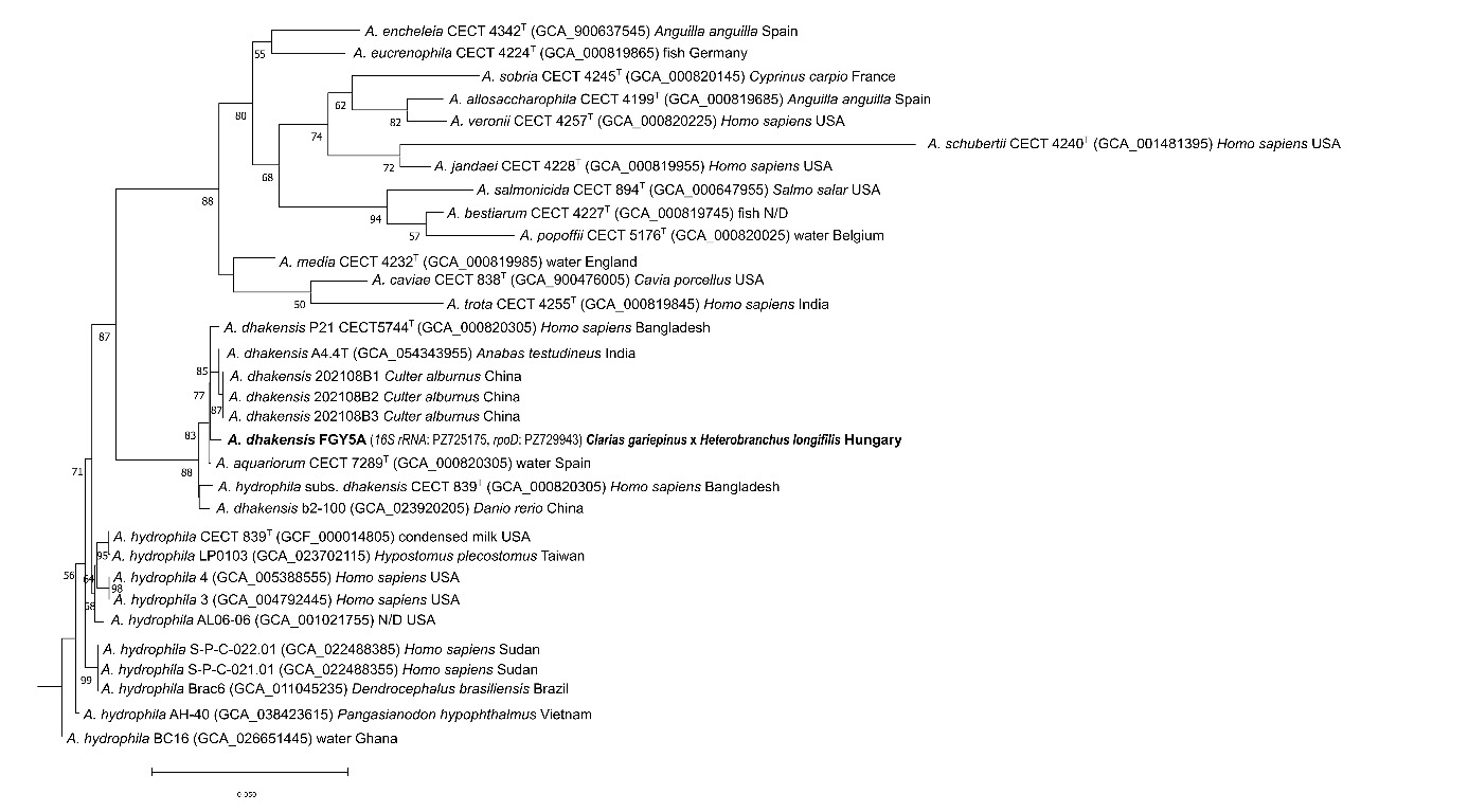
The phylogenetic tree was inferred based on a 2056-bp concatenated nucleotide alignment of partial 16S rRNA and rpoD gene sequences. Reference sequences were selected from the NCBI GenBank database using BLASTn homology searches, complemented by additional phylogenetically relevant sequences, including isolates of fish origin. The nucleotide alignment was generated using MAFFT version 7.450 (Katoh and Standley 2013), and the evolutionary model selection was performed with ModelTest-NG version 0.2.0 (Darriba, Posada, et al. 2020), which identified the TIM1+I+G4 model for 16S rRNA sequences and TrN+G4 for rpoD as the best fit for the nucleotide alignment. A maximum likelihood phylogenetic tree was inferred using RAxML-NG version 1.2.2 (Kozlov, Darriba, et al. 2019); branch support was assessed using the transfer bootstrap expectation (TBE) (Lemoine, Domelevo Entfellner, et al. 2018) approach with 1000 replicates. TBE support values below 50% were cut off. The phylogenetic tree was visualised using MEGA version 11 and further edited in Inkscape version 1.3.2. (Tamura, Stecher, et al. 2021). Vibrio cholerae strain RFB16 was chosen as an outgroup.
Results
Clinical observations
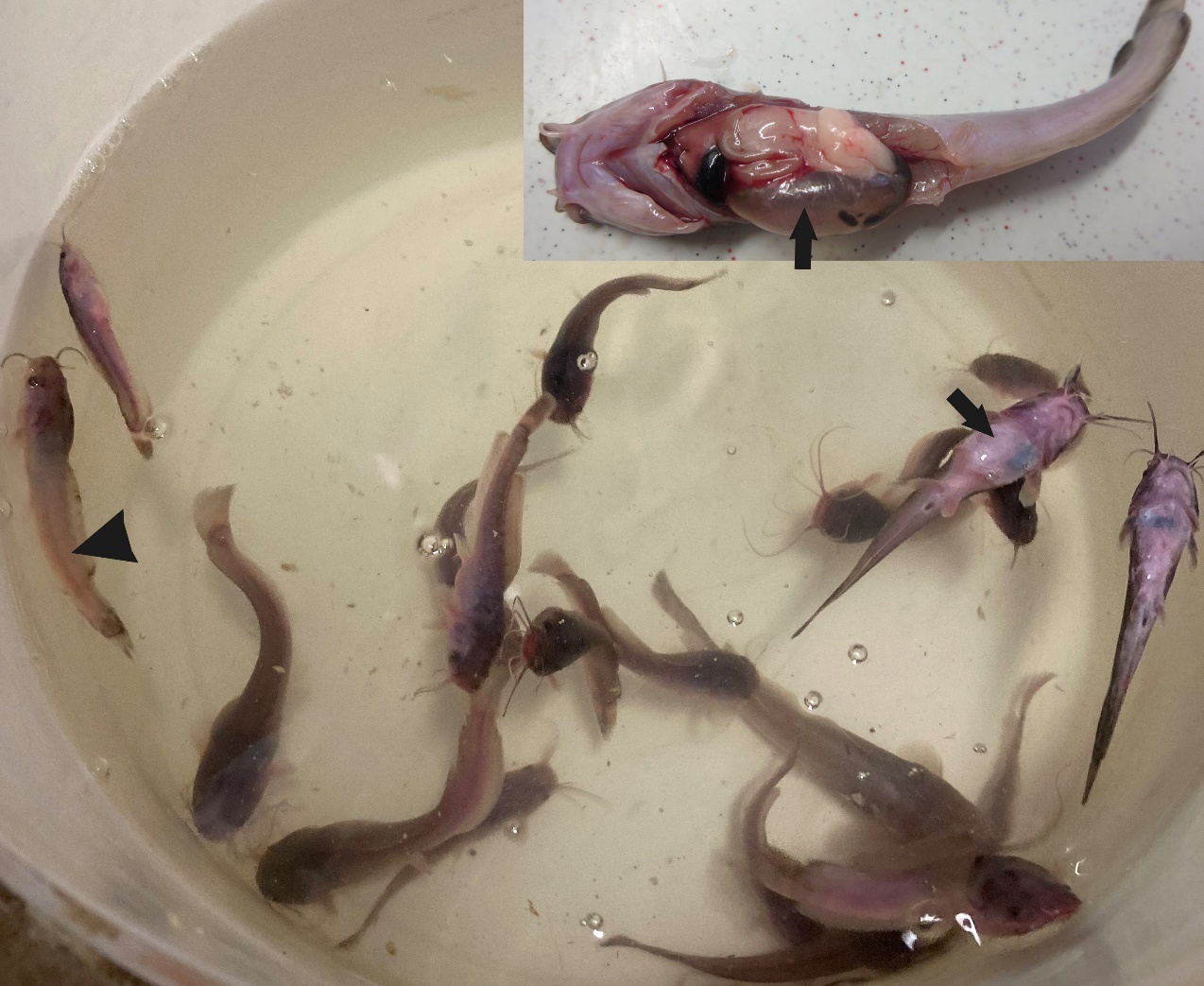
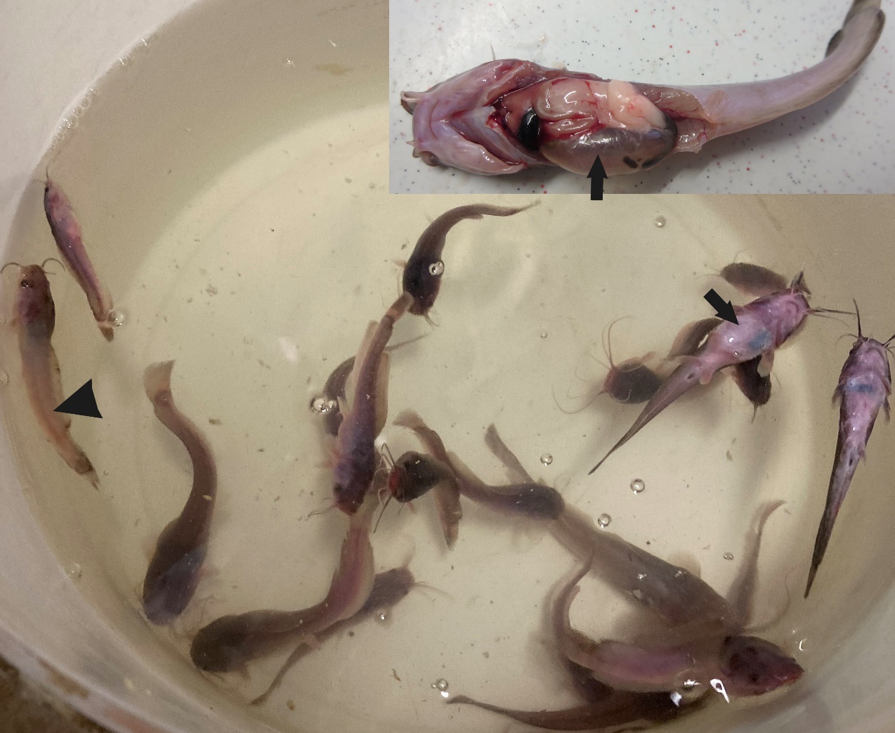
Gross examination revealed marked intestinal distension, multifocal petechial haemorrhages of the intestinal wall, and extensive inflammatory changes affecting a large portion of the intestine (Figure 1). The presence of feed material within the intestinal lumen indicated that the fish had continued to ingest feed before necropsy. No parasites were detected in the examined organs, including the gills, skin, and internal organs.
Characterisation of bacterial isolates
Bacteriological examination of intestinal mucosal samples yielded abundant bacterial growth under both aerobic and anaerobic incubation conditions. Based on colony morphology, three distinct colony morphotypes were identified. Gram staining and oxidase test confirmed that all three isolates are Gram-negative, oxidase-positive rods. The predominant colony morphotype (>90% of colonies) consisted of rapidly growing, grayish-white, pigmented, haemolytic colonies, exhibiting identical colony morphology and growth characteristics under both incubation conditions. In addition, two less abundant morphotypes, each representing <10% of the total colony population, were observed. One consisted of slow-growing, non-pigmented, haemolytic colonies, whereas the other comprised slow-growing, pigmented, non-haemolytic colonies. The biochemical profiling using API 20 NE system did not provide definitive species-level identification of the bacterial isolates. The haemolytic isolates exhibited a biochemical profile similar to that of the Aeromonas hydrophila/caviae complex, although the dominant morphotype differed in its negative L-arabinose (ARA) reaction. The biochemical profile of the third isolate showed similarity to, but did not fully correspond with, that of the Shewanella putrefaciens group (Table 1).
The consensus sequences obtained from the three isolates representing the dominant bacterial colony morphotype were identical. Sequence analysis tentatively identified all three isolates as Aeromonas dhakensis. The 16S rRNA gene analysis showed 99.85% identity (query coverage, Q: 100%, E-value 0.0) to the A. dhakensis type strain P21ᵀ (CECT 5744; NCBI Acc. No. CDBH01000037). The consensus sequence obtained in the present study (PZ725175) contained an ambiguous nucleotide (R = A/G) at position 963, which was counted as a mismatch by BLASTn. Apart from this ambiguous position, the sequence differed from the type strain by only one nucleotide, corresponding to an effective sequence identity of 99.93%. Similarly, the 99.93% BLAST identity obtained with several other A. dhakensis isolates resulted solely from the ambiguous nucleotide at position 963; otherwise, the sequences were identical. These included A. dhakensis strain B88 (NCBI Acc. No. KU041796) from a striped catfish (Pangasianodon hypophthalmus) in Vietnam, A. dhakensis strain b2-100 (ON533364) from Danio rerio in China and A. dhakensis strain AHAPRM2 (PZ263896) from a diseased fish in India. The rpoD gene analysis confirmed complete identity (Q: 100%, E-value 0.0) with A. dhakensis VA54 (AP027936) from Anabas testudineus (Teleostei) in Vietnam and A. dhakensis strain UGH41 (LC547029) from a human clinical case from Japan. The A. dhakensis type strain P21T CECT5744 (NCBI Acc. No. EF465510) was 99.2% identical (Q: 100%, E-value 0.0) with A. dhakensis FGY5A (PZ729943). The only A. dhakensis strain isolated from African catfish to date is strain C83c (NCBI Acc. No. OK058317) from Nigeria. Its 811-bp partial fragment exhibited 99.38% 16S rRNA gene sequence identity to A. dhakensis strain FGY5A (PZ725175) identified in the present study.
Regarding the other two colony-morphotypes, the haemolytic one was identified tentatively as Aeromonas veronii. Based on its 16S rRNA gene sequence, it was 100% identical to A. veronii strain AV2009 (OP476500) from China, to Aeromonas sp. A8-52 isolated from the digestive tract of freshwater fish (KM222609) in China, and to A. veronii strain H8 (MN007151) from the digestive tract of Silurus lanzhouensis from China. Based on rpoD, the A. veronii isolate from the present study was 99.62% identical to A. veronii strain A7 (MW838176) from a human faeces sample in Australia. The third colony-morphotype was identified tentatively as Shewanella xiamenensis based on the rpoD gene, and it was 99.7% identical to S. xiamenensis strain FH-1 (CP106874) from the intestine of Danio rerio from the USA.
Phylogenetic results
Phylogenetic analysis showed that A. dhakensis strain FGY5A grouped with the A. dhakensis type strain P21T and several fish-derived isolates, including strain A4.4T from Anabas testudineus, strains 202108B1, 2020108B2 and 202108B3 from Culter alburnus, and strain b2-100 from Danio rerio, with strong bootstrap support (Figure 2). Aeromonas hydrophila formed a separate sister lineage, clearly distinguishing the two species in the phylogenetic tree. In addition to the type strain, A. dhakensis P21T CECT 5744 / CIP 107500, originally isolated from a human patient in the USA, the A. dhakensis clade also included strains previously classified as A. aquariorum and A. hydrophila subsp. dhakensis, both of which are now regarded as synonyms of A. dhakensis (Beaz-Hidalgo, Martínez-Murcia, et al. 2013).
Antimicrobial susceptibility testing (AST) results and treatment suggestions
Disc diffusion AST test revealed that the A. dhakensis isolates were susceptible to examined antibiotics except to sulphonamides (S3300/SMX300) for which the bacterial strain was resistant, and erythromycin (E15) with intermediate susceptibility (Table 2). Based on the AST profiles and considering the severity of the bacterial outbreak, the affected fish stock was treated under veterinary supervision with a combination of neomycin and oxytetracycline (Neo-Te-Sol AUV), administered through medicated feed at a dosage of 100 mg/kg body weight for five consecutive days. At the initiation of treatment, fish continued to exhibit clinical signs and mortality persisted. However, mortality decreased markedly within two days after the start of treatment and subsequently ceased. No fish exhibiting abdominal distension were observed one week after treatment initiation.
Discussion
Aeromonas dhakensis is a facultatively anaerobic, Gram-negative, rod-shaped bacterium commonly found in warm freshwater environments. It was previously misidentified as Aeromonas hydrophila due to phenotypic similarities (Beaz-Hidalgo, Martínez-Murcia, et al. 2013; Chen, Lamy, et al. 2016). The species exhibits optimal growth at temperatures between 28 and 37 °C, with maximal proliferation occurring at 35–37 °C, and is considered markedly more virulent than many other Aeromonas species (Bartie and Desbois 2024). A. dhakensis was first described in 2002 based on isolates recovered from stool samples of children and environmental water samples collected in Dhaka, Bangladesh, the location from which the species name originates (Huys, Kämpfer, et al. 2002).
In aquaculture, A. dhakensis represents a significant pathogen, particularly affecting warm-water freshwater fish species under elevated water temperature conditions. Besides A. hydrophila, A. dhakensis is commonly associated with Motile Aeromonas Septicaemia (MAS), a disease characterised by high morbidity and mortality rates (Bartie and Desbois 2024). In humans, A. dhakensis is considered an opportunistic pathogen causing gastroenteritis, primarily affecting immunocompromised individuals and children. Transmission occurs predominantly via water, either through direct contact with contaminated water or through the consumption of contaminated food (Chen, Lamy, et al. 2016). The bacterium is also capable of causing wound infections when damaged skin comes into contact with contaminated water and may lead to secondary bloodstream infections. In rare cases, respiratory tract infections, including pneumonia, or urinary tract infections have been reported (Chen, Wu, et al. 2014; Jiang, Zhao, et al. 2024). In fish, A. dhakensis most commonly induces haemorrhagic septicaemia, which represents the most severe clinical manifestation of infection caused by this species (Mulia, Pratiwi, et al. 2023). Additionally, ulcerative dermatitis may develop following superficial skin injuries or stress-related immunosuppression, facilitating bacterial invasion. Less frequently, A. dhakensis has been associated with meningoencephalitis in fish (Bartie, Ngô, et al. 2023; Hassan, Ali, et al. 2025; Wang, Feng, et al. 2025).
In addition to A. dhakensis, two other potential pathogen bacterial strains were detected. Although pathogenic strains of A. veronii are well documented and are most commonly associated with bacterial septicaemia and skin ulceration in freshwater fish (Behera, Parida, et al. 2023; Li, Raza, et al. 2020), some strains, such as A. veronii A7, have also been reported to cause gastroenteritis in humans (Yuwono, Wehrhahn, et al. 2021). Regarding S. xiamenensis, the third bacterial species identified in the present study, a recent report described the isolation of this species from an infected koi (Cyprinus rubrofuscus) exhibiting ulcerative dermatitis (Rich and Naguib 2024). In contrast, S. xiamenensis strain FH1 (CP106874), which was closely related to our S. xiamenensis isolate (99.7% sequence identity), was considered non-pathogenic and described as a gut-associated bacterial strain of Danio rerio (Grenfell, Intile, et al. 2023). In the present study, both A. veronii and S. xiamenensis were isolated from only one of the five examined fish, whereas the A. dhakensis strain was consistently recovered and represented the predominant isolate.
The potential pathogenic role of A. dhakensis in African catfish and their hybrids is supported by previous findings (Adah, Saidu, et al. 2024) who isolated A. dhakensis from African catfish exhibiting intestinal congestion, gastrointestinal fluid accumulation, fin erosion, and skin haemorrhages. In the present case, pronounced gross intestinal lesions were observed, and A. dhakensis was consistently recovered as the dominant bacterial species from the affected intestinal tissue, supporting a possible association between A. dhakensis infection and gastrointestinal pathology. However, several limitations preclude confirmation of a direct causal relationship. Histopathological examination was not performed and, therefore, the nature and extent of the intestinal lesions could not be further characterised microscopically. In addition, bacteriological examination was restricted to the sites of the observed clinical and pathological alterations, and no samples were collected from the kidney or other internal organs. Consequently, systemic dissemination of A. dhakensis could not be assessed or confirmed. Thus, while the predominance of A. dhakensis in the affected intestinal tissue, together with the observed lesions and previously reported findings in African catfish, supports its potential involvement in the disease, a definitive causal association with enteritis cannot be established from the present findings alone.
The source and route of introduction of A. dhakensis into the production system also remain unknown, although anthropogenic introduction cannot be excluded. To the best of our knowledge, this is the first reported detection of A. dhakensis in a hybrid African catfish and the first record of its occurrence in Hungary.
Acknowledgements
We thank Győző L. Kaján for his helpful advice regarding the phylogenetic analysis.