
Introduction
Amoebic gill disease(AGD) caused by Paramoeba perurans (synonym Neoparamoeba perurans), a member of the Sarcomastigophora (Paramoebidae) family (Crosbie et al. 2012), has a significant global impact on the Atlantic salmon (Salmo salar) aquaculture industry (Oldham et al. 2016). Economic losses due to AGD-related mortalities have been estimated at USD 12.55 million in Norway in 2006 and USD 81 million in Scotland in 2011 (Shinn et al. 2015). The increasing prevalence and economic impact have positioned AGD as one of the most economically important parasitic diseases affecting marine-farmed salmon globally. Infected salmon develop mucoid, proliferative gill lesions characterised by lamellar epithelial hyperplasia and variable fusion of adjacent lamellae (Adams et al. 2004; Slinger et al. 2021), resulting in impaired oxygen exchange (Herrero et al. 2018) and, if untreated, potential mortality (B. L. Munday et al. 2001). Affected fish display pronounced respiratory distress, characterised by increased opercular movements and surface-oriented swimming behaviour.
Since its initial description in Tasmania in the 1980s (B. Munday 1986), AGD has been reported across all major salmon-producing regions and is now considered endemic in salmonid aquaculture in countries including Australia, New Zealand, the USA, Scotland, Ireland, the Faroe Islands, Japan, Spain, Norway, and Chile (B. L. Munday et al. 2001; Bustos et al. 2011; Rodger 2014; Oldham et al. 2016; Blindheim et al. 2023).
To mitigate the impact of AGD, in vitro culture methods for P. perurans have been developed (Crosbie et al. 2012), providing a platform to study the parasite’s biology and behaviour under controlled conditions. These methods allow both field isolates and in vitro clones derived from recovered isolates to be maintained in culture (Dahle et al. 2020; Blindheim et al. 2023) . Amoebae exhibit a high tolerance to suboptimal environmental conditions, surviving at low temperatures (2–4°C) and across a wide salinity range (20–50‰), while still maintaining colony growth(Collins et al. 2019) . However, their virulence appears to decline following prolonged clonal culture, in some cases resulting in failure to induce disease in aquarium challenge models, likely due to downregulation of virulence-associated gene expression (Smith et al. 2022), which limits the reproducibility of such studies. Additionally, maintaining a viable isolate of P. perurans requires considerable time and attention. Due to the growth characteristics of P. perurans, continuous in vitro culture requires frequent passaging, as interruptions rapidly result in culture collapse and loss of isolates. Given the substantial labour required for amoeba maintenance and evidence that prolonged in vitro culture can reduce virulence, we conducted a literature review and consulted with other research groups working with amoebae, who reported encountering similar challenges. Previous attempts to cryopreserve P. perurans using protocols developed for other fish-associated amoebae have produced inconsistent and isolate-dependent outcomes, and in our case the strain could not be recovered following freezing. Based on the literature review, we adapted existing cryopreservation approaches from other amoebae for application to P. perurans and aimed to share these findings with the wider research community. Accordingly, the objective of this study was to evaluate four cryopreservation methods using two polyclonal field isolates to identify a strategy for the long-term storage of P. perurans.
Materials and Methods
Experimental design
Cryopreservation experiments were conducted as an exploratory pilot study. For each isolate and each freezing method, six independent cryovials were prepared and thawed at two time points (15 and 90 days post-freezing), resulting in six biological replicates per condition and time point. The study was not designed or powered to compare freezing methods statistically, and outcomes were evaluated descriptively using culture recovery and qualitative viability assessment.
Maintenance P. perurans culture
Two polyclonal P. perurans isolates (“Bears” and “Toms”) were kindly provided by Dr M. McCormack (Atlantic Technological University, Ireland). Detailed information regarding passage number prior to receipt of the isolates was not available. Upon arrival, both cultures were actively growing and were maintained under standard conditions for approximately 16 weeks prior to cryopreservation.
They were cultured at 17°C in unvented T75 cell culture flasks (Corning™ Cell Culture Treated Flasks, Corning Inc., NY, USA) using 34‰ salinity, 0.1 % w/v sterile Malt-Yeast Broth (MYB), which was filtered using 0.2 µm pore filters (Corning® bottle-top vacuum filters, Corning, Tewksbury, USA) then sterilised by autoclaving. Amoebae were cultured until each isolate reached approximately 1 × 10^7 cells, achieved using 6–7 culture flasks, with cell numbers quantified using a Neubauer counting chamber (BRAND™ Blaubrand™ Neubauer Improved Counting Chamber; Sigma-Aldrich, Darmstadt, Germany). Amoebae were removed from the flasks using cell scrapers (Sarstedt, Sarstedt Inc., NC, USA), collected in 50 mL centrifuge tubes (Falcon™ 50 mL High Clarity Conical Centrifuge Tubes, Fisher Scientific, Corning Inc., NY, USA) and centrifuged at 600 RCF for 10 min. After removing the supernatant, the pellets were collected, resuspended by vortexing and divided into 49 aliquots of approximately 2x10^5 cells per aliquot. An aliquot from each isolate was used to confirm amoeba species identity and to verify the pre-freezing concentration in each vial using a species-specific RT-qPCR assay.
Real-Time qPCR for P. perurans
Real-time quantitative PCR targeted the 18s rRNA gene of P. perurans as previously described (Downes et al. 2015). Briefly, genomic DNA was extracted from the amoebae using a commercial kit (DNeasy® Blood & Tissue Kit) according to the manufacturer’s instructions. The qPCR master mix was composed of 10 μL PerfeCTa qPCR ToughMix, Low ROX (2X) (Quantabio, Manchester, UK), 300 nM final concentration of each primer (forward primer 5’- AAAAGACCATGCGATTCGTAAAGT -3’[MGEQ] and reverse primer, 5’-CATTCTTTTCGGAGAGTGGAAATT-3’), 200 nM final concentration of fluorescent probe (TaqMan® probe, [FAM] 5’-ATCATGATTCACCATATGTT-3’[MGBEQ]) and 2 μL DNA in a final volume of 25 μL. The thermal cycling conditions were 95°C for 30 seconds, followed by 45 cycles of 95°C for 5 seconds and 60°C for 30 seconds using a Quant Studio 5 Real-Time PCR System (Thermo-Fisher, London, UK), following the manufacturer’s standard protocols. Each sample was tested in triplicate. DNA from a confirmed case of AGD was used as a positive control for each round. The standard curve was generated using serial dilutions (10 to 10^7 copies of the 18S rRNA gene target) using a gBlocks® Gene Fragments (integrated DNA Technology, London, UK); sequence: CATGGTAGTCCTCTATACTACCATCGAAAGTTGTTAGGGCAGATATTTGAATGAACTATCGCGGCACAAAAGACCATGCGATTCGTAAAGTTATCATGATTCACCATATGTTAAATTTCCACTCTCCGAAAAGAATGGCATTGGCTTTTGAATCTAATAAATACATCCCTCCCAGCTCCCGAAAGAACCAGTCAAGATTTTATGCATGTATTAGCTCTAGATTTACCACGGTTATCCAAGTAGTAAAGAGACATCAAATAA.
Cryopreservation of amoebae cultures
The remaining 48 aliquots from each isolate were resuspended in four different cryoprotectant solutions (12 aliquots per solution) and transferred into sterile 1.8 mL cryogenic storage vials at a volume of 0.5 mL per vial (Cryogenic Vials; Starlab International GmbH, Hamburg, Germany). Samples were placed in a controlled-rate freezing container (Mr Frosty™ Cryo 1 °C Freezing Container; Thermo Fisher Scientific Inc., Waltham, USA) containing 500 mL isopropyl alcohol and cooled at −70 °C overnight prior to long-term storage at −150 °C.
Each isolate was frozen in four different freezing solutions based on their effectiveness with other amoeba species (Fahy et al. 1984; Kilvington and White 1991; Jessu et al. 2024) These include M1) Sucrose (0.5 M) in sterile-filtered, autoclaved 36‰ salinity seawater (SW), M2) 10% dimethyl sulfoxide (DMSO) in 36‰ SW; M3) cell freezing medium (CFM)-DMSO 1× (Sigma-Aldrich, Saint Louis, USA) and M4) 10% DMSO in foetal bovine serum (FBS) + 36‰ salinity SW (1:1 solution).
Thawing of amoebae
The thawing of the samples was performed at 15 and 90 days after freezing (daf), to test short- and long-term survival of the amoebae. The cryotubes were removed from the freezer and rapidly thawed in a water bath at 50℃ for 5 seconds. Immediately afterwards, the semi-frozen pellet was transferred to a 50 mL Falcon tube containing 20 mL of sterile MYB, vortexed, and incubated at 17°C in a T75 cell culture flask. The culture was maintained in the dark for 12 weeks, with passages performed every other week. For each passage, the flask was scraped, and the entire contents were collected into a 50 mL centrifuge tube. The tube was then centrifuged at 600 RCF for 10 min, and the supernatant was removed. The pellet was resuspended in 20 mL of fresh MYB and transferred to a new flask.
Viability testing
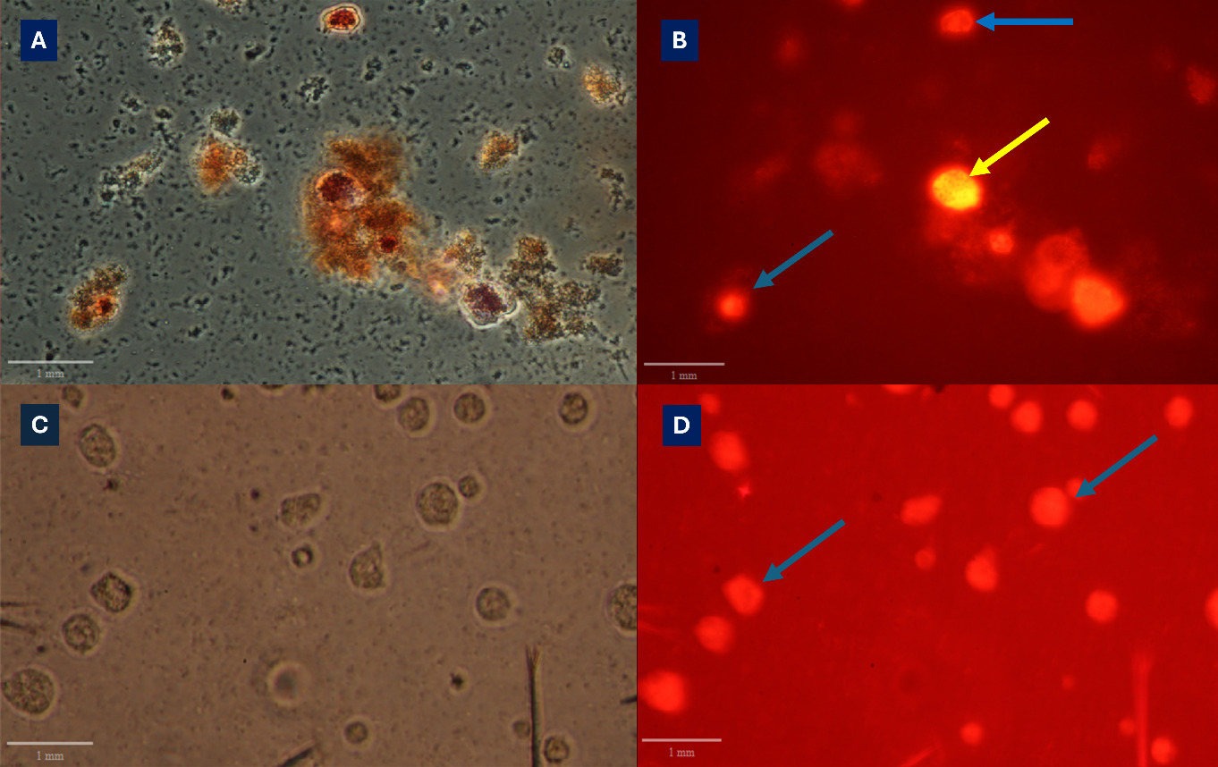
An aliquot (100 µL) of each frozen sample was thawed and placed in a 6-well plate containing 2 mL of 36‰ SW, then stained with 200 µL per well of a 0.33% Neutral Red solution (Sigma-Aldrich, Gillingham, UK). After 2 h, the plates were centrifuged at 1000 RCF, and the supernatant was removed. The plates were washed three times with sterile phosphate-buffered saline (PBS). After the last wash, 2 mL of 36‰ SW was added, and each well was observed under a fluorescent microscope (absorbance at 540 nm and 630 nm). As the Neutral Red dye is taken up only by live amoebae, which fluoresce when stimulated with UV, the number of fluorescent amoebae was used to identify viable cells (presence or absence).
Results
For the two strains, the initial vial contents were quantified by qPCR (in copies of the 18S rRNA gene target), yielding 90,637 and 106,428 amoebae/vial for Bear and Toms, respectively. The viability of the samples was also assessed by testing individual vials for the presence of live amoebae using Neutral Red. This was performed by observing different areas within each well and identifying fluorescent amoebae (Fig. 1). Although the amoebae in most groups were dead, live amoebae were detected in the groups from which viable amoeba cultures were subsequently obtained (M3 and M4).
Viable amoebae were recovered following cryopreservation using two of the four methods tested. For method M3, viable cells were successfully recovered from all six vials (100%; 6/6) for both the Bear and Toms isolates at 15 and 90 days after freezing (daf). Method M4 showed reduced recovery rates, with 100% viability for both isolates at 15 daf, which decreased to 83% (5/6) for Toms and 67% (4/6) for Bear at 90 daf. All vials from which live amoebae were recovered subsequently gave rise to established cultures. No viable amoebae were recovered using methods M1 or M2. These results are summarised in Table 1.
Discussion
Against this background, the present study aimed to explore whether simple cryopreservation approaches could support long-term storage of viable P. perurans cultures. Despite these encouraging observations, this study has several important limitations that must be acknowledged. The work was designed as a pilot, exploratory investigation rather than a statistically powered comparison of cryopreservation methods. Although six cryovials per isolate and method were thawed at each time point, no formal experimental design was implemented to support statistical comparison among freezing protocols, and no quantitative statistical analyses were performed. Consequently, the results should be interpreted as preliminary evidence of feasibility rather than definitive proof of method superiority. Although this was a pilot study with a limited number of replicates, the results were considered sufficiently relevant to justify dissemination.
Cryopreservation of Paramoeba perurans has previously been reported by Tröße et al. (2021), who successfully recovered four of five clonal isolates following freezing in 10% DMSO and storage in liquid nitrogen. However, the authors also reported very low estimated recovery rates and highlighted the need for further optimisation of cryopreservation methodologies. The present study complements and extends those findings by evaluating additional cryoprotective formulations under laboratory conditions and demonstrating recovery of viable amoebae following cryopreservation using multiple approaches. While the current work remains preliminary and was not designed to quantify post-thaw survival, the results provide independent confirmation that cryopreservation of P. perurans is feasible and support continued development of practical long-term preservation strategies for AGD research.
Importantly, the aim of this study was not to optimise cryopreservation conditions or to statistically compare freezing methods, but rather to assess feasibility using straightforward protocols that could be readily adopted by laboratories working with AGD. In this respect, the observation that viable amoebae could be recovered using simple DMSO-based approaches supports further investigation into cryopreservation as an alternative to continuous passage. The use of a qualitative Neutral Red viability assay also proved useful as a rapid screening tool, as detecting metabolically active amoebae shortly after thawing was consistent with eventual recovery of in vitro cultures.
Given reports that only a small proportion of cells survive cryopreservation, with conservative estimates of recovery rates ranging from 0.0002% to 0.0005% (Tröße et al. 2021), detecting even a single viable amoeba remains a considerable challenge. However, viability assessment was qualitative rather than quantitative. The Neutral Red staining approach used here was applied to determine the presence or absence of metabolically active amoebae shortly after thawing, rather than to estimate the proportion of cells surviving cryopreservation. While this approach proved useful as a rapid screening tool that correlated with subsequent culture recovery, it does not provide information on recovery efficiency or percentage survival, and differences among freezing methods cannot be quantified. We were able to identify viable amoebae in 4/6 to 6/6 cryotubes, in which we could initiate a new culture. The results showed that recovery after thawing occurred only in vials containing viable amoebae, indicating that the viability test reliably predicts post-thaw survival. This approach offers a practical method for evaluating the viability of cryopreserved samples without requiring visible growth in culture. Additionally, centrifuging the culture to pellet the amoebae at the bottom of the plate resolves the challenge of distinguishing between floating and adherent forms. This approach ensures that all amoebae, regardless of their original location in the culture, are collected and assessed. It allows for the identification of metabolically active cells, which are expected to be the ones capable of initiating growth in a new culture. However, future studies should incorporate direct cell counts before and after freezing or alternative quantitative viability assays to enable a more robust evaluation of recovery rates.
Cryopreservation is essential for maintaining amoeba isolates with their original phenotypic traits, but effects on virulence were not assessed in the present study. Although loss of virulence during prolonged in vitro culture is widely reported in the literature and was cited as a motivation for developing cryopreservation strategies, the present work did not include in vivo challenge experiments or other functional assays to assess whether cryopreserved and recovered amoebae retain their pathogenic potential. Accordingly, no conclusions can be drawn regarding preservation of virulence, and this remains a critical topic for future investigation.
Morphological assessment of P. perurans trophozoites before and after cryopreservation was limited. Microscopy images presented here are illustrative to show criteria and were not intended to provide a detailed comparative analysis of trophozoite morphology or attachment behaviour following thawing. The absence of systematic morphological analysis and quantitative image data represents a further limitation of this study.
Although some variation in post-thaw recovery times was observed, there was no evidence that extending storage duration to 90 days at −150 °C significantly reduced recovery efficiency. Future studies should evaluate the maximum duration of cryopreservation at different storage temperatures, including −70 °C and −150 °C, to determine their effects on long-term amoeba viability. Although culture conditions following thawing were standardised, detailed historical information on passage number prior to receipt of the isolates was not available. Variation in culture history may influence cryotolerance and recovery, warranting consideration in future work with fully documented isolates. In addition, a quantitative assessment of post-thaw live-cell recovery should be undertaken. Repetition of the most effective cryopreservation methods with increased numbers of biological and technical replicates would enable robust statistical estimation of recovery rates and cell survival, thereby improving confidence in the reproducibility and reliability of the protocol.
In summary, this study provides preliminary evidence that viable P. perurans can be recovered following cryopreservation using relatively straightforward, accessible methods. While the findings support the feasibility of long-term storage as an alternative to continuous culture, they should be interpreted with caution, given the qualitative nature of the data and the lack of statistical analysis. Further work incorporating increased replication, quantitative viability measurements, virulence assessment, and extended storage durations will be required before robust, standardised cryopreservation protocols for P. perurans can be established.
Conclusion
In this study, we have described two cryopreservation approaches that showed potential for recovering viable P. perurans as an alternative to successive, time-consuming passages in standard in vitro culture. While previous studies have demonstrated the possibility of cryopreserving P. perurans, the present work provides further evidence that viable cultures can be recovered using relatively simple and accessible cryopreservation protocols.
Informed Consent Statement
Not applicable.
Data Availability Statement
Not applicable.
Ethics Statement
Not applicable.
Conflicts of Interest
“The authors declare no conflict of interest.”
Acknowledgements
The authors would like to thank the “NeoGIANT: The Power of grape extracts” project, funded by the European Union’s Horizon 2020, for financing this study. Also, thanks to Dr Michelle McCormack (Atlantic Technological University, Ireland) and Dr Catherine Collins (INRAE, Université Paris-Saclay, France) for their support with amoeba culture and diagnosis, and Dr Stephen Fitzgerald (Moredun Research Institute, UK) for his assistance in designing the gBlock.