The horse mackerel fish belongs to the genus Trachurus within the Carangidae family. In the Turkish seas, these fish are represented by two species, namely, Trachurus mediterraneus and Trachurus trachurus. These fish have the ability to adapt well to their culture environment and are, therefore, used in aquaculture, taking fish from the wild and reared then in cages. Successful results have been reported in fattening trials with this fish reared in Turkey’s Eastern Black Sea region (Başçınar et al. 2015). Bacterial diseases occurring in both wild and cultured fish may lead to serious ecological and economic losses. High levels of mortality and the cost of chemicals used for treatment have caused economic losses for fish farmers, particularly in intensive aquaculture systems. There are a few reports with regard to disease problems in horse mackerel obtained from wild stocks, particularly bacterial diseases. Severe skin lesions caused by Vibrio sp. and Pseudoalteromonas haloplanktis have been reported in wild horse mackerel (Kayış and Er 2015). A study investigating the bacterial pathogens from horse mackerel during the fattening period reported on the isolation of Aeromonas hydrophila, Chryseobacterium indologenes, Vibrio vulnificus, Bulkholderia cepacia, Photobacterium damselae damselae, and Vibrio alginolyticus from the fish (Boran et al. 2013). Some studies have also reported the presence of disease symptoms associated with certain bacteria in the horse mackerel fish in wild conditions or during the fattening period. In particular, some characteristic bacterial disease symptoms, including skin lesions (Kayış and Er 2015), darkened skin, ascites, and petechial bleeding in the abdomen, have been reported (Boran et al. 2013), although not completely elucidated. Previous studies concerning this topic have remained limited to screening studies. However, in the present study, the pathogenic effect of Vibrio vulnificus, previously isolated from the skin lesions of the horse mackerel (T. mediterraneus) caught from the Black Sea was investigated in an experimental infection trial, which showed that this bacterium caused an infection with significant symptoms in the fish, which ultimately resulted in large numbers of fish mortalities.
The fish used in the present study (T. mediterraneus) were line-caught fish from the coast of Trabzon within the coast of the Eastern Black Sea Region of Turkey. Subsequently, the fish were brought to the Fisheries Faculty in the Toxicology Laboratory of Recep Tayyip Erdoğan University and maintained in oxygenated tanks at a temperature of 18 ±2 °C under 12:12 light/dark conditions for a week prior to the experiment for adaptation. In this period, Chloramine-T, 15 mg/L was applied to the fish for 30 min to eliminate any detrimental bacterial flora that might have occurred on the external surface of fish due to the capture conditions. In addition, bacterial examination was conducted in the liver, spleen, and kidney tissues of the fish.
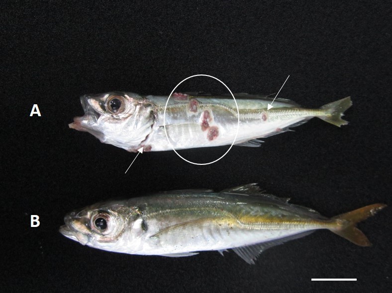
The trial setup consisted of glass tanks, which provided a static system with sufficient oxygen in a volume of 50 L. The experiment was conducted in triplicate. The fish groups used in the experiment and the average fish weight values are provided in Table 1. Vibrio vulnificus isolated from mackerel in the Eastern Black Sea region of Turkey in 2017 by our researchers was used for infection. Serious skin lesions were observed in the fish from which the pathogen was isolated (Figure 1). Tryptic Soy Agar (TSA) medium supplemented with 1.5% sodium chloride was used for the isolation and identification of bacteria. The medium was incubated at 20±2°C for 24h. The genomic DNA was obtained from pure colonies using the boiling method (Dashti et al. 2009). The primers used for the PCR reactions specific to the 16S rRNA region were 27 Fwd 5’-AGA GTT TGA TCC TGG CTC AG-3 ’ and 1492 Rev 5’-GTT TAC CTT GTT ACG ACT T-3’ (Kayiş et al. 2015). The genetic material obtained from PCR was sent for sequencing. The identification of the bacteria was performed by comparing the results obtained in the present study with the previously reported data (Accession number of the bacteria in NCBI, MW295498).

In the experiment, pure bacterial culture was inoculated on Tryptic Soy Broth (TSB) medium containing salt. Subsequently, the cultured bacteria were suspended in physiological saline water (PSW) and 0.1 mL of the solution was administered intraperitoneally to fish using an insulin syringe. The number of bacteria to be administered to the fish was determined in Plate Count Agar (PCA) medium using the dilution method. The number of bacteria administered to the fish is provided in Table 1. Sterile physiological saline water (PSW) was administered to the control group fish. In the initial stage of the experiment, the fish were not fed for the first three days and after the third day, they were fed with commercial sea bass feed in amounts up to 1% of their body weight. The bacterial examination was performed on the fish exhibiting disease symptoms. During the experimental period, the tanks were cleaned regularly, and about 50% of the tank water was changed every three days. The pH, temperature, and oxygen values of the water were measured during the experiment, and the values are provided in Table 1. The salt water used in the experiment was obtained from the Rize coast of Black Sea and its salinity was 0.17%.
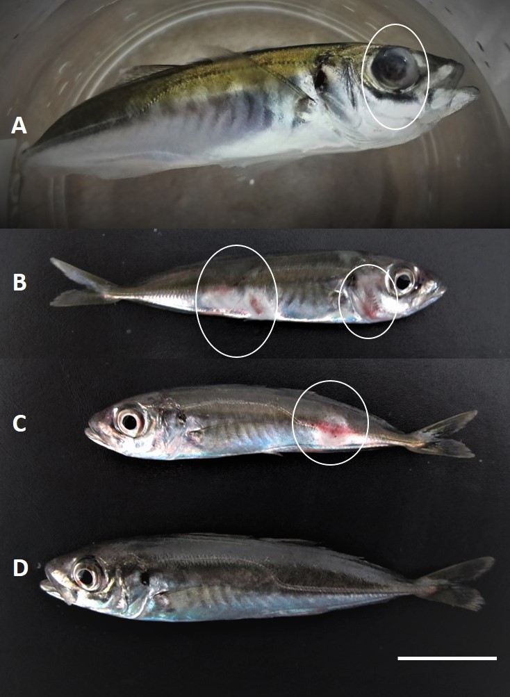
The mortality rates in the three experimental replicates were determined to be 50%, 60%, and 70% at the end of three-week infection trial. No deaths were recorded in the control groups. The infected fish were inclined to swimming at the bottom of the tank which began towards the end of 6th day post-infection. The fish in the control group, on the other hand, demonstrated normal swimming behavior within the tank. Bilateral exophthalmos in the eye was detected in a few fish in the experimental groups, while lesions on the skin were observed in several fish (Figure 2). Vibrio vulnificus was isolated from the liver, kidney, and spleen tissues of the symptomatic dead fish.

In the present study, the effects of Vibrio vulnificus infection on the horse mackerel were examined. A mean mortality of 60 % occurred in the horse mackerel infected with V. vulnificus. It’s possible that moralities resulting from infection with this pathogen in the farming situation may be prevented or reduced if the culture environment is improved, the onset of disease is detected at an early stage, appropriate antibiotics can be administered in time, and if the fish are vaccinated beforehand. In summary, the present study reports the problems associated with V. vulnificus infection in wild horse mackerel caught from the natural marine environment when the fish were infected with an isolate obtained from a natural disease outbreak in this species. This included the symptoms associated with the infection and the level of mortality that occurred. This study provides useful information to help identify and predict future disease problems with V. vulnificus in this fish species.