Introduction
Myxozoans are morphologically reduced and genetically derived cnidarian parasites that have acquired annelids and bryozoans as their primary hosts, and vertebrates, predominantly fish, as their secondary hosts. Several myxozoans are known pathogens impacting freshwater and marine aquaculture production systems, with some species classified as emerging pathogens promoted by climate change processes. Despite the obvious need, there is currently no general legalized treatment or vaccine for myxozoans. Solutions are delayed due to quirks in the biological and molecular characteristics of myxozoans, and by the lack of tools and consensus in research approaches used to tackle the most economically impactful myxozoans.
The myxozoan research community is composed of several laboratories from around the globe dedicated to research on this group of parasites, as well as many other research institutions that contribute occasionally to scientific outputs. The community holds a workshop at each international EAFP meeting, a tradition maintained also during the virtual conference in 2021. A major aim of this workshop is to encourage the formation of informal groups of laboratories that coordinate their activities and align their research strategies to produce the data that is needed to advance research in the myxozoan field. The aim is to unify species descriptions, optimize research methodologies and produce comparative results in different host-parasite systems, which can be used towards the design of common antiparasitic strategies and vaccines.
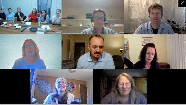
Similar to previous EAFP meetings, the online 2021 Myxozoan workshop collected information on recent progress and opinions providing a framework for future studies, as summarized below. The workshop was held on 22nd September at 19:00 CET with the aim of including a wide spectrum of experts worldwide to facilitate in-depth discussions. The meeting was informal and held over coffee or cocktails, depending on time zone (Figure 1). This approach gave rise to excellent discussions (Figure 2), making significant headway in addressing some of the major issues facing myxozoan research.
Myxozoan laboratory models, experimental infections, and in vitro cultures
Myxozoans have a complex life cycle and alternate between an invertebrate (annelid or bryozoan) and a vertebrate (generally fish). Only approx. 60 myxozoan life cycles are known to date, and laboratory research models are scarce: permanently maintained only for Myxobolus cerebralis, M. pseudodispar, Ceratonova shasta, Entermoyxum leei, E. scophthalmi, Tetracapsuloides bryosalmonae and Sphaerospora molnari). With parasite development in each host generally encompassing a period of 6 weeks to 3 months, laboratory models are time- and labour-intensive to maintain. The laboratory life cycle is shortened only for Enteromyxum spp., which can transmit from fish to fish within sloughing cells of infected intestinal epithelia (Sitjà-Bobadilla and Palenzuela 2012), and S. molnari and C. shasta, where proliferative stages from blood or ascites can be transferred by intraperitoneal injection (Holzer and Pimentel-Santos 2021; Ibarra, Gall, and Hedrick 1991). In these cases, research progress is accelerated by short circuiting myxozoan life cycles. However, there are clear disadvantages of such approaches, including potential degradation of the parasite laboratory lines owing to adaptation to the absence of the invertebrate host (Gorgoglione and Kotob 2021). This may inevitably lead to loss of virulence, including production of spores and other transmission stages that has been seen after only 5 passages from fish to fish (S.D. Atkinson, pers. comm. 2021, for C. shasta).
While in vivo laboratory propagation is performed at least for some species, there is presently a complete lack of in vitro models of myxozoan parasite stages for research. Only recently, important progress was made at least regarding the isolation of myxozoan proliferative stages from fish blood [100% purity; (Born-Torrijos et al. 2021)]; first presented at EAFP 2021 during the Myxozoa session II. In vitro cultivation trials have not yet succeeded in maintaining myxozoans in cell culture for longer than 48 hrs (mainly for S. molnari and C. shasta). During the workshop, it was recommended that future approaches to in vitro cultivation should focus on better understanding the nutritional requirements of these parasites, by targeting pathways in transcriptome data involved in metabolism and studying mass-spectrometric profiles of harvested conditioned cell media. Furthermore, it was suggested that some myxozoan parasites, especially those inhabiting the gut (C. shasta and Enteromyxum spp.) appear to require cells as substrates (comment by A. Sitja-Bobadilla and J. Bartholomew). Kudoa septempunctata sporoplasms have been shown to invade Caco-2 human intestinal cells (Ohnishi et al. 2013). The intestinal rainbow trout cell line, RTG, from the Centro de Investigación en Sanidad Animal (C. Tafalla) or the Scottish Fish Immunology Research Centre (J. Holland) was recommended as a substrate for parasite culture. This cell line was adapted to saltwater conditions for E. leei, but more time is required for optimization of culture conditions. Other possibilities for live parasite culture may be bioprints or organoids, which have so far not been tested in myxozoans yet may be successful due to their physical and biochemical similarity to real fish host tissues.
Myxozoan genomics and transcriptomics
There has been significant research activity in the sequencing and assembly of myxozoan genomes and transcriptomes, although all assemblies to date require further refinement. This will be facilitated by overcoming technical difficulties in handling myxozoans, and by development of consensus assembly pipelines with each point discussed in depth during the workshop.
Myxozoans possess extremely small genomes (e.g., Tetracapsuloides bryosalmonae, 12.9 Mbp; Buddenbrockia plumatellae, 27.7 Mbp; Sphaerospora. molnari, 40 Mbp; as presented during the EAFP 2021, Myxozoa session I) and so great care must be taken to prevent issues arising from over sequencing. For genome sequencing, combining long read chemistries, such as PacBio, Nanopore, or 10X Genomics, with short read technologies (e.g., Illumina) was found to be the best approach for myxozoan nuclear and mitochondrial genome assembly. This overcomes the issue posed by the extensive repeats found in myxozoan assemblies, since short read sequence technologies are unable to accurately sequence through repeat regions without including long read datasets. Despite such optimization, overcoming host and environmental DNA contamination remains one of the biggest issues in myxozoan assemblies. This is confounded by large genome size and polyploidy exhibited by fish hosts (Alama-Bermejo and Holzer 2021). Filtering out fish-derived reads with fish 'omic assemblies offers one solution, although this may lead to the removal of horizontally transferred genes from myxozoan datasets. Segregation of host and myxozoan reads could be based on codon usage differences with many myxozoan protein-coding genes favouring AT-rich codons (Faber et al. 2021). However, it is likely that not all myxozoan genes follow this rule, especially those encoding proteins enriched in amino acids using GC-rich codons, such as e.g., minicollagens. Ultimately, ultrapure myxozoan spores are required to develop host-free genome assemblies. Individual spores should be “polished” using mild trypsinization to remove host adherent cells prior to library preparation and sequencing.
Even in the absence of host or environmental sequence contamination, a single host tends to be infected with a myxozoan metapopulation, which leads to a great deal of read variation. This issue is likely to preclude a clear understanding of myxozoan biology via transcriptomic profiling. This could, at least in part, be resolved by performing fish challenges using single myxospores or (clonal) spore sacs, an approach that seems to be feasible for some myxozoans. This links to the issue surrounding the multicellularity of myxozoans. To fully understand the biological meaning behind gene expression changes during myxozoan infection and development will require teasing apart the transcriptome fingerprint of individual myxozoan cell types using 10X Genomics single cell sequencing pipelines. This is a currently being pursued by some myxozoan researchers, although attempts so far have been unsuccessful.
Even if further improvements to myxozoan assemblies are implemented, one of the future priorities will be to unify bioinformatic/annotation pipelines and experimental approaches, especially given the current conflictions in the reported size of intergenic repeat units and overall size of myxozoan genomes. Furthermore, new knowledge concerning the fallibilities of automated sequence filtering tools, for example in removing mitochondrial sequences from raw reads and mitochondria isolation protocols will be hugely beneficial to the field. Ultimately, such know-how needs to be captured and consensus pipelines and guidelines for manual curation developed for future use, including a novel database of all identified myxozoan protein-coding genes. As part of this process, it is important to carefully assess myxozoan gene expression profiles in different hosts and developmental stages throughout infection. This can be highly informative when targeting antigens for therapeutic intervention. Importantly, biologically meaningful myxozoan or host transcript changes are not necessarily restricted to high fold changes. Low fold changes in highly abundant transcripts are also likely to be biologically meaningful. Parasite gene expression studies, to date, have used reference genes to aid transcript normalization, although myxozoan replication determined via the Trinity pipeline may also act as a suitable proxy for normalization. Ultimately, host and parasite gene expression changes will require verification at the protein level, although very few antibodies are currently available, especially against myxozoan antigens. Comparative approaches between myxozoan species are currently absent. Thus, harnessing current and future progress towards consensus 'omic approaches will greatly facilitate this area of myxozoan research and may highlight unified ways to control myxozoan-borne diseases via vaccines and other therapeutics.
Investigating host-parasite interactions and development of therapies/vaccines
A thorough understanding of the molecular mechanisms driving myxozoan interactions with their hosts is an important prerequisite to pinpointing future immune therapeutic strategies. Building on studies establishing a strong baseline understanding of the immunology in fish species of high economic importance, early myxozoan studies were based on targeted host gene expression studies. Such work has highlighted key molecular immune mechanisms that could be targeted therapeutically to rebalance fish responses towards reduced disease pathology and fish mortalities. Early approaches were superseded by transcriptome wide studies providing a much deeper understanding of immune modulation during myxozoan diseases. Some key immune effector proteins were shown to be heavily influenced by myxozoan infections, including IgT, the putative mucosal immunoglobulin. IgT is strongly upregulated in response to infection with intestinal myxozoans (Zhang et al. 2010) and in non-mucosal tissues, such as kidney, in T. bryosalmonae infection (Gorgoglione et al. 2013; Abos et al. 2018). The anti-inflammatory effector proteins, IL-10 and SOCS-3 are strongly influenced by numerous myxozoans and at early stages of gill tissue invasion in the case of SOCS-3 and T. bryosalmonae (summarized in Holzer et al. 2021). Both IL-10 and SOCS-3 are considered important therapeutic targets in human medicine providing the impetus to explore their therapeutic intervention in treating myxozoan diseases.
In contrast to host studies, very little progress has been made concerning the identity of myxozoan virulence factors driving immune modulation in infected hosts, which is a crucial step in the development of effective vaccines. Transcriptome studies focusing on expression of myxozoan genes have recently emerged. As in other parasites, myxozoan proteases and the lectin-like chaperone, calreticulin are likely to play prominent roles in virulence and some, at least, will lend themselves to therapeutic intervention. Secretory antigens encoded by orphan or unknown genes are abundant in myxozoan datasets. The retention of such novelty, relative to downsized development and metabolic pathways, could be indicative of the importance of these antigens in myxozoan virulence especially those highly expressed in infected fish.
Despite progress to date, there is a need to develop unified technical approaches to study host-myxozoan interactions so that datasets from different experiments and, indeed, different myxozoan species can be compared. This will facilitate closing current gaps in knowledge between different fish and myxozoan species, allowing the identification of common immune evasion strategies and other key biomarkers of infection and protection. As part of this process, it was suggested that linking myxozoan infection models based on the type of infection (e.g., common routes of infection, balance between local mucosal versus systemic immune responses, tissue tropisms, and infective/spore forming stages) would greatly facilitate comparative studies.
There needs to be a unified rationale driving antigen choice for vaccine development. Bioinformatic pipelines to identify surface expressed antigens and to select B cell epitopes, host and stage-specific expression profiling of secretory antigens, and antigenic variation over time are all important considerations in pinpointing therapeutic targets. Equally important is the use of sera from challenged and re-challenged fish to identify antigens eliciting strong antibody responses. This approach has been successfully implemented in E. leei studies where passive immunization elicits protective immunity (Picard-Sánchez et al. 2020). So far, several immunodominant antigens have been identified by mass spectrometry and await studies to determine their protective efficacy. In driving forward common links in fundamental and applied research, we must work towards unified approaches in developing novel antibodies of immune and antigen biomarkers of infection and protection and in the production of recombinant proteins. This will be crucial in undertaking functional studies in the future.
Whilst progress, to date, is encouraging, we are ultimately limited to the selection of antigens provoking B cell responses given the absence of empirically determined MHC peptide repertoires in any fish species. This hampers the selection of T cell epitopes in fish pathogen antigens, which could prove key in tackling myxozoans given that they exhibit both intra- and extracellular stages in fish hosts. For vaccine studies, protective efficacy via classical (intraperitoneal or intramuscular) vaccine delivery would need to act as a prerequisite to in-feed/oral approaches using nanoparticle (NP) or microparticle (MP) formulations. Oral delivery is the goal all fish immunologists will be striving for given its cost-effectiveness and positive impact on fish health and welfare relative to injection-based approaches.
Research in the development of fish NP or MP vaccine approaches is rapidly growing in momentum (summarized by Jazayeri et al. 2021). Nevertheless, much work still needs to be done to ensure vaccines can survive the harsh environment of the intestine, can effectively take up antigen presenting cells, and induce mucosal and systemic immune responses by breaking oral tolerance. Whilst NPs and MPs have some intrinsic adjuvant effect, novel molecular adjuvants, such as pathogen-associated molecular patterns (PAMPS), may also be required to optimize immune recognition of vaccine formulations. Indeed, the identification and characterization of myxozoan PAMPs would greatly assist this aspect of myxozoan vaccine development.
eDNA as a tool to reveal myxozoan diversity
eDNA metabarcoding of myxozoan spores present in aquatic environments is a non-invasive method for assessing myxozoan biodiversity and monitoring the geographical distribution and densities of myxozoans in the environment. The methodology involves environmental sampling of sediments or water, extraction of DNA, PCR amplification of a selected marker, and an appropriate bioinformatics pipeline for high throughput sequencing data processing and analyses. For myxozoans, only a single study has been published so far (Hartikainen et al. 2016) but ongoing efforts are focusing on a range of marine and freshwater habitats.
Marker selection is an important prerequisite of eDNA studies with the most taxonomically diverse sequence dataset for Myxozoa being derived from the18S rDNA gene. Several variable regions are in this gene with the V4 region considered the most suitable for population studies given its high variability and coverage. However, it is also highly variable in length (ranging from 120 bp to 2 kb), which needs to be accounted for in the study design and metabarcoding sequencing approach. Data processing of myxozoan eDNA sequences has used the UPARSE pipeline (clustering-dependent method based on clustering reads with a > 97% similarity to a centroid read), which clusters reads into operational taxonomic units (OTUs). This threshold is, however, artificial and may not be representative of species divergence in different lineages of myxozoans where such margins can vary considerably (discussed by Bartošová-Sojková et al. 2018). Clustering-independent methods such as DADA2 or USEARCH-UNOISE3, that group reads according to amplified sequence variants (ASVs), are an alternative method for biodiversity reconstruction. The number of ASVs and OTUs in the same sample do not normally coincide. It is, therefore, important to consider which method yields the most realistic estimate of diversity in a group of highly derived and fast evolving parasites such as the myxozoans. In general (outside Myxozoa), eDNA developments favour the use of unclustered amplified sequence variance (ASV). Biases that affect inferred sample composition, such as systematic under- or overestimation of certain taxa, can be a problem for both methods, especially when influenced by factors that vary between samples (e.g., read quality). Future studies assessing myxozoan biodiversity with eDNA should examine these issues in more detail. Based on comparative analyses and samples of known diversity (spore composition), future studies should facilitate the development of an optimized methodological pipeline.
The workshop highlighted the need for a new database to encompass taxonomic assignment and storage of myxozoan eDNA data. A global database for taxonomic assignment of metabarcoding reads should include at least one sequence for all available myxozoan species and to be curated to ensure new sequences satisfy several criteria, including: 1) quality; 2) sufficient length (>500bp, the longer the better); and 3) provision of correct and complete taxonomic annotation. Such a database is currently being built using the EukRef pipeline (del Campo et al. 2018) which will be available as part of the PR2 database and as standalone (I. Martinek, pers. comm. 2021). To improve taxonomic annotations in the future, it is important that newly published sequences cover the whole length of the 18S rDNA gene and that they are incorporated into such a database.
Unified marker regions and pipelines are essential to be able to make global comparisons of data from different studies. In the long term, myxozoan biodiversity estimates could inform on the distribution and diversity of species in different habitats and latitudes and how these may change over time especially in the context of environmental changes provoked by climate change or pollution.
Final remarks
The participants of the virtual EAFP 2021 Myxozoan workshop actively discussed innovations, methodological approaches, problems, and pitfalls, and they provided recommendations for future research in this group of difficult to tackle economically important fish parasites. We are looking forward to seeing everybody again in 2023, and we wish everybody good luck in the design and execution of myxozoan research projects over the next two years.


