Introduction
White spot syndrome virus (WSSV) is the causative agent of White Spot Disease (WSD) in crustaceans, an epizootic disease listed in the legal framework of the EU regarding the control and prevention of listed and emerging diseases. WSSV is an enveloped double-stranded DNA virus and belongs to the genus Whispovirus, family Nimaviridae (Vlak et al. 1999; van Hulten et al. 2001; Cavalli et al. 2011; Lo et al. 2012, OIE Aquatic Manual). Some WSSV strains seem to exhibit different virulence levels and induce different immune responses (Gao et al. 2014). According to the OIE World Animal Health Information System (WAHIS) WSSV is widely distributed in parts of Asia, the Americas and Africa. In the EU, WSD was only confirmed in shrimp farms in Greece, Italy and Spain (Stentiford and Lightner 2011) between 1995 and 2001 while later reports are missing. WSSV was first identified in penaeitBd shrimp in 1992 in Taiwan (Chen 1995) and in the Fujian province of China in 1991/1992 (L. Jiang et al. 2017). The first WSSV infection in crayfish was reported in 1995 in the National Zoo of Washington D.C., USA (Richman et al. 1997).
Outbreaks of WSD typically occur during warm season with water temperatures of about 20°C and above. In signal and red swamp crayfish, both introduced from Northern America into Europe, WSSV replicates best and causes highest mortalities at temperatures above 20°C. Korkut, Noonin, and Söderhäll (2018) recorded no mortalities at 6°C, but high mortalities at 22°C, while N. Jiang et al. (2019) showed that WSSV does not replicate at 15°C, but reaching peak levels at 25 °C. Environmental contaminants such as antibiotics can enhance crayfish susceptibility to WSSV. Recently, it was shown that sulfamethoxazole may increase the susceptibility of signal crayfish to WSSV (Hernández-Pérez et al. 2020).
WSSV has an ovoid or ellipsoid to bacilliform shape with a regular symmetry and measures 120-150 nm in diameter and 270-290 nm in length. Most notable is the thread- or flagellum-like extension (appendage) at one end of the virus (Lo et al. 2012, OIE Aquatic Manual). The prevalence of WSSV varies from less than 1% in infected wild populations to 100% in crustaceans kept in aquaculture establishments (World Organisation for Animal Health (OIE), n.d.-a). WSSV is the most important infectious agent in crayfish aquaculture exhibiting cumulative mortalities of up to 100% in highly susceptible populations resulting in serious economic losses (Maeda et al. 2000; Escobedo-Bonilla et al. 2007; Sánchez-Paz 2010; Stentiford et al. 2012). In cultured shrimp from coastal regions in Bangladesh, prevalences of 25-79% have been reported on the farm level (Hossain et al. 2015).
The range of host animals is extremely wide. WSSV can obviously infect numerous crustacean species of the order Decapoda from fresh-, brackish- and marine waters (Maeda et al. 2000). To date, no decapod crustaceans were reported to be resistant to the disease (Lightner 1996; Flegel 1997; Lo and Kou 1998; Maeda et al. 2000; Stentiford, Bonami, and Alday-Sanz 2009, OIE Aquatic Manual). Corbel et al. (2001) succeeded to experimentally infect eight European marine and freshwater crustacean species and found high mortality rates between 7 to 21 days post-infection in Liocarcinus depurator, Liocarcinus puber, Cancer pagurus, Astacus leptodactylus, Orconectes limosus, Palaemon adspersus, and Scyllarus arctus. In a study performed by Bateman et al. (2012) the spectrum of susceptible hosts was extended to three other marine species: Homarus gammarus, Nephrops norvegicus and Carcinus maenas; the estuarine species: Eriocheir sinensis and two freshwater species: Austropotamobius pallipes and Pacifastacus leniusculus, with the tested crayfish species being identified as highly susceptible. For that reason, all crustaceans of the order Decapoda are listed as susceptible species for infections with WSSV within the EU according to the Regulation (EU) 2018/1882, adopted on the basis of the Regulation (EU) 2016/429 (“Animal Health Law”). Transmission of WSSV is horizontal, and it has been suggested that transmission also occurs between crayfish and shrimp (L. Jiang et al. 2017). Oidtmann and Stentiford (2011) have highlighted the importance of trade with unprocessed frozen commodities of infected crustaceans and the associated risk that the infection may become established in crustaceans of the importing country. This highlights the epizootic potential of this virus regarding decapods. Although in the present case report, imported commodities where cooked and considering that WSSV is easily heat-inactivated (OIE Aquatic Manual), insufficiently processed commodities or contaminated containers could still pose a risk of introducing infective virus into disease free areas.
White spots embedded within the exoskeleton are the most commonly observed clinical signs. In most shrimp species, these spots range from barely visible up to 3 mm in diameter, and they sometimes coalesce into larger plaques (OIE Aquatic Manual). The major target tissues are those of ectodermal and mesodermal origin, especially the cuticular epithelium and subcuticular connective tissues (Momoyama et al. 1994; Wongteerasupaya et al. 1995). However, the presence of white spots on the outer skeleton is not pathognomonic, because environmental factors or bacterial diseases are able to cause white spots as well. Moreover, some crustaceans, like most crayfish, are often reported to show no signs of white spots when infected with WSSV. This underlines the importance of differential diagnostics including virus DNA detection. In infected shrimp, additional clinical symptoms of WSD such as anorexia, lethargy, colour changes of the exoskeleton to pink or brown, and disturbances in behaviour like swimming around the edges of ponds/tanks and at the water surface may occur (OIE Aquatic Manual).
Materials and Methods
Diagnostic samples
Red swamp crayfish (Procambarus clarkii) wild caught in Asian inland waters were cooked, frozen, packed and labelled before they were imported into an EU member state and from there moved to Germany in March 2015. After the food batch was put on the market by a food business operator, a package of crayfish was collected by an official food inspector during an official routine sampling. This food sample was sent to the competent federal state laboratory to test for pharmacologically active substances.
As part of routine diagnostics, food packages containing crayfish underwent sensory (adspectory, olfactory, palpatory) investigation upon arrival according to the Guidelines for the Sensory Evaluation of Fish and Shellfish in Laboratories (CAC-GL 31-1999; FAO) in connection to the German official methodology described in § 64 LFGB (Lebensmittel- und Futtermittelgesetzbuch, German Food and Feed Code) L 00.90-6.
Some red swamp crayfish showed typical white spots on their cuticula and subsequently tissue samples of five individuals with white spots were subjected to molecular analysis for the detection the WSSV DNA in the competent federal state laboratory.
In January 2021, and independently from the red swamp crayfish samples, the German National Reference Laboratory (NRL) received samples from another six individuals of signal crayfish (Pacifastacus leniusculus) (NRL registration numbers K 95 – K 100, see Table 1) from a small river in south-west Germany showing white spots in the cuticula and which were thus suspected to be infected with WSSV. Signal crayfish samples were collected during the cold season (water temperature around 12°C) in a wild stream of south-west Germany, cooked and preserved in 95% ethanol until DNA extraction in the NRL was performed as described below.
DNA extraction and PCR screening
About 40 mg of the exoskeleton showing white spots were homogenised in 360 µL T1 buffer with 50 µL Proteinase K solution (NucleoSpin® Tissue Kit, Macherey-Nagel, Düren, Germany) in a TissueLyser® (Qiagen, Hilden, Germany). The homogenate was incubated for 90 min. at 55°C. Following centrifugation, the DNA was extracted from 200 µL supernatant with the NucleoSpin® Tissue Kit on a Microlab Star® liquid handling workstation (Hamilton, Martinsried, Germany) according to the manufacturer´s instructions. For both red swamp and signal crayfish DNA, PCR was performed according to OIE Aquatic Manual with slight modifications. Briefly, samples were screened with a WSSV-specific PCR described by Lo et al. (1996; 1996) amplifying a 1,447 bp product with primer pair 146F1/146R1 (5´-ACT ACT AAC TTC AGC CTA TCT AG-3´; 5´-TAA TGC GGG TGT AAT GTT CTT ACG A-3´), followed by nested PCR yielding a 941 bp product when amplified with primer pair 146F2/146R2 (5´-GTA ACT GCC CCT TCC ATC TCC A-3´; 5´-TAC GGC AGC TGC ACC TTG T-3´). Amplification products were analysed on a 2% agarose gel stained with HDGreen Plus DNA Stain (Intas, Göttingen, Germany).
Along with extracted DNA further carapaces and pleopod samples from the red swamp crayfish individuals were sent to the German NRL for WSD (NRL registration numbers K 54 – K 63). The results shown in this paper originate from these samples. All samples are listed in Table 1.
Sequencing of PCR products
In the case of red swamp crayfish where samples appeared to be PCR-positive, DNA was extracted from one of the bands at the expected size of 1,447 bp using the NucleoSpin Extract II Kit (Macherey-Nagel) following the original protocol. Four sequencing reactions using the primers for nested PCR mentioned above were prepared using the BigDye® Terminator v1.1 Cycle Sequencing Kit (Applied Biosystems, Darmstadt, Germany). Reaction conditions were 1 min at 96°C, 10 sec at 96°C, 5 sec at 50°C and 4 min at 60°C. Before sequencing, products were further purified using Nucleo SEQ Columns (Macherey-Nagel).
Sequences were recorded by an ABI 3130XL genetic analyser (Applied Biosystems), aligned using Genious 8.1.3 software and blasted against known WSSV sequences using the NCBI/ BLAST homepage.
Results and Discussion
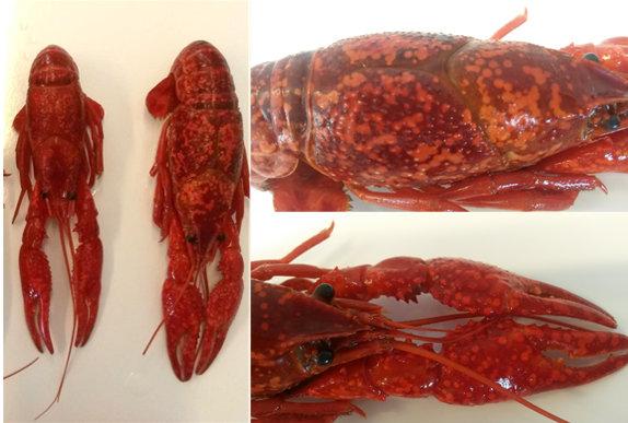
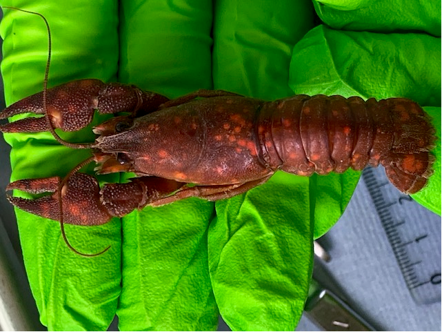
The investigated red swamp crayfish had a total length of between 15.7-20.6 cm and a total weight of 44.0-63.7 g. Animals were cooked and frozen before export and therefore had an intense red coloration of the exoskeleton. During sensory evaluation numerous circular to ellipsoid and some confluent spots with a diameter of 1-3 mm on the cuticula of the carapace, the abdomen, the telson and the claws were observed (Figure 1). White spots were also found on the inner site of the carapace. This led to the assumption of a strong suspicion of WSSV as the causative agent of clinical signs. In the case of the signal crayfish similar clinical signs were observed (Figure 2). However, no obvious mortality was recorded in the corresponding river system.


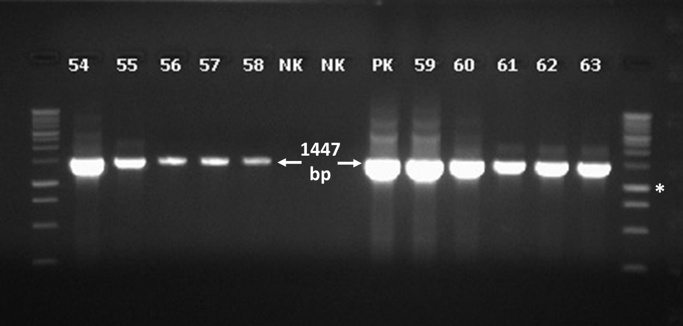
To confirm WSD suspicion in red swamp crayfish (Procambarus clarkii) imported from Asia, specimens were subjected to molecular analyses. WSSV-specific DNA bands could initially be detected after PCR analysis in all samples investigated by the competent federal state laboratory. Further molecular investigations at the NRL for WSD confirmed the positive PCR results. Subsequent sequencing of the 1,447 bp PCR product corroborated the detection of WSSV DNA. Sequence alignment showed 100% identity with DNA sequences deposited in the gene bank (Figure 3). Amplification of WSSV-specific DNA from cooked crustaceans is possible and has been shown by others previously (Devivaraprasad Reddy, Jeyasekaran, and Shakila 2011).
_of_wssv-dna_on_a_2__agarose_gel_stained_with_hdgreen_plus.png)
In contrast to the positive results obtained with red swamp crayfish, both PCRs and nested PCRs with DNA samples from suspected signal crayfish (Pacifastacus leniusculus) were negative. This result was not surprising, since outbreaks of WSD have never been reported in Germany. Also, at the recorded temperatures of approx. 12 °C replication of WSSV is retarded and clinical manifestation are unlikely to occur. Unfortunately, the reason for white spots could not be determined and white spots were likely associated with environmental issues such as high alkalinity or bacterial disease.
The affected food batches from the positive red swamp crayfish were evaluated as not in compliance with article 14 2b) of the Regulation (EC) No 178/2002. According to this article “food shall be deemed to be unsafe if it is considered to be: … (b) unfit for human consumption.” and therefore the batches were destroyed.
Although WSSV genomic DNA has been detected in red swamp crayfish products in Germany, an outbreak of WSD, for legal reasons, was not officially declared, because the samples did not derive from an aquaculture establishment or open waters in the EU, but from a fishery product stored by a food business operator in a food establishment.
To our knowledge this is the first report on the detection of WSSV genomic DNA in Germany. This case report demonstrates the potential risk of the introduction of WSSV into countries outside its endemic distribution. Moreover, it shows a potential risk arising from food imports apart from the risk resulting from movements of live aquatic animals. Especially raw or insufficiently heated fishery products can be a risk for spreading crustacean diseases like WSD. Thus, imported fishery products may still pose a risk to wild and cultured decapods in the countries of destination. The disease risks associated with the import and release of live non-native crayfish species is of course much higher and was assessed by Longshaw et al. (2012). The authors found a shipment of live red swamp crayfish imported from Singapore into UK to be infected with WSSV. Also, it cannot completely be ruled out that crayfish are not sufficiently cooked or that packaging materials and chilled commodities are secondarily contaminated by the virus. Such material could potentially be used as bait in angling or contaminate waste water during processing, respectively, and thus still pose a risk to the environment.
While there is sufficient information on the susceptibility of certain shrimp and crayfish species available, information about the susceptibility of noble crayfish (Astacus astacus L.) and brown shrimp (Crangon crangon) is scarce. Noble (or European) crayfish is among the most widely distributed native crayfish species to Europe (Kouba, Petrusek, and Kozák 2014). Stocks are declining since the introduction of crayfish plague through American crayfish species. Brown shrimp is endemic in the North-East-Atlantic and represents one of the most important crustacean species for the ecosystem as well as for the coastal fisheries of the Netherlands and Germany. A potential WSSV susceptibility of most decapods provides indications of a serious environmental and economic impact to wild native and introduced decapods. Thus, further research should concentrate on the WSSV susceptibility of European crustacean species that have not yet been investigated so far taking environmental aspects e.g. water temperature into account.
According to legal EU animal health requirements adopted on the basis Council Directive 2006/88/EC resp. Regulation (EU) 2016/429, health certificates have to be issued for importing products of animal origin from aquatic animals. Aquatic animals and products of animal origin from aquatic animals may only be introduced into the EU if the animals don’t show any (clinical) signs of (listed) diseases. However, consignments of aquatic animals or products thereof must only be randomly examined at the border control post (BCP) at the place of entrance. If the clinical signs of white spots would have been noticed during a control check of the red swamp crayfish consignment at the BCP, the consignment would have been rejected.
According to legal EU food and feed safety requirements primarily adopted on the basis of the Regulations (EU) 2017/625 and (EC) No 178/2002 food and feed imported into the EU for placing on the market within the EU shall comply with the relevant requirements whereby certificates have to be issued for importing into the EU.
To efficiently control spreading of listed aquatic animal diseases associated with food for human consumption, surveillance and control measures need to be combined at three levels: (1) at the point of occurrence; (2) at the point of the exporting and the importing countries with respect to live animals, animal by-products and food for human consumption; and (3) at the point of the retailing level. More intense training of professionals regarding aquatic animal diseases at all levels – from farm to fork – could be a possible tool to observe and further raise the awareness for control measures especially for diseases associated with food for human consumption.
Acknowledgments
We thank Heike Lünsmann, Selver Rudi, Marie Keitel, Sandra Schöbel, Anja Zander, Susann Schares and Günter Strebelow for their expert technical support.
Data Availability Statement
All necessary data generated or analysed during this study are included in this manuscript. Original datasets generated during and/or analysed during this study are available from the corresponding authors on request.
Conflict of interest
The authors declare no competing financial interests.