Introduction
Abnormal colourations, such as albinism, leucism, xanthochromism, melanism, ambicolouration, metachromism, and polychromism, may occur in wild fish specimens (Quigley et al. 2018). Fish have multiple pigment cell types known as chromatophores such as black melanophores, and red erythrophores among others (Mills and Patterson 2009), which retain their pigments intracellularly rather than transferring their pigments to other cell types.
Albinism and leucism are disorders of the melanin pigmentary system which is genetically determined. Leucism (also called “partial albinism”) is described as an “individual with reduced or absent integumentary pigment, but with pigmented retinas, resulting in diminished or no body colouration and darkly pigmented eyes” (Muto et al. 2013). This condition is controlled by a single recessive allele and, unlike albinism, it affects not only melanin but all types of skin pigment (Quigley and Wallace 2013).
Helicolenus dactylopterus (Delaroche, 1809) belongs to the family Sebastidae comprising 133 species and 7 genera (Froese and Pauly 2021). Despite the many records of albinism and leucism in both Actinopterygii and Chondrichthyes (Follett and Dempster 1966; Veena et al. 2011; Muto et al. 2013; Quigley and Wallace 2013; Megarajan et al. 2018; Quigley et al. 2018; Bañon, Cerdeira, and Carlos 2020; González-Ortegón et al. 2020), leucism had been reported in more than 20 and 36 wild fish species of Actinopterygii and Chondrichthyes, respectively, until 2018 (Megarajan et al. 2018), a low number compared to the 35,588 fish species described so far (Fricke, Eschmeyer, and van der Laan 2020). In the Sebastidae family, only three cases of albinism and leucism were reported, namely leucism in Sebastes melanostomus (Eigenmann and Eigenmann, 1890) and albinism in Sebastes paucispinis Ayres, 1854 (Follett and Dempster 1966) and Sebastes pachycephalus Temminck and Schlegel, 1843 (Muto et al. 2013) and no bibliographic reference was found for genus Helicolenus Goode and Bean, 1896. Although malpigmentations have been studied in aquaculture, there is little known from wild fish specimens (Muto, Takayama, and Kai 2016).
H. dactylopterus is described in literature as having a large head without tabs or tentacles with the following spination: nasal spine present, low preopercular, supraopercular and post-ocular, longer parietal than nuchal spine, 1 or 2 in the second suborbital bone, the second preopercular spine longest. It has a nape that is relatively steeply inclined. It has a large mouth, darkly colored inside with villiform teeth in both jaws, the dorsal fin has 11-13 spines more commonly 12 and 10-14 rays more often 11-13, the pectoral fin as 17-20 rays of which 8 to 9 of the lowermost are free. The anal fin has 3 spines and 5 rays. It has ctenoid scales in 55-80 vertical rows, chest, cheeks and maxilla usually has scales but snout and ventral part of the head naked, in the lateral line presents tubular scales. H. dactylopterus has back and sides of the flank red and a pink belly, presents 5 to 6 dark bands below the anterior, middle and posterior dorsal fin spines, below soft dorsal rays and in the base of caudal fin (Hureau and Litvinenko 1986; Quéro, Porche, and Vayne 2003). This paper reports the first record of leucism H. dactylopterus.
Material and Methods
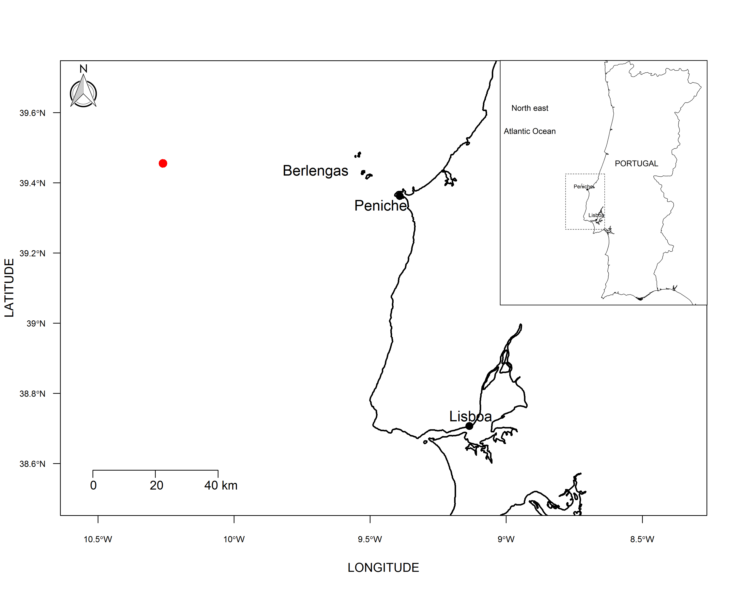
On 25 of July of 2018 a male leucistic specimen of Helicolenus dactylopterus was caught by a commercial fishing vessel using longline targeting demersal fish species namely wreckfish Polyprion americanus (Bloch and Schneider, 1801), blackspot seabream Pagellus bogaraveo (Brünnich, 1768) and blackbelly rosefish Helicolenus dactylopterus at a depth of 329 meters (Figure 1). The specimen was carefully examined for morphological characters following the previous descriptions of Hureau and Litvinenko (1986) and Quéro, Porche, and Vayne (2003).

The specimen was photographed and preserved in 10% buffered formaldehyde solution and stored at IPMA.
To validate the taxonomic identification, a sample of the specimen was analysed using genetic tags for species identification, using DNA barcoding based on mitochondrial DNA gene cytochrome c oxidase I (5’-COI). For this purpose, a sample muscle tissue was collected and preserved in 96% ethanol for molecular analyses. Total genomic DNA was extracted from muscle tissue using magnetic bead separation technology from MPure Tissue DNA Extraction Kit (MP Biomedicals, USA), following the manufacturer’s protocol. The 652 bp barcode region of the mitochondrial DNA gene cytochrome c oxidase I (COI) was subsequently amplified and sequenced following published protocols (Ivanova et al. 2007).
After assembling and editing the bidirectional sequences, a sequence of 652 bp fragment was obtained. The haplotype was deposited in BOLD with process ID IPMAF016-21. The sequence of H. dactylopterus under consideration was submitted to NCBI BLAST® (http://blast.ncbi.nlm.nih.gov/Blast.cgi) and BOLD Systems (http://v4.boldsystems.org/index.php/IDS_OpenIDEngine) to search for matching sequences.
To determine if the malpigmentation poses some disadvantage to the individual, condition factors (Fulton factor) (Costa 2013) were determined for the individual and compared with Portuguese wild population of H. dactylopterus. The normal condition indexes for wild populations were determined with data collected for the species onboard surveys carried out by IPMA in Autumn (PT-IBTSQ4) from 2014 to 2017 under the National Programme for Biological Sampling (PNAB-DCF) (ICES 2017).
Condition factors indexes used were:
i) Condition factor obtained with total weight:
CFt=Total weightLength3 ×100
ii) Condition factor obtained with gutted weight
CFg=Gutted weightLength3 ×100
The condition factors were determined only for mature males.
Results
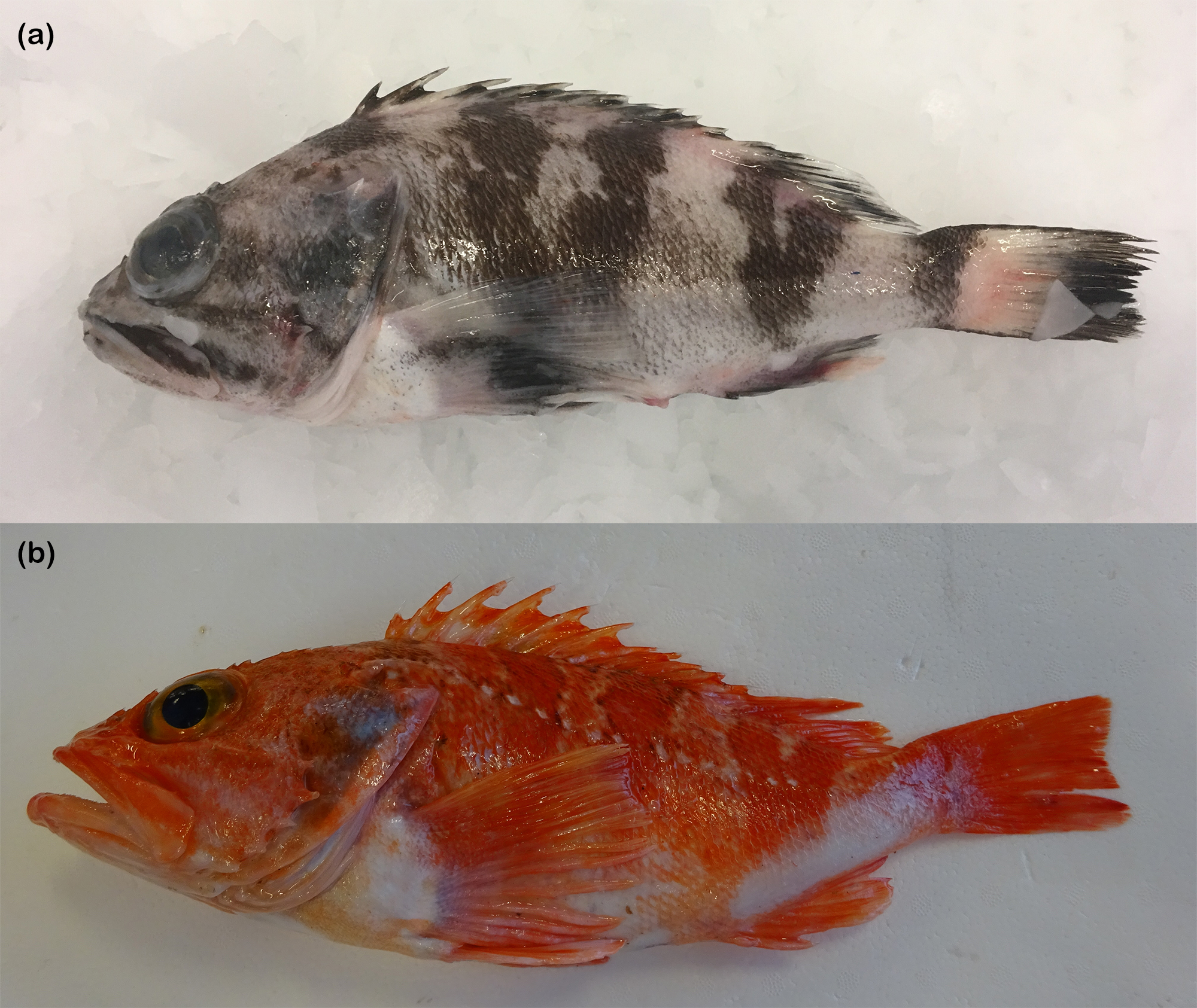
Morphometric characteristics, meristic counts and body proportions of the leucistic specimen of H. dactylopterus are presented in Table 1. The abnormal pigmentation was evident on the head, back, sides and belly which were white, with 6 dark bands below the anterior, middle and posterior dorsal fin spines, below soft dorsal rays and in the base of caudal fin, and black stripes on the head. The eye colour was normal (Fig. 2a). The normal pigmentation of the species is a pink belly and red back and flank sides (Fig. 2b).

Similarities of more than 98% were found with different species from the Helicolenus genus, both in Genbank and in BOLD Systems database, including several matches with H. dactylopterus, Helicolenus barathri (Hector, 1875), Helicolenus percoides (Richardson and Solander, 1842) and Helicolenus hilgendorfii (Döderlein, 1884). Despite no specific genetic identification being possible, the specimen clearly belongs to the Helicolenus genus. The condition factor indexes for leucistic specimens and normal wild specimens are described in Table 2.
Discussion
The leucistic specimen of H. dactylopterus morphological characters, meristic and morphometric characters matched with the previous descriptions of the species by Hureau and Litvinenko (1986) and Quéro, Porche, and Vayne (2003).
The genetic analysis of this sample and the sequences publicly available in the databases show that the gene regularly used for the genetic identification of marine organisms, and in particular of marine fish, failed to make the specific distinction in this genus. This unusual trait may suggest one of two things: the gene used may suffer from selective pressure in this atypical genus that does not allow its natural evolution or the different species described for this genus are actually one. Only with more genetic data, including data on other genes, will it be possible to identify the reason why the intragenic genetic divergence is abnormally low.
The disorder that affects the specimen appears to be only related with the erythrophores, since when compared with a normal specimen only the red colouration was lost, the dark bands and stripes associated to the melanophores are present being even more visible because the lack of the red pigment. Leucism may affect other pigmentation besides melanin (Mills and Patterson 2009) which is this case.
Besides the abnormal colouration the specimen here described showed no other abnormality and seemed to have been perfectly fit for the natural environment. The condition factor of the specimen showed normal values when compared with mature males of the Portuguese population and both condition factors were slightly above the mean and the median of the natural population. In addition the fish had no signs of disease nor infection by parasites.
ACKNOWLEDGMENTS
This study was financed by the Portuguese National Programme for Biological Sampling (PNAB) under the EU Data Collection Framework (DCF). The genetic analysis was financed by the National Programme for Bivalve Mollusc Monitoring (SNMB). We are grateful to Claudio Formiga skipper of the vessel “Monte do Senhor” for bringing and offering the specimen.