INTRODUCTION
In recent years, some bacterial fish pathogens have been difficult to treat effectively due to the presence of multidrug-resistant bacteria in aquaculture systems and the low efficacy of antimicrobials in vivo against disease outbreaks. Antibiotics are often used randomly without antibiotic sensitivity testing being carried out during an epidemic. Thus, antibiotic-resistant bacteria and resistance genes can spread in the aquatic environment (Cabello et al. 2013). In recent years, alternative treatment methods to antibiotics, such as phage therapy have been investigated for the treatment of bacterial diseases (Richards 2014).
Bacteriophages (phages) are bacterial viruses discovered in the early 1900s and have been successfully used in the treatment and prophylaxis of bacterial diseases since then. Although they were first used in eastern European countries such as Georgia, Russia, and Poland, they have also been used in some European countries and more recently America (Sulakvelidze, Alavidze, and Morris 2001). Although the use of phages for therapeutic purposes began to decrease with the discovery of antibiotics, interest in phages has increased again more recently with the emergence of multidrug-resistant bacteria (Aydoğan and Hadımlı 2016).
Aeromonads are Gram-negative bacteria with straight rods, are facultative anaerobes, and motile via a flagellum that exist worldwide in fresh and salt water, and have wide host ranges. Aeromonas hydrophila along with A. sobria, A. veronii, and A. caviae are known to cause Motile Aeromonas Septicemia (MAS) (Lewis and Bender 1960). Aeromonas hydrophila is considered an opportunistic pathogen and could frequently be found in gut flora. Hence, stress and water quality play a key role in disease occurrence (Holt et al. 1994). Aeromonas hydrophila is an important risk for aquaculture facilities worldwide. It has been reported as the causative agent of various diseases in a variety of cultured fish species, including sea bream (Sparus aurata), carp (Cyprinus carpio), sturgeon (Acipencer gueldenstaedtii), rainbow trout (Oncorhynchus mykiss), and sea bass (Dicentrarchus labrax) (Ture et al. 2018). Recently, a phage called Aeromonas phages-T65, which can infect A. hydrophila under in vitro conditions was isolated in our laboratory.
The Double-Layer Agar (DLA) technique called a plaque assay is extensively used in phage research for the isolation, enumeration and identification of phages (Adams 1959; Kropinski et al. 2009). Phages that form large and well-defined plaques can be easily observed and enumerated using the DLA technique. However, some phages form small and turbid plaques that are quite difficult to detect and count. To overcome this problem, the use of antibiotics and other chemicals has been suggested (Santos et al. 2009). A significant increase in the size and visibility of the phage plaques have been reported when bacteriophages infect bacteria, by addition of small doses of antibiotics and some chemicals to the different layers of media used in the assay (Hadas et al. 1997; Islam et al. 2012; Kamal and Dennis 2014). This phenomenon is called phage-antibiotic synergy (PAS) (Kim et al. 2018; Gu Liu et al. 2020; Morrisette et al. 2020).
In the present study, the effect of different classes of antibiotics, glycerol and antibiotic-glycerol complex on phage plaque size and enumeration was investigated. Moreover, the effect of a combination of penicillin-streptomycin (pen-strep) on plaque size was assessed for the first time for A. hydrophilia.
METHOD
Culture Media in the DLA technique
Tryptic Soy Agar (TSA, Merck, Germany), prepared according to the manufacturer’s instruction, was used as the bottom agar, whereas, TSA prepared at a final concentration of 50 % was used as the top agar layer. Glycerol (Applichem, Germany) was added at the desired concentration to the top, bottom, or both layers after sterilisation by autoclaving. Sterilised pen-strep (Lonza) was purchased and added to the top, bottom, or both layers. Ampicillin was provided by the Biology Department of Karadeniz Technical University and added to the top, bottom, or both layers after sterilisation.
Phage and Bacteria
The phage, Aeromonas phages-T65, used in this study has been shown to infect A. hydrophila (data not shown). Isolation and preparation of the phage were described by Ture et al. (2021). This phage was isolated from sewage and purified by several passages of a single plaque and is a member of Myoviridae family. The strain of A. hydrophila used in the study was strain T65 and was supplied by our laboratory.
Phage Titer Determination
The phage titer, described as plaque forming units (PFU)/mL of solution, was determined using the DLA technique defined by Sambrook and Russel (2001) with modifications. Firstly, phage was diluted from 100 to 108 and then 104 to 108 dilutions were used in the DLA technique. Each dilution of the phage (80 μL) was added to 80 μL of a bacterial suspension at a multiplicity of infection (moi):1 (108 CFU/mL bacteria and 108 PFU/mL phage) and incubated overnight at 25°C, 120 rpm. This mixture was added to 5 mL top agar (50% TSA), gently homogenised, and poured on pre-poured 10 mL bottom agar (TSA). Antibiotics and/or glycerol were added at the desired concentration to the bottom top or both layers as described by Santos et al. (2009). The plates were dried for 10 min at room temperature and then incubated at 25°C overnight.
Phage plaque size
Pictures of the plates were taken with a Cannon 700D camera on a black background to prevent distortion and to provide equal light exposure and contrast conditions in all photographs. Brightness, contrast, or colour was not modified on the photographs. The diameter and area of the plaques were measured from photographs using the computer image analysis program ImageJ (Schneider, Rasband, and Eliceiri 2012) (https://imagej.nih.gov/ij/) to obtain accurate dimensions. Each value represents the average of ten plaque measurements. Area increase was calculated as the ratio between the average diameter of plaques obtained with each treament compared to the control plates (untreated agar) (Santos et al. 2009).
Statistical Analysis
All data was demonstrated as the means ± standard deviations (SDs) from independent experiments. Kruskal Wallis test was conducted to determine statistical significance and a P- value of <0.05 represented significance.
RESULTS
In this study, the DLA method was modified by adding different concentrations of ampicillin, pen-strep antibiotics and glycerol to augment the size of the plaques obtained. Ampicillin was added to both layers at different concentrations (2, 4 and 8 mg/L). After incubation, plaques were not detected on plates except in the control plate, which contained no antibiotics. When ampicillin was added to only the top layer at different concentrations (2, 4, and 8 mg/L), plaques were still undetectable.
When a mixture of pen-strep antibiotics was added to both the top and the bottom layers at different concentrations (40 and 80 U/µL) no plaques were detected on the plates except for the control plates. When pen-strep antibiotics were only added to top layer at the same concentrations, the plate containing 40 U/µL pen-strep showed an notable increase in plaque size and area compared to the control plate. The size of the plaque did not appear to increase when 80 U/µL pen-strep was added to top layer compared to plaques seen on the control plate (Table 1).
The effect of glycerol on plaque size was analysed using two final concentrations in both layers and compared with a control plate containing no glycerol. The best enhancement in both plaque size and enumeration was achieved with 5% glycerol. However, 10% glycerol resulted in a decrease in plaque size. Therefore, further experiments were conducted using 5% glycerol with different concentrations of pen-strep (Table 1).
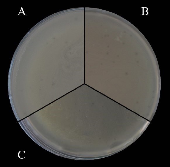
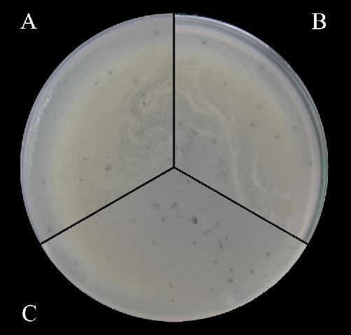
Finally, the combined effect of the pen-strep antibiotic mixture and 5% glycerol was evaluated using different combinations (Figure 1 and 2). On the first trial, 5% glycerol was added to both layers while antibiotics were added only to the top layer. Plates with 40 U/µL pen-strep and 5% glycerol in the top layer displayed plaques with nearly the same size as the control plates, while 80 U/µL pen-strep in top layer with the addition of 5% glycerol to both layers resulted in a decrease in plaque size. Based on these results, 5% glycerol and antibiotics were only added to the top layer, and the difference in plaque size was analysed. A combination of 5% glycerol with 40 U/µL pen-strep in the top layer increased the size of the plaque compared to the control plates, but this increase was low compared to their impact when used separately (Table 1). On the other hand, a combination of 5% glycerol with 80 U/µL pen-strep in the top layer showed an increase in plaque size that was higher when only 80 U/µL pen-strep was added to the top layer, but was not significantly higher compared to only the addition of 5% glycerol to both layers.


Furthermore, phage titers were calculated and compared between the classical DLA procedure and the newly improved method outlined here in order to ensure that the glycerol and antibiotics did not cause a reduction in the number of plaques. Phage titer was determined to be 1-2 x 10⁸ between groups, therefore the phage titers did not appear to differ between the classical DLA procedure and newly improved method.
DISCUSSION
During our studies, a lytic phage was isolated against A. hydrophila by our laboratory from a natural source to develop an alternative treatment for disease in fish caused by this bacterium. However, the phage formed tiny plaques that were difficult to observe so that they can not be enumerated when plated using the DLA technique. To overcome this problem, glycerol, ampicillin, and pen-strep (separately and in combination) were added to the culture media to try to increase the plaque size and area.
It has been reported that some chemicals stimulate bacteria to produce phages. In a study that tested the effect of glycerol at three different concentrations (5%, 10% and 20%), the best improvement in plaque observations was achieved with 5% glycerol (Santos et al. 2009). Similarly, the impact of glycerol at two different concentrations (5% and 10%) were analysed and the best enhancement in plaque size was obtained with 5% glycerol in our study. Results showed that glycerol had a significant role in plaque size enhancement and this could be the result of improved phage diffusion by the presence of the glycerol. Also, it is a nonfermentative carbon source for these bacteria and its presence could result in a delay in the onset of the stationary phase as mentioned by Santos et al. (2009).
Antibiotic additives can increase plaque size and the final titer of phages in solid media. The effects of different antibiotic concentrations were analysed, and the following optimal concentrations were determined: 0.5 mg/L ampicillin, 0.06 mg/L cefotaxime, 1.5 mg/L tetracyclines, and overdose penicillin (Santos et al. 2009). As a result, it has been reported 1.5 mg/L tetracyclines and 5% glycerol complex significantly improved both the plaque size and contrast. In contrast to that study, our study showed that the pen-strep combination had an impact on plaque size not using an overdose, but ampicillin did not have any effect on plaque size. The difference between the studies could be due to differences in the host and phage characteristics used in the two studies.
When the phages infect bacteria in the presence of sublethal doses of some antibiotics, the sizes of the phage plaques are significantly increased. In a previous study, it was reported that sub-lethal concentrations of cefotaxime and cephalosporin, increased the production of a phage against Escherichia coli by more than 7-fold. A similar effect was also observed with mitomycin C. Common features of these antibiotics are that they inhibit bacterial cell division and trigger the phage production system (Comeau et al. 2007). Similar results were obtained in our study with the application of 40 U/µL pen-strep in top layer of agar.
ß-Lactams, including penicillins, are a group of antibiotics targeting the cell wall of the bacterial cell (Rawat and Nair 2010). On the other hand, aminoglycosides including streptomycin inhibit protein synthesis in bacterial cells (El Salabi, Walsh, and Chouchani 2013). Aminoglycoside and penicillin were used in combination to prevent antibiotic resistance and their synergistic effect results from facilitating the entry of aminoglycoside into the bacterial cell by penicillin (Marothi, Agnihotri, and Dubey 2005). Given the synergistic effect between penicillin and streptomycin in our study, penicillin could have contributed to the entry of streptomycin and phage into bacterial cells, and streptomycin could then have caused stress on bacteria by inhibiting protein synthesis. This stress could contribute to phage production in bacterial cells and finally applying 40 U/µL pen-strep to the top agar layer augmented the plaque size. Antibiotics targeting protein syntheses, such as tetracycline and kanamycin, have been analysed for plaque size improvement (Łoś et al. 2008). The mechanism of plaque size at low concentration of these antibiotics was explained by partially slowing down but not fully halting protein synthesis of the host bacteria, and host cells in this altered state might be more accessible to attack by phages.
In one study, the application of ciprofloxacin induced higher levels of phage production against the cystic fibrosis epidemic strain of Pseudomonas aeruginosa (Fothergill et al. 2010). In another study, a modified DLA method was described that allowed easy detection and accurate enumerate plaques of Sp5, a bacteriophage of E. coli O157 strain. In this modified method, the top agar of agar was supplemented with mitomycin C and Ca2+ or Mg2+ in which mitomycin C prevented the lysogenisation of phages and/or enforced the lytic cycle. Also, the divalent minerals produced a synergistic effect in combination with mitomycin by improving phage adsorption to the host cells (Islam et al. 2012).
One of the important problems encountered during the isolation of enteric phages includes the formation of pinpoint or tiny plaques. In another study, the phage MR-5, formed tiny plaques against its host S. aureus, making its detection and enumeration more difficult. The classical DLA method was developed for increasing the plaque size. For this purpose, sublethal concentrations of three different classes of antibiotics were used. As a result, the β-lactam and quinolone antibiotics employed for increasing the plaque size did not show any significant effect on the plaque size in line with our results for ampicillin. In that study, linezolid, tetracycline, and ketolide antibiotics brought significant enhancement in the plaque size (Kaur, Harjai, and Chhibber 2012).
In conclusion, in the present study, the effect of glycerol, ampicillin, and pen-strep for improving the plaque size and area were tried separately and in combination. A combination of 5% glycerol with 40 and 80 U/µL pen-strep in the top layer of agar contributed to an increase of plaque size compared to the control (which was non-significant), but this increase was low compared to their impact when used separately, i.e. 40 U/µL pen-strep or 5 % glycerol used seperated resulted in a statistical significant increase in plaque size. These results indicate that modifications of the traditional DLA method with glycerol and pen-strep supplementation can be use in the isolation of new virulent phages.
ETHICAL APPROVAL
Ethical approval was not required.
FUNDİNG INFORMATİON
This project was funded by the Republic of Turkey Ministry of Agriculture and Forestry (TAGEM/HSGYAD/B/21/A5/P4/2505).
AUTHOR CONTRIBUTION
Mustafa Türe and Ayşe Cebeci were responsible for the experimental design, analyses, and interpretation of the data. Ayşe Cebeci, Elif Aygür, Furkan Balcı, Nihal Çalışkan, and Esen POLAT were responsible for the conduction of the experimets and supplying resources. Mustafa Ture and Ayse Cebeci drafted the manuscript. All authors have read and agreed to the published version of the manuscript.
CONFLICT OF INTEREST
The authors declare that there are no conflicts of interest.
ACKNOWLEDGMENTS
We would like to thank İlyas Kutlu for his assistance during solution preparation.