1. INTRODUCTION
Over the past years, although a large portion of the international aquaculture output has been devoted to food products, the global trade in ornamental fish has grown rapidly, and nowadays, fish culture has become a significant part of international aquaculture industries in most countries all over the world (Winfree 1989; Monticini 2010; Noga 2010; Shokrpoor et al. 2022). The popularity of ornamental fish has led to advances in techniques of culturing and breeding in captivity (Wilson et al. 2001). However, there is still a great global demand for ornamental fish species that are difficult to breed. In recent decades, the trade, culturing and breeding of ornamental fish has made significant progress in Iran, and has been met with a great response from enthusiasts after being introduced as an economic and profitable profession (Mood et al. 2010; Mousavi, Sheikhzadeh, and Marandi 2020, 2021; Rahmati-Holasoo et al. 2022). Currently, more than 150 species of freshwater ornamental fish are bred successfully in Iran.
Green terror (Andinoacara rivulatus Günther, 1860) is a freshwater cichliform ornamental fish, which belongs to the cichlidae family and is farmed and bred as a desirable commercial species in Iran. The origin of fish is native to rivers of Ecuador and Peru in South America (Lewbart 1998). Basically, the name of green terror (green Jewel) is attributed to this fish species, because of the bright coloration of its fins and body. The omnivorous green terror (A. rivulatus) is quite tolerant to undesirable environmental conditions and the male is territorial. However, the female is responsible for the protection, maintenance and breeding of larvae after spawning (Lewbart 1998; Reis et al. 2003).
In recent years, green terror farming has been accompanied by a considerable development and this fish species is propagated in some provinces of Iran. However, a significant portion of green terrors are imported. In the past, a few reports of infections have been studied in this fish species in the country. Also, research on metazoan parasites of freshwater ornamental fish, is limited in Iran. The occurrence of massive losses in fish may be considered as one of the most important factors in imposing severe economic costs on breeding centers. The aim of this study was therefore to investigate the cause of losses of green terrors in an ornamental fish breeding center in Alborz province, Iran.
2. MATERIALS AND METHODS
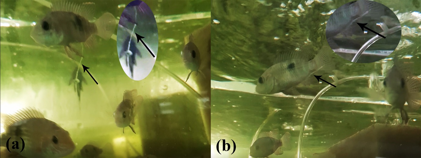
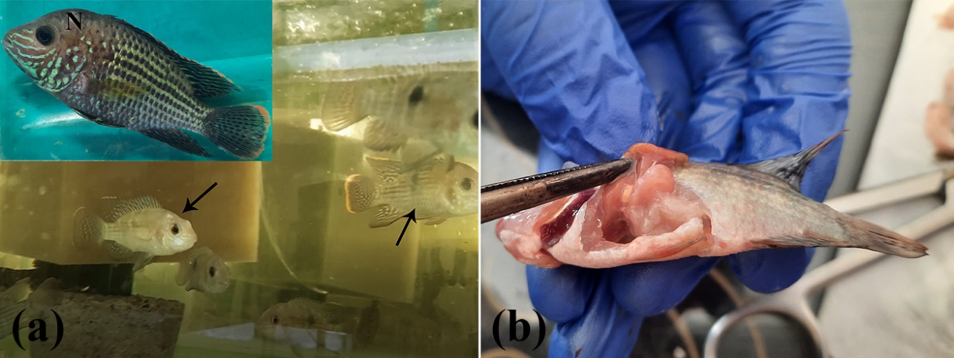
In June 2021 and July 2021, following the appearance of clinical signs including white hanging feces (Figure 1a, b) and paleness of the body (Figure 2a), in conjunction with continuous mortalities of green terrors in an ornamental fish breeding center in Nazarabad, Alborz province, fish were sampled. For this purpose, 30 ornamental green terrors (5% of total) were packed in specific polyethylene bags, supplied with oxygenated water and transported for supplementary analysis to the ornamental fish clinic, Faculty of Veterinary Medicine, University of Tehran. Subsequently, the samples were transferred to pre-prepared aquariums and monitored for deviations in appearance. On gross examination, the fish measured 10 cm mean total length. Investigations were then undertaken in order to study various parasitic infections, thus wet smears were prepared from the skin, fins, and gills of the fish and examined carefully under a light microscope. Additionally, in order to accurately investigate the bacterial and parasitic infections, the fish were euthanised and necropsied under aseptic conditions (Figure 2b). Aerobic and anaerobic bacterial cultures from the liver, spleen, and anterior kidney were streaked on blood agar and incubated at 25°C. Additionally, the gastrointestinal tract was removed. After incubation of the standard culture media for 72 h, the gastrointestinal tract of fish was examined using a light microscope (Nikon E600, Japan) and a stereomicroscope (Olympus SZ60, Japan). Subsequently, nematodes were counted at 40X magnification (4X objective and 10X eyepiece) beginning with the first dot. Gastrointestinal nematode parasites were fixed in 70% ethanol and purified with glycerol for analysis. In order to treat the rest of the fish, levamisole was administered orally at a dose of 2 mg/g of food for 10 days. Additionally, it was administrated for 48 h every 7 days for 28 days. The mortalities completely stopped and following re-examination after 28 days, no infection was observed.


3. RESULTS
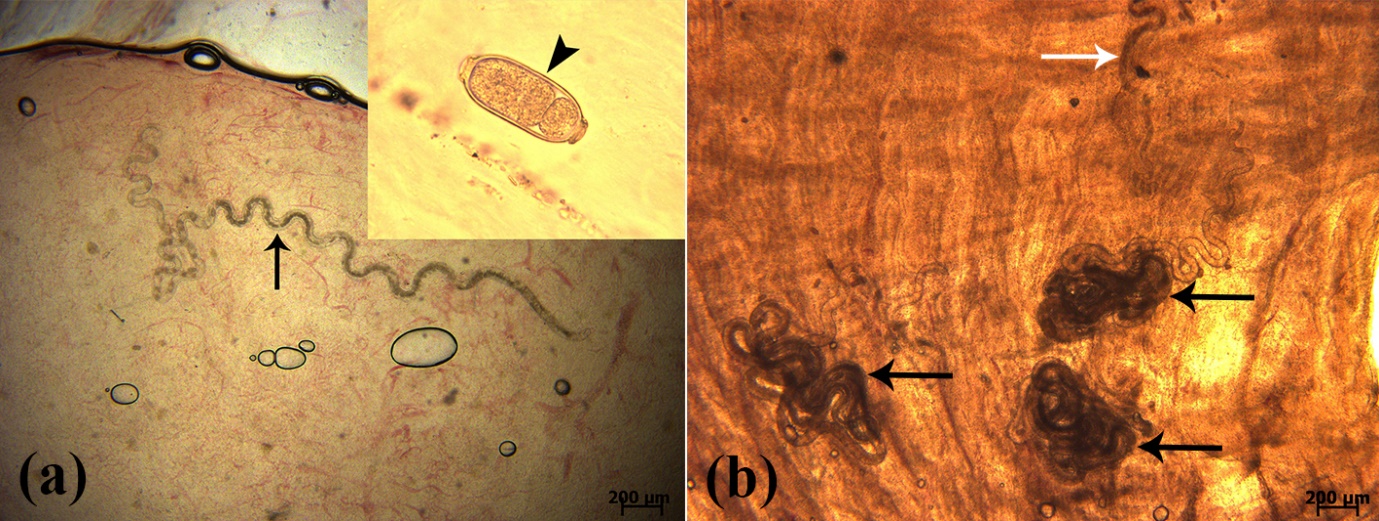
In the elementary stages of macroscopic examination, the thinness and paleness of the fish was quite evident. No external parasites were observed in green terrors examined by light microscopy and under a stereomicroscope. Also, no trace of bacterial growth was observed on standard culture media. However, a metazoan nematode parasite Capillaria sp. was observed under a light microscope during examination of the digestive tract of the fish (Figure 3a, b). Both males and females of Capillaria sp. were identified. However, the number of male nematodes were relatively lower than that of female nematodes. Between 4 and 8 male Capillaria sp., and between 16 and 20 female Capillaria sp. were identified in the studied green terrors (per fish). The barrel-shaped eggs of the female nematodes and the spicules of the male nematodes were evident. A large number of parasite eggs were found in the intestines of fish under a light microscope (Figure 3a). Diagnosis of metazoan nematode Capillaria sp. was based on the morphological characteristics of the nematodes as well as the eggs.

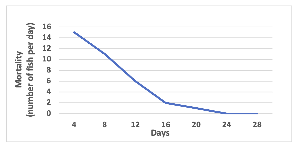
Oral administration of the anti-nematode levamisole at a dose of 2 mg/g of food for 10 days and 2 mg/L of water for 48 h (treatment repeated once a week for 4 weeks) was effective and the fish losses, which amounted to 15 per day, stopped completely following treatment (Figure 4).

4. DISCUSSION
Recent studies conducted on parasitic infections of ornamental fish are limited in Iran, and the need for further research in this field is evident. In addition, most previous studies have focused on external parasites, and research based on internal parasites have often been neglected. For example, in a study that investigated the prevalence of dermal parasitic infections of ornamental fish in Isfahan province, some protozoans and monogenean trematodes were identified in green terrors (A. rivulatus) (Salemi, Taghavi, and Abedi 2013). Also, in another study which investigated infections with various internal and external parasites in three species of ornamental fish in Tehran province, several ectoparasites including Ichthyobodo sp., Ichthyophthirius multifiliis and Gyrodactylus sp., as well as only one species of internal parasite Capillaria sp. were isolated from green terrors (Rahbari 2016). However, in a study conducted on the parasitic infections of ornamental fish in Khorasan-Razavi province, no parasites were detected in green terrors (Gharavy et al. 2017). In Iran, metazoan nematode Capillaria sp. has been isolated from discus (Symphysodon aequifasciatus) (Rahmati-Holasoo et al. 2010) and angelfish (Pterophyllum scalare) (Rahmati-Holasoo et al. 2022). Far greater documented reports of internal parasitic infections caused by Capillaria sp. have been noted in other countries compared to Iran. In a study which focused on the isolation and identification of ornamental fish parasites in Turkey, Capillaria sp. was isolated successfully from discus S. aequifasciatus (Koyuncu 2009). Also, in another study which was conducted on freshwater ornamental fish parasitic infestations in Sri Lanka, Capillaria sp. was isolated from two ornamental fish species including guppy (Poecilia reticulata) and angelfish (P. scalare) (Thilakaratne et al. 2003). Additionally, in a study of ornamental fish parasitic infestations in the Madan area of North Sumatra, Indonesia, goldfish was identified as a host for Capillaria sp. (Dewi and Fadhilla 2018).
As like as many ornamental fish species, green terror (A. rivulatus) originates from the tropical regions of the world. Capillaria sp. is a well-known parasite which can infect both marine and freshwater ornamental fish (Moravec, Orecchia, and Paggi 1988). As with all parasites which have a direct life cycle and do not need an intermediate host, these fast-spreading spawning nematodes represent one of the most problematic parasites (Wildgoose and British Small Animal Veterinary Association 2001), and are capable of inducing significant mortalities in various freshwater ornamental fish species such as cichlids due to the intestinal invasions (Wildgoose and British Small Animal Veterinary Association 2001; Moravec, Orecchia, and Paggi 1988; Rahmati-Holasoo et al. 2022). Adult worms often settle in the lumen of the small intestine. Female nematodes produce the first-stage larvae 13 to 14 days after infection. These larvae remain in the small intestine and often differentiate into the second stage larvae within 10 days. After the female worms produce barrel-shaped eggs, these eggs are released out of the fish’s body along with the feces, and following the ingestion of parasite eggs by fish, parasitic contamination spreads to the aquatic environment (Ko 2006). In addition, there is evidence that being ovo-viviparous and larvae producing by some female nematodes can also lead to auto-infection (Cross, Banzon, and Singson 1978). One of the most important routes of occurrence of capillariasis in ornamental fish breeding centers is via entry of parasites following poor quarantine measures, i.e. for the arrival of new fish (Adel et al. 2013). It should be noted that some points during fish production, such as division of the farm into isolation units, increasing the length of quarantine period, using a specific-pathogen-free (SPF) water source, incorporation of more immunostimulant compounds such as probiotics and prebiotics into feed formulations of fish (Rahmati-Holasoo et al. 2022), and finally avoiding use of live foods such as oligochaete worms (e.g., tubifex worms), as possible carriers (Yanong 2002), have had a substantial impacts on prevention of the presence of parasites and concerning diseases more efficiently. In the cases of infections caused by Capillaria sp. as well as other nematode parasites, various anti-nematode chemical compounds such as levamisole (Concurat®, Tramizol®, Nimizol®, and Repercol®), mebendazole, albendazole and thiabendazole may be used to successfully control and treat infected fish (Bassleer 2004). In this study, oral administration of the anti-nematode levamisole at a dose of 2 mg/g of food for 10 days and 2 mg/L of water for 48 h (treatment repeated once a week for 4 weeks) was used effectively leading to complete cessation of mortalities. Also, minced garlic and hexane extract at a dosage of 200 mg/L showed 100% effectiveness against capillarids. However, addition of ammonium-potassium tartrate and aqueous extract of garlic gave 86% and 0% anthelmintic effectiveness, respectively (Peña, Auró, and Sumano 1988).
The present study showed that the entry of nematode parasites and/or new parasite-infected fish into the aquatic environment may cause significant losses in ornamental fish farms and also threaten the viability of some valuable native species, indirectly. Therefore, due to the well-known economic importance of ornamental aquaculture, the application of comprehensive and codified health and management protocols, along with the regular periodic monitoring of fish health, and accurate timely diagnosis for controlling nematode infections in a wide range of ornamental fish such as cichlids, may play an important role in preventing the occurrence of disease-related mortalities and inflicting significant economic losses for ornamental fish breeders.
Conflict of interest
The authors declare that there are no conflict of interests.
Funding
The authors declare that no funds, grants, or other support were received during the preparation of this manuscript.
Author contributions
All authors contributed to the study conception and design. Material preparation, data collection, and analysis were performed by Hooman Rahmati-Holasoo, Amin Marandi, and Hosseinali Ebrahimzadeh Mousavi. The first draft of the manuscript was written by Amin Marandi and all authors commented on previous versions of the manuscript. All authors read and approved the final manuscript.