.tiff)
INTRODUCTION
Rainbow trout Oncorhynchus mykiss are susceptible to numerous skin conditions (Diler, Görmez, et al. 2019), like infectious diseases caused by Aeromonas spp., Flavobacterium spp., Saprolegnia spp., Ichthyobodo spp. or Ichthyophthirius multifiliis (Bruno, Noguera, and Poppe 2013). However, in the past decades, skin conditions of unknown aetiology have emerged in rainbow trout (Oidtmann et al. 2013; Peeler et al. 2014; Verner-Jeffreys, Pond, et al. 2008).
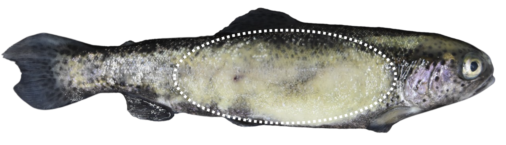
In 2002, a new disorder affecting rainbow trout was reported in fish farms in the United Kingdom (UK) for the first time. Since then, the number of cases has increased dramatically, and in 2014, 43% of the UK farms had already reported the occurrence of the disease (Diler, Görmez, et al. 2019; Peeler et al. 2014). Rainbow trout from still water fisheries are also affected (Maddocks et al. 2015). Fish show white to grey patches on the skin, typically on the flanks, which extend to up to 90% of the body surface (Cano, Verner-Jeffreys, et al. 2016; Diler, Görmez, et al. 2019; Peeler et al. 2014). The grey patches represent areas of excess mucus production and thickened epidermis with raised scales. Due to these distinctive lesions, the disease was named puffy skin disease (PSD). Affected fish may also show inappetence and emaciation (Peeler et al. 2014). Even if morbidity is high, mortality often remains limited to few animals or even no mortality (Christie, van Aerle, et al. 2018; Maddocks et al. 2015; Peeler et al. 2014). Histologically, pathognomonic findings include epidermal hyperplasia, spongiosis, and oedema of the dermal stratum spongiosum, including scale pocket oedema (Cano, Verner-Jeffreys, et al. 2016).
The prevalence and severity of PSD have strong seasonality, increasing during the summer and autumn months (13°C – 15°C) (Cano, Verner-Jeffreys, et al. 2016; Diler, Görmez, et al. 2019) and decreasing over winter (Maddocks et al. 2015; Peeler et al. 2014). The disease seems to be horizontally transmitted, as it can be transmitted between tanks and farms by affected animals (Cano, Verner-Jeffreys, et al. 2016). Therefore, infectious aetiology has been suggested as the most probable cause. However, an aetiological agent remains undetermined (Maddocks et al. 2015).
Despite the low mortality rates, PSD impairs fish health and well-being while making them unappealing to consumers, leading to high economic losses. In severe cases, culling and downgrading of carcasses may even be necessary (Maddocks et al. 2015; Peeler et al. 2014).
Following the first description of PSD in the UK and Turkey, this is the first report on PSD in Switzerland (Diler, Görmez, et al. 2019; Peeler et al. 2014).
MATERIAL AND METHODS
In February 2022, 21 rainbow trout from a private aquaculture facility in the Swiss Alpine region were submitted alive for diagnostics to the Institute for Fish and Wildlife Health, University of Bern, Switzerland. Fish were presented with inappetence and white to grey patches on the skin with raised scales. Mortalities reached 0.1-0.2%/tank/day.
The affected fish farm comprises a hatchery, a pre-ongrowing and a grow-out facility with a separate water supply. The farm produces 80-90 tonnes of trout per year. The hatchery is supplied with 20 L s-1 spring water at a temperature of 5 to 14°C. In the pre-ongrowing facility, juveniles, up to 50 g, are reared in separate troughs and concrete channels. Larger animals are then transferred to the grow-out facility, which is supplied with 80 L s-1 of groundwater and 20-30 L s-1 of spring water with a water temperature of 4 to 14 °C. The farm usually purchases eggs or 50 g trout from two other Swiss aquaculture facilities. In addition, live trout are occasionally purchased in the European Union, most recently from Austria in September 2018 and January 2019.
The affected rainbow trout originated from different tanks of the grow-out facility. The temperature was 6-7° C, and the density was 30 kg fish/m3. The tanks were partly arranged in series and partly in parallel. The animals were preventively treated with Wofasteril (2mL/m3 for 30 min) every Monday and Thursday. Shortly before sending the animals for examination, fish were treated with formalin (200mL/m3, 30-min immersion, three consecutive days).
Fish sent for diagnostics were euthanised with 150 mg/L buffered 3-aminobenzoic acid ethyl ester (MS 222®, Argent Chemical Laboratories). Fish were measured, and a complete necropsy was performed. All specimens were tested for the presence of external and intestinal parasites by direct microscopic examination of wet mounts of skin, first left branchial arch, and intestinal content. To examine the presence of bacteria, spleen, liver, and kidney samples were cultured on blood agar plates (Biomerieux, Switzerland) and bromthymolblue-lactose-agar plates (Merck, Germany) supplemented with 0.5% sucrose for 48 h at 22°C. The growth of bacteria was checked daily. In addition, the presence of Flavobacterium spp. was investigated on special agar plates to enhance the growth of flavobacteria for five days at 15°C (Anacker and Ordal 1955).
Samples from affected skin areas were placed in 10% neutral buffered formalin. Formalin-fixed tissues were processed routinely for paraffin embedding, sectioned at 4 µm, and stained with haematoxylin and eosin (HE) for histologic processing and pathological assessment.
RESULTS
Rainbow trout measured between 23.5 and 28 cm. On post-mortem examination, 7/21 fish showed unilateral or bilateral, focally extensive, white to grey, rough patches affecting up to 75% to 90% of the skin surface (Figure 1). These lesions extended horizontally from the caudal aspect of the operculum to the anal fin and vertically from the dorsal to the ventral fin. The scales in those areas were raised, and the skin appeared thickened and oedematous. No parasites were detected by direct microscopic examination of wet mounts of skin.

Histologic evaluation of the skin samples revealed moderate to severe multifocal epidermal erosion and ulceration, often with complete loss of the epithelium, focally extensive, severe, thickening of the epithelium by epidermal hyperplasia with the formation of finger-like projections, intercellular (spongiosis) and intracellular oedema (hydropic degeneration) (Figure 2). In the dermis, severe scale pocket oedema with mild, multifocal infiltration by degenerated neutrophils and lymphocytes, necrosis, and cellular debris displacing the scales was evident. Multifocally, blood vessels within the affected sites were lined by mild to moderate hypertrophied endothelium (reactive). These findings were compatible with PSD.
.tiff)
Concurrent infections were identified in several affected fish (i.e., mild infection with Flavobacterium spp. of gills and spleen, a mild mixed bacterial infection of liver and spleen, mild to moderate infection with Spironucleus spp. of the intestine, infection with amoeba of the gills).
DISCUSSION
The skin is one of the largest organs in fish and a primary barrier to the external environment (Noga 2010). For that reason, it plays an important role in the first immune defence mechanisms (Roberts 2012). Disruptions of the skin, such as erosions or ulcerations, are often correlated with loss of barrier functions and disease development (Sveen, Karlsen, et al. 2020).
In farmed fish, skin lesions are frequently related to mechanical trauma, nutritional imbalances, pathogen exposure, or other dermatological conditions (Bruno, Noguera, and Poppe 2013; Sveen, Karlsen, et al. 2020). In recent decades, new skin conditions such as PSD and Red Marked Syndrome have emerged in rainbow trout (Peeler et al. 2014; Verner-Jeffreys, Pond, et al. 2008).
Considering the horizontal transmission of PSD, an infectious aetiology has been suggested (Peeler et al. 2014) but no infectious agent has been identified so far. Ichthyobodo necator and Ichthyophthirius multifiliis have been occasionally observed (Cano, Verner-Jeffreys, et al. 2016; Peeler et al. 2014). However, both pathogens are endemic in the trout industry and frequently occur without specific skin lesions (Cano, Verner-Jeffreys, et al. 2016). Alternatively, intracellular bacteria-like organisms have been suggested as a potential aetiological agent for PSD (Maddocks et al. 2015). Yet, this observation could not be confirmed (Cano, Verner-Jeffreys, et al. 2016). To date, diagnosis is based on gross and histological findings after the exclusion of known aetiologies.
Even though PSD has been detected throughout the year, the prevalence and severity of the lesions typically increase from late spring to early autumn (13°C – 15°C) (Diler, Görmez, et al. 2019; Maddocks et al. 2015). Conversely, in the present report, PSD was observed in February with an average water temperature of 6-7°C. The detection of clinically affected fish during the winter indicates that other factors may trigger the onset of the disease (Maddocks et al. 2015). Stocking density, water quality, genetics (Peeler et al. 2014) and even concurrent diseases may play a significant role in developing the clinical lesions
No treatment has been identified so far. Although antibiotics, brief salt dips (3%), formalin and increased levels of vitamins in the diet have been used, no specific effects were observed (Bruno, Noguera, and Poppe 2013; Maddocks et al. 2015). Hence, further work is needed to determine the aetiology and develop effective control strategies to reduce the impact of this condition in rainbow trout aquaculture (Cano, Verner-Jeffreys, et al. 2016).
CONCLUSIONS
PSD is a transmissible skin condition of unknown aetiology affecting rainbow trout Oncorhynchus mykiss. Even though PSD does not typically cause high mortality rates, variable levels of morbidity are observed (Peeler et al. 2014), leading to a reduction in feed intake and skin lesions associated with high economic loss (Cano, Verner-Jeffreys, et al. 2016; Maddocks et al. 2015; Peeler et al. 2014). Therefore, conclusive and comprehensive identification of PSD in farmed rainbow trout is critical since it is the most important aquaculture species in Switzerland and the whole of Europe (Delalay, Berezowski, et al. 2020; Directorate-General for Maritime Affairs and Fisheries 2021).
ACKNOWLEDGMENTS
We gratefully acknowledge technicians from the Institute for Fish and Wildlife Health for their assistance during the necropsies and from the Institute of Animal Pathology for processing the histological samples. We also thank the private fish farm for providing the necessary information.