Introduction
Gills in fish are delicate, intricate structures, intimately exposed to the environment and vulnerable to damage (Ferguson 2006; Roberts 2012). They are frequently involved in disease processes and often sampled for histology. Accurate interpretation of fish histopathology can be particularly challenging for a number of reasons. Since they come from a broad range of disciplines, that include aquatic animal biologists, research physicians, ecotoxicologists, anatomic pathologists, and veterinary diagnosticians, many professionals who publish findings are not specifically trained in histopathology interpretation. Another contributing factor is a paucity of specific reference material and educational opportunities dedicated to fish histopathology as a discipline in itself. Finally, there is a huge diversity of fish species, diseases, toxins and environmental conditions that can all contribute to the challenge of distinguishing true pathological change from artefacts and post-mortem autolysis (Wolf et al. 2015).
Artefacts can originate from several sources, particularly sampling methods, fixation, or post-mortem changes, all of which potentially interfere with accurate interpretation of gill tissue (Wolf et al. 2015). Gill tissue has a limited range of pathological responses to injury, the main alterations including epithelial lifting, lamellar cell degeneration or necrosis, epithelial cell hypertrophy, oedema, lamellar apposition, chloride and mucous cell hyperplasia, inflammation, dysplasia and vascular lesions (Ferguson 2006; Roberts 2012; Strzyzewska, Szarek, and Babinska 2016; Wolf et al. 2015). These can occur either singly or as any combination, and in both focal and diffuse forms. Distinguishing pathology from artefact is critical for accurate histopathological interpretation. This is particularly true for farmed Atlantic salmon as there is an increasing range of complex presentations, due both to environmental and infectious aetiologies (Boerlage et al. 2020), and more recently as a direct result of treatments and handling techniques (Østevik et al. 2022). Accurate diagnosis of gill pathologies are crucial for optimal fish health management.
Artefacts in gills can arise from several sources, and in particular from inappropriate euthanasia and sample collection. Percussive stunning, one of the methods of euthanasia employed when sampling fish for histopathology, can give rise to haemorrhages leading to lamellar aneurysms or telangiectasis (Herman and Meade 1985). Gill aneurysms, with lamellar thrombi, can be a significant pathological finding, potentially arising from physical trauma, exposure to harmful phytoplankton, or from farm management processes including bath treatments and net cleaning (Østevik et al. 2021, 2022). In the process of gill sample collection, steel forceps or other similar instruments may damage gill filaments and lamellae during removal of tissue and transfer to fixative solution. Further sources of artefacts are those induced by fixation and processing. In particular, epithelial separation and cellular swelling can arise if there is a time delay prior to fixation (Speare and Ferguson 1989). Artefacts can also be attributed to sectioning and staining of gill arches, for instance inappropriate plane of sectioning may appear to present as hypercellular lamellae (Wolf et al. 2015). Finally, post-mortem changes occur very rapidly in fish tissues, with gills and intestines identified as the first organs in fish to experience autolysis (George et al. 2016). As time delays between death and tissue fixation increasingly make interpretation of gill tissue difficult for both clinical diagnostics and research, it is essential that the process of sampling gill tissue is optimised to minimise sampling artefacts. This study aimed to assess potential artefacts in gills of freshwater Atlantic salmon created from three potential sources: time interval post-mortem prior to fixation, diagnostic sampling techniques and euthanasia by percussive stunning.
Materials and Methods
Thirty farmed freshwater Atlantic salmon pre-smolts from a lake site in Co. Galway, Ireland with an average weight of 70g, were submitted to the Fish Vet Group, Ireland, for routine health assessment prior to seawater transfer. There was no history of clinical disease in this stock. As these fish were due to be euthanised post examination, they were identified as suitable for use in a small study to investigate gill artefacts related to sampling. Three common potential sources of gill tissue artefact were chosen: method of euthanasia, techniques used for handling gill tissues during histology sampling and time interval post-mortem to sample fixation.
The pre-smolts were overdosed in a buffered anaesthetic bath with tricaine methane sulfonate (Tricaine Pharmaq, 250mg/l solution). Opercular movements ceased completely at approximately 3 min post immersion and the fish was assumed to be fully euthanised at 10 min post immersion. This timepoint was designated as time zero for the experiment. According to the sampling groups described below, the fish were removed from the bath and the second gill arch on the left-hand side was excised for histology. Apart from group 3, this was done by gently gripping the cartilaginous part of the second arch with forceps, cutting the arch with sharp scissors in 2 places to excise the intended section, and transferring the section into 10% neutral buffered formalin solution. The sampling group protocols were as follows:
Group 1 – Effects of various time delays to fixation
Varying delays in time from fish death to gill fixation were applied to 15 fish. Gill arches from each of five fish, 10 min, 20 min, and 60 min post-mortem, were transferred to the fixative solution. Fish remained immersed in freshwater prior to designated sampling timepoints.
Group 2 – Effects of percussive stunning as a method of euthanasia
Five fish received a sharp blow to the head at time zero with a metal priest as would typically be carried out for percussive stunning in the field. Gill arches were transferred immediately to the fixative solution.
Group 3 – Assessment of mechanical damage induced by gripping gill tissue with forceps
Blunt, serrated, stainless-steel forceps were used to grip gill filaments from three fish during excision for transfer to the fixative.
Group 4 – Assessment of mechanical damage induced by swabbing gill filaments for RT PCR
The second gill arch from two fish was swabbed with a cotton-wool tipped swab prior to removal and transfer to the fixative solution. This is a typical non-lethal method of sampling used routinely for pathogens such as Neoparamoeba perurans (Downes et al. 2017).
Control group – Gills, visceral organs, skin and muscle sampled to screen for underlying disease
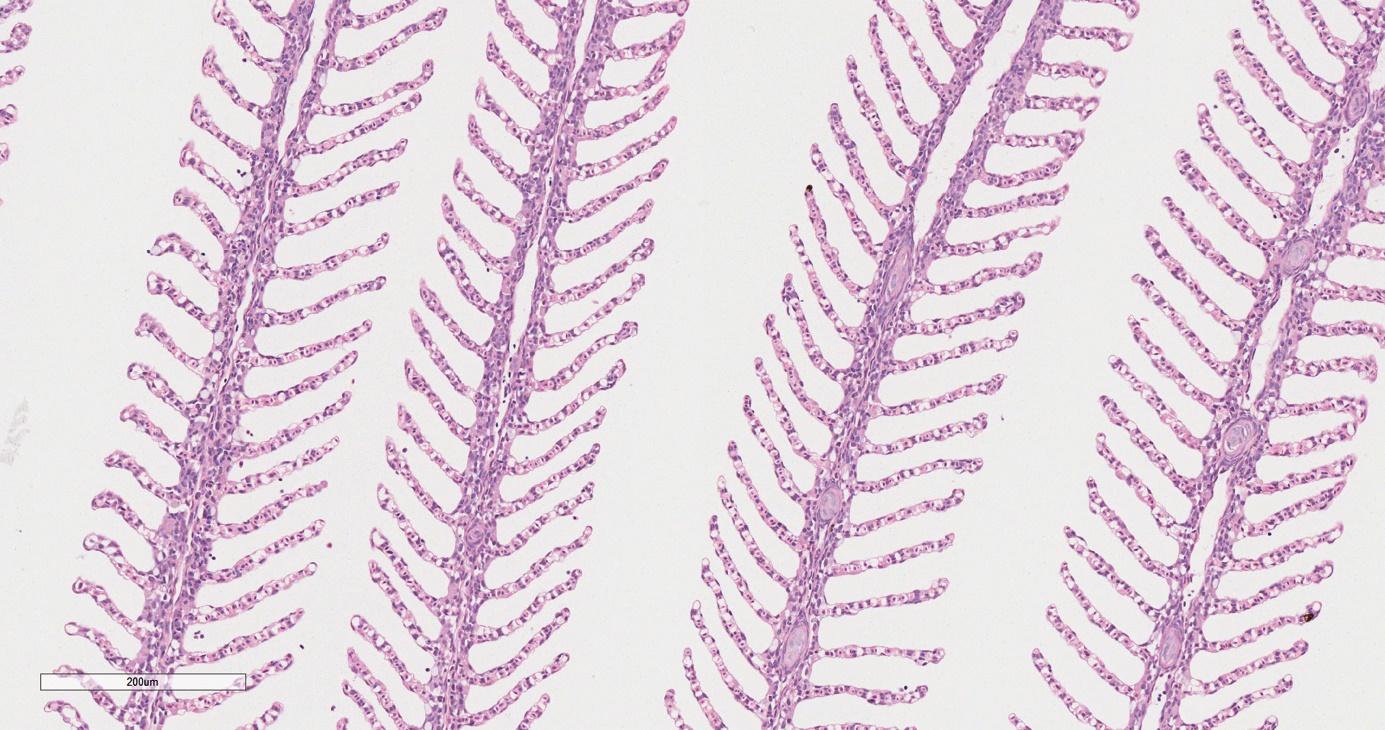
Gills from five fish were sampled at time zero to act as a normal gill tissue control. Other tissues from these fish (skin, muscle, spleen, kidney, liver, pyloric caecae, pancreas and heart) were sampled in accordance with best practice as recommended for fish diagnostic investigation (Noga 2010) to screen for any underlying abnormality in the stock.
All fish were processed for histology using standard procedures in a commercial laboratory (Fish Vet Group, Inverness, Scotland) where tissues were sectioned (3 to 4μm), stained with haematoxylin and eosin (H & E) and the slides digitally scanned (Leica Aperio) and assessed using Aperio ImageScope software (version 12.3.3).
Results and Discussion
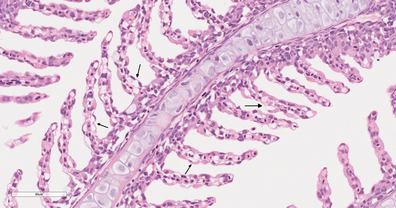
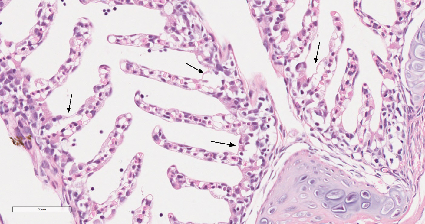
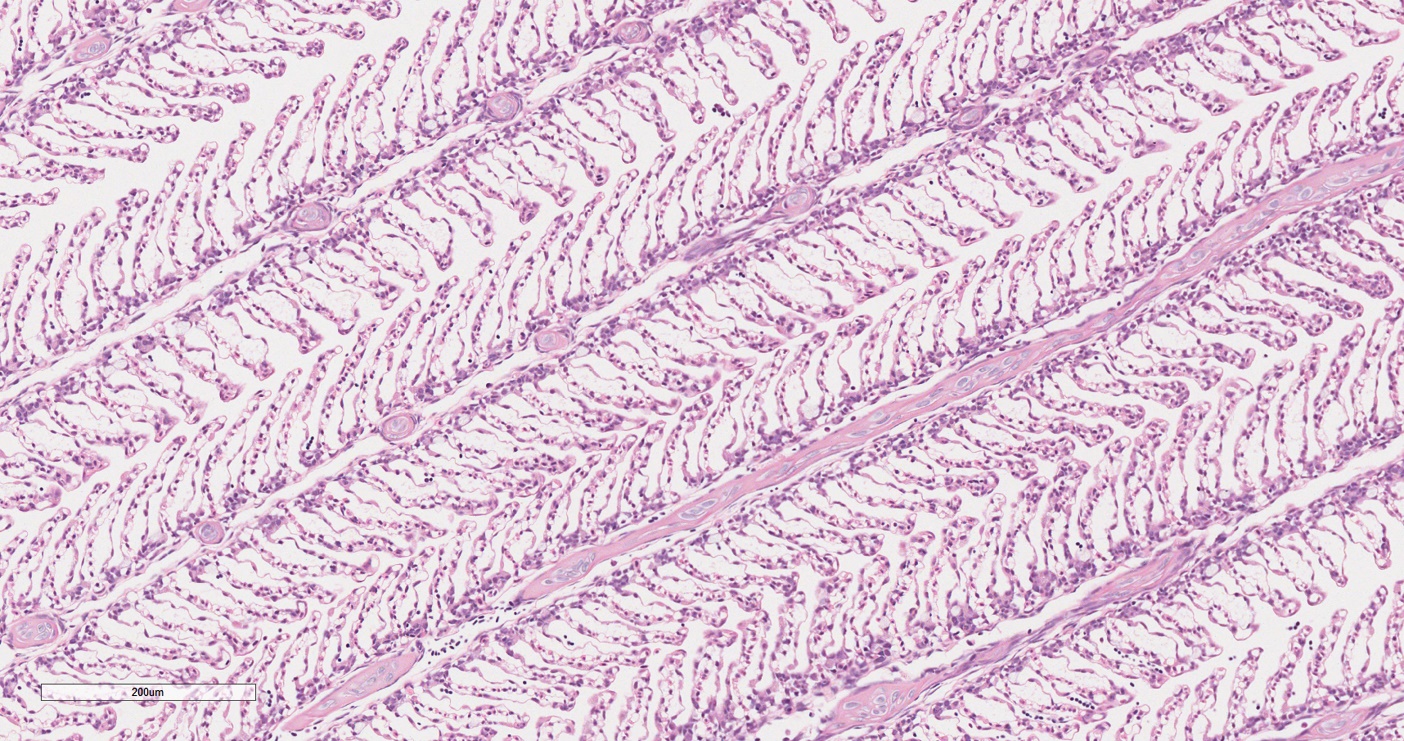
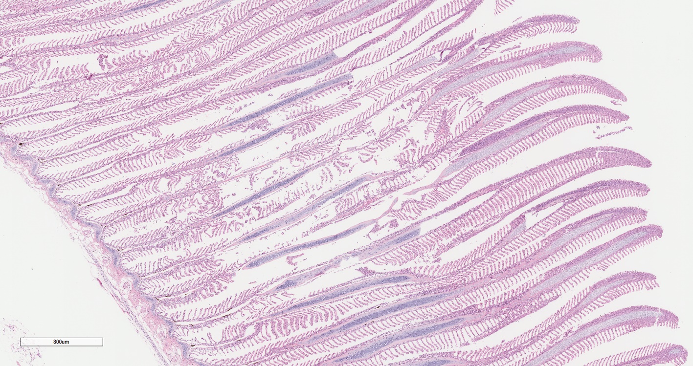
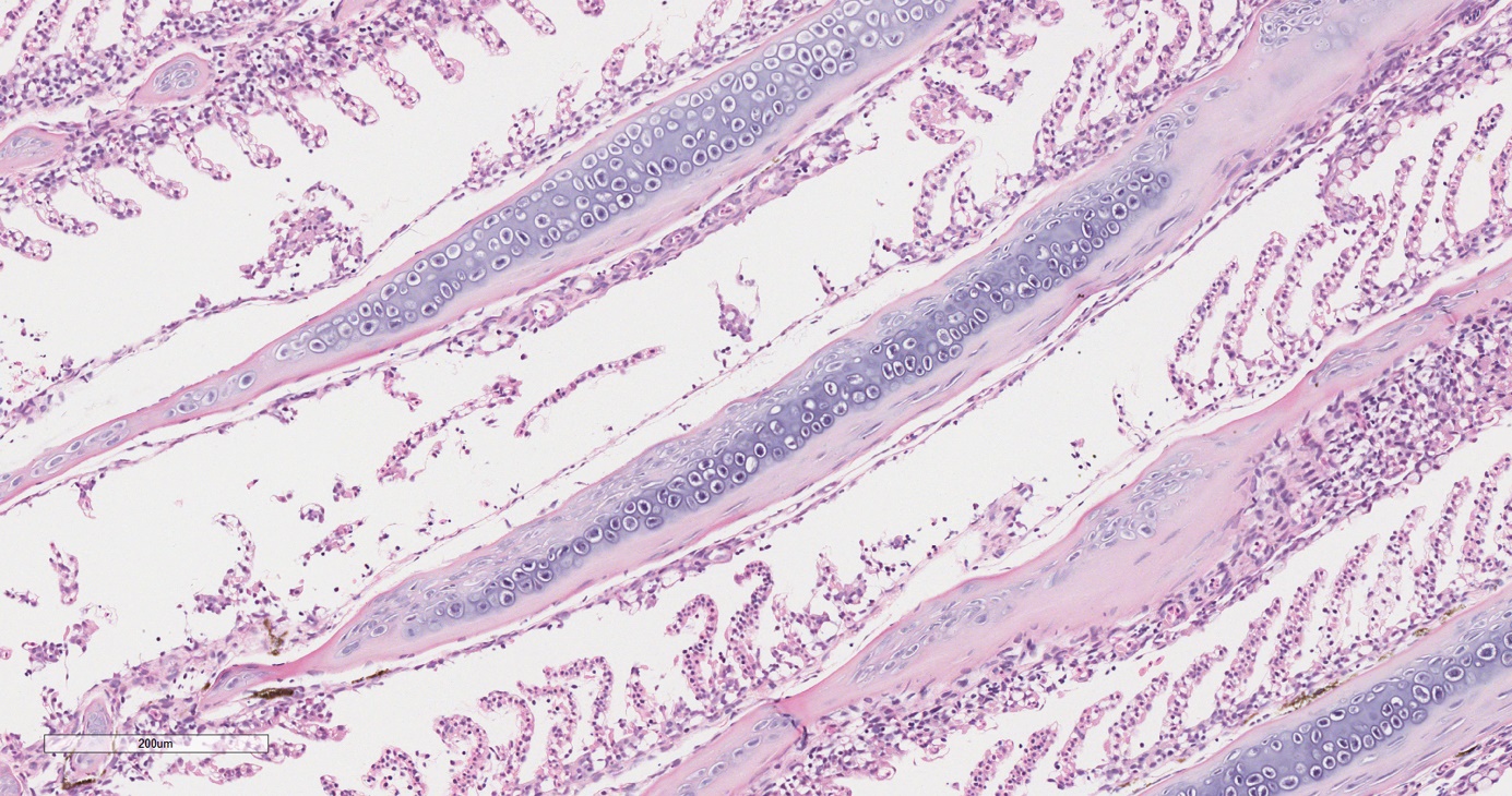
The effects of delayed fixation on histology were assessed at 10, 20 and 60 min post-mortem. At 10 min, mild, focal lamellar epithelial cell separation was present in 15 to 30% of the gill lamellae (Figure 1). At 20 min, changes were more extensive, with up to 50% of gill lamellae presenting with epithelial cell separation (Figure 2) and up to 100% of lamellae similarly affected at 60 min (Figure 3).


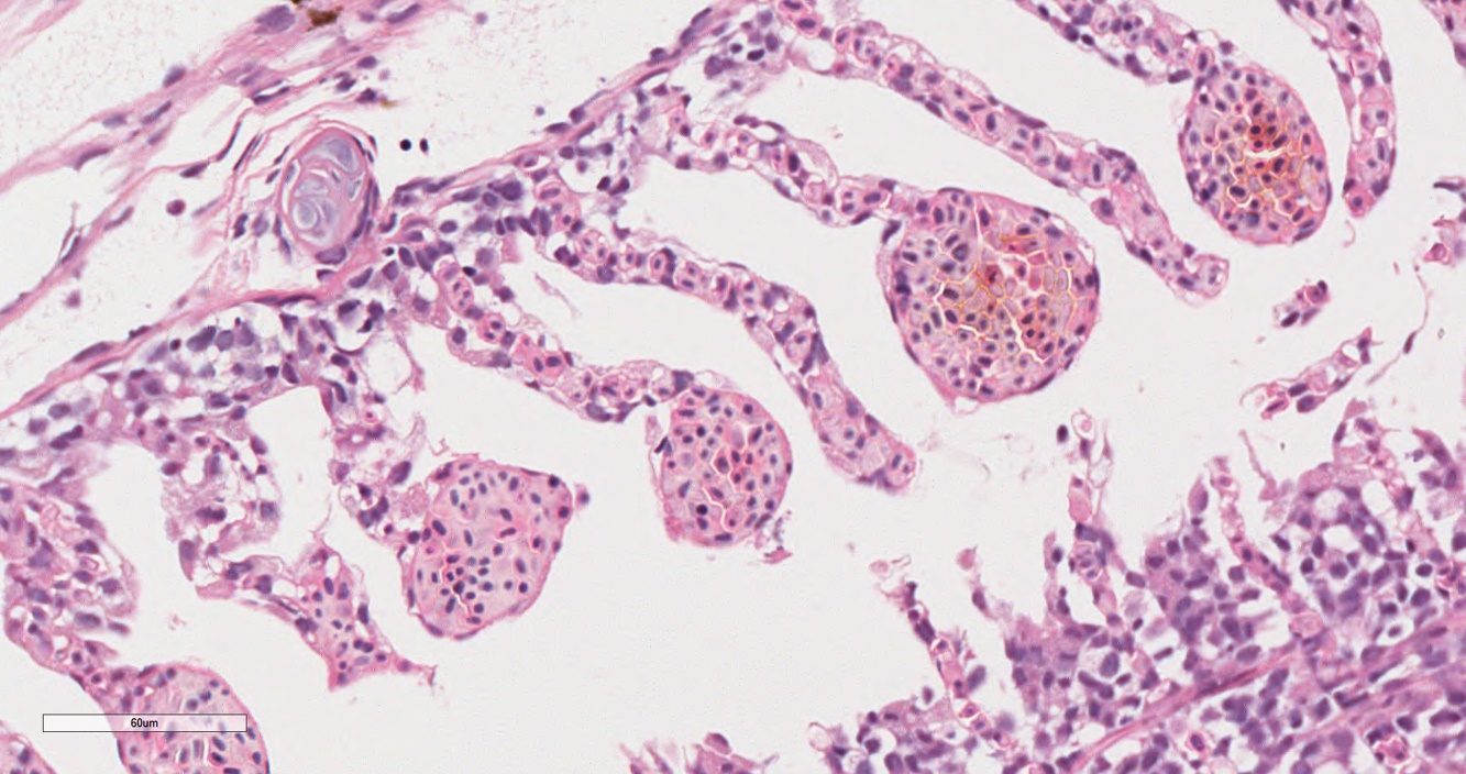
Percussive stunning resulted in the presence of multifocal lamellar aneurysms or telangiectasis (Figure 4). No such artefacts were observed in the control fish that were killed with anaesthetic overdose (Figure 7).

There were not sufficient fish available to test five fish in each of the groups 3 and 4, where potential direct trauma from sampling methods was being assessed. However, these techniques, as anticipated, resulted in dramatic alterations in the filaments so larger group numbers were not required to demonstrate an effect. The use of forceps to transfer the gill into the fixative resulted in fractured or abnormal filaments with focal epithelial cell sloughing and loss of lamellae (Figure 5). Finally, swabbing of gill surfaces with cotton-wool tipped swabs caused substantial focal loss of lamellae and sloughing of epithelial cells (Figure 6).


Fixation delays, even of a relatively short duration, appeared to create significant alterations. If a study design requires large numbers of fish to be sampled simultaneously, there could potentially be variable delays in fixation depending on the rate and order of sampling. In a review of gill histology changes induced in irritant and toxin studies Mallatt (1985) reported that lifting or separation of gill epithelium was the most frequent finding in the 104 papers selected. Mallatt (1985) also recorded that lifting of the gill epithelium was less frequently reported in saltwater (39% of saltwater studies) compared to freshwater (69% of freshwater studies) and he suggested that the osmolarity of the medium was influential in the lesion development. Munday and Jaisankar (1998) in their study of rainbow trout (Oncorhynchus mykiss) gills in freshwater and seawater also confirmed that trout gills in seawater maintained their integrity for longer than those in freshwater. Changes were reported at two h postmortem in seawater (described as oedema in the interlamellar region of the filaments). It further emphasizes that rapid fixation of tissues should be ensured to prevent compromised samples, especially in freshwater.
In this study the salmon euthanised by a percussive stun exhibited lamellar aneurysms consistent with those reported by Herman and Meade (1985) and were distinct from those considered pathological with associated lamellar thrombi such as when fish are treated with mechanical or thermal delousing methods or exposed to effects of in-situ net cleaning (Østevik et al. 2021, 2022). It is therefore recommended that percussive stunning is not utilised for euthanasia if gills are a focus for study via histology. The forceps-induced damage in gills has not previously been reported and, despite it being considered an infrequent diagnostic finding, it can be minimised by avoiding the filaments when sampling with forceps which may be applied to the cartilaginous arch, or alternatively rubber-tipped forceps could be used. The finding of cotton-wool tipped swabs inducing focal gill histopathology has also not been previously reported. When fish are sampled on sites for both histopathology and RT PCR, as is frequently the case, it is important that the gill arch for histopathology is taken from the opposite side to the side swabbed for RT PCR. Recently, improved PCR diagnostic sensitivity has been shown with calcium alginate fibre-tipped or viscose/rayon-tipped type swabs by Fernandez-Senac et al. (2020), and it would be of interest to assess histology associated with these types of swabs. It was reported by Fernandez-Senac et al. (2020) that reduced fish stress and improved diagnostic sensitivity could be achieved by sampling only the third or fourth gill arches, rather than all arches as is commonly practiced in European salmon farming.
It has been considered that formalin may not be the best fixation medium for gill tissues and alternatives such as Davidson’s or Bouin’s solution preferable by some workers (Speare and Ferguson 1989; Wolf et al. 2015). However, in many cases, 10% neutral buffered formalin is the most practical choice especially when a range of organs are being sampled simultaneously and it has the advantages of being inexpensive, readily available, and it has broad utility. It is, however, hazardous, slower to fix tissues and limited for detection of intact DNA or RNA for further analysis (Cadoret et al. 2013).
This study illustrates how easy it is to unintentionally create artefacts in gill tissues sampled for histology. The changes can appear similar to genuine histopathology, from which they can be difficult to distinguish. It is hoped publishing this study might raise awareness of the potential issues that can arise from sampling and processing, and workers who are sampling in the field will take the necessary precautions to avoid creating artefacts when sampling gills. A list of best practice recommendations are provided below. It is also hoped that describing these common and easily created artefacts will help to reduce the misinterpretation of gill lesions and misdiagnosis when reading histopathology. The ability to distinguish what is artefact from genuine pathology is critical in both clinical diagnostics and research, particularly when planning experimental design, as well as during investigations of disease pathogenesis and aetiology, and finally when providing advice on prognosis or treatments.
Recommendations for field sampling to minimise artefacts when sampling gill tissue
-
Anaesthetic overdose is preferable to percussive stunning when sampling gills.
-
Time interval should be as short as possible from euthanasia to gill excision and immersion in fixative.
-
Sharp scissors or scalpel should be used to cut through the gill arch while removing the gill section of interest to minimise tissue disruption at the edge of the tissue.
-
Gentle tissue handling using blunt or rubber-tipped forceps should be employed. Only the gill rakers or gill arch should be gripped avoiding any contact with the gill filaments and lamellae.
-
If swabbing gill arches for RT PCR, the opposite side of the fish should be used to excise gill tissue for histology.
In summary, a standardised approach to post-mortem technique, with chemical euthanasia, rapid fixation and gentle tissue handling can minimize or eliminate artefact and contribute to optimal gill histology for diagnostic interpretation and research applications.