Loading [Contrib]/a11y/accessibility-menu.js
Skip to main content
null
Bulletin of the EAFP
  • Menu
  • Articles
    • Case study
    • Expert Opinion
    • Method
    • Note
    • Research article
    • Review
    • Workshop Report
    • All
  • For Authors
  • Editorial Board
  • About
  • Issues
  • search

RSS Feed

Enter the URL below into your favorite RSS reader.

http://localhost:19994/feed
Research article
Vol. 43, Issue 2, 2023May 26, 2023 CEST

Survey on Betanodavirus in Wild Fish Species in the Turkish Coastlines of the Mediterranean Sea and Molecular Characterization: detecting different genotypes from different fish in different locations

M. Kaplan, K. Pekmez, A.A. Çağırgan, B.K. Tekelioğlu, U. Oğuz, B. Kafa, F. Arslan, G. Kalaycı,
BetanodavirusMediterraneansurveillanceTurkeywildlife
Copyright Logoccby-4.0 • https://doi.org/10.48045/001c.74914
Photo by Sebastian Pena Lambarri on Unsplash
Bulletin of the EAFP
Kaplan, M., K. Pekmez, A.A. Çağırgan, B.K. Tekelioğlu, U. Oğuz, B. Kafa, F. Arslan, and G. Kalaycı. 2023. “Survey on Betanodavirus in Wild Fish Species in the Turkish Coastlines of the Mediterranean Sea and Molecular Characterization: Detecting Different Genotypes from Different Fish in Different Locations.” Bulletin of the European Association of Fish Pathologists 43 (2): 73–92. https:/​/​doi.org/​10.48045/​001c.74914.
Save article as...▾
Download all (4)
  • Figure 1. Location of the collected samples for assessing the prevalence of NNV
    Download
  • Download
  • Download
  • Appendix 1.
    Download

Sorry, something went wrong. Please try again.

If this problem reoccurs, please contact Scholastica Support

Error message:

undefined

View more stats

Abstract

Viral nervous necrosis is a viral disease caused by betanodavirus causing deaths and epidemics in wild or farmed sea fish, especially in juveniles. Betanodavirus has been detected in farmed European seabass and gilthead seabream in Turkey. However, there are no studies on the presence of betanodavirus in wildlife along the Turkish Mediterranean coast. This study aimed to investigate the presence of betanodavirus in wild fish along the Turkish coast of the Mediterranean and to characterize isolates using molecular tools. Hence, 400 fish belonging to 27 different wild species were caught and tested using real-time RT‒PCR. The betanodavirus genome was detected in red mullets (25/50) and garfish (2/4) and identified as RGNNV and RGNNV/SJNNV genotype, respectively according to partial genome sequencing of the RNA1 and RNA2 segments. Phylogenetic analysis showed that isolated viruses were similar to isolates collected from other parts of the Mediterranean Sea and China, India, and Japan. The results of the studies show that different genotypes are circulating in the studied region in different species. These findings indicates that more research on wildlife, both around the farm and in other regions, are needed to prevent spreading of the virus to aquaculture facilities.

Introduction

Viral nervous necrosis (VNN) is a significant viral disease caused by betanodaviruses, particularly in larvae and juveniles and rarely in adults. VNN is depicted as one of the most economically threatening problems for marine aquaculture (Fernández Sánchez et al. 2022). With the industry beginning to culture different fish species, VNN outbreaks cause concerns about sustainability (Costa and Thompson 2016; Thiéry et al. 2011; Panzarin et al. 2010; Toffan et al. 2017).

Betanodaviruses are classified in the Nodaviridae family and have an icosahedral structure and a round morphology, are non-enveloped with a diameter of approximately 25 nm (Breuil et al. 1991). Their genomes are single-stranded with positive polarity and two segments, RNA1 and RNA2 (Johnson, Price, and Ball 2003). The RNA1 (3.1 kb) is responsible for the synthesis of RNA-dependent RNA polymerase (RdRp) and is involved in the adaptation of the virus to temperature (Ahlquist et al. 2003; Gallagher and Rueckert 1988; Hata et al. 2010; Nagai and Nishizawa 1999; Panzarin et al. 2014). During the replication of RNA1, subgenomic RNA3 transcript production is directed by RdRp and contributes to the replication process (Fenner, Goh, and Kwang 2006; Iwamoto et al. 2005; Johnson, Price, and Ball 2003). The RNA2 (1.4 kb) segment encodes the capsid protein and is responsible for host tropism and immunoreactivity of different genotypes (Ito et al. 2008; Iwamoto et al. 2004; Johnson, Price, and Ball 2003; Nishizawa et al. 1995; Panzarin et al. 2016).

According to phylogenetic analysis of the T4 variable region of the RNA2 segment, betanodaviruses are grouped into four major genotypes, including striped jack nervous necrosis virus (SJNNV), tiger puffer nervous necrosis virus (TPNNV), barfin flounder nervous necrosis virus (BFNNV), and red-spotted grouper nervous necrosis virus (RGNNV) (ICTV 2021). Rather than being species-specific, genotypes require a water temperature at which they develop optimally (Costa and Thompson 2016; Mori et al. 2005). Because of the segmented nature of the virus, the formation of reassortant strains has thus far been identified in the SJNNV and RGNNV genotypes, and RGNNV/SJNNV or SJNNV/RGNNV reassortants have been reported many times in sea bass, sea bream and sole (Bitchava et al. 2019; He and Teng 2015; Kaplan, Pekmez, Cagirgan, et al. 2022; Toffan et al. 2017; Volpe et al. 2020).

Betanodaviruses are commonly referred to as nervous necrosis viruses (NNV). So far, at least 173 different cultured or wild fish and invertebrate species have been susceptible to NNV and the virus has caused epidemics in at least 62 of them (Bandín and Souto 2020). Worse still, this number increases day by day. NNV typically causes outbreaks in farmed fish, but severe outbreaks have also appeared in wild fish, particularly grouper (Gomez et al. 2009; Valencia et al. 2019; Vendramin et al. 2013). Asymptomatic NNV infections have also been detected in wild fish (Baeck et al. 2007; Barker et al. 2002; Ciulli et al. 2007; Gomez et al. 2004, 2008b; Liu et al. 2014; Panzarin et al. 2012). Bivalve mollusks or crustaceans can also play a role in the spread of various NNV strains (Berzak et al. 2019; Ciulli et al. 2010; Gomez et al. 2008a; Kim et al. 2017; Panzarin et al. 2012).

In recent years, research on different geographical regions has shown that RGNNV is common not only in farms but also among wild fish in the Mediterranean basin and on the coasts of Asia and Australia (Bandín and Souto 2020). The lack of effective treatment for the disease and the introduction of applied management such as monitoring the spread of the virus helps to reduce the effects of the disease (Doan et al. 2016). For this reason, surveys of farm and wild fish in different regions provides important information, increasing the capacity for early warning systems and awareness, and beyond that increasing biosecurity, including implementation of vaccination regimes. This study aimed to investigate the presence of betanodavirus in wild fish along the Turkish Mediterranean coast, where betanodaviruses are endemic, and to characterize those circulating isolates.

Materials and methods

Sampling

Along the Mediterranean coast of Turkey, 400 wild fish belonging to 27 species were caught by fishing between 2019 and 2021 (Table 1). Samples were collected from five different regions of the Mediterranean coast of Turkey, which were Antalya Bay, Mersin Bay, Iskenderun Bay, and Finike and Manavgat coasts (Figure 1).

Table 1.Fish species, locations and epidemiological data of collected samples.
Common name Latin name Location Sampling date Water temperature Sample size Tissue Positivity
Atlantic chub mackerel Scomber colias Gulf of İskenderun 05.10.2019 25 2 Brain-eye 0/2
Lizzard fish Saurida lessepsianus Gulf of İskenderun 05.10.2019 25 5 Brain-eye 0/5
Sea bream Sparus aurata Gulf of İskenderun 05.10.2019 25 1 Brain-eye 0/1
Bogue Boops boops Gulf of İskenderun 05.10.2019 25 3 Brain-eye 0/3
Goldbanded goatfish Upeneus moluccensis Gulf of İskenderun 05.10.2019 25 4 Brain-eye 0/4
Round herring Etrumeus golanii Gulf of İskenderun 05.10.2019 25 5 Brain-eye 0/5
Garfish Belone belone Gulf of İskenderun 05.10.2019 25 4 Brain-eye 2/4
Sea bream Sparus aurata Gulf of İskenderun 20.09.2020 30 5 Brain-eye 0/5
Grey mullet Mugil cephalus Gulf of İskenderun 20.09.2020 30 5 Brain-eye 0/5
Red mullet Mullus barbatus Gulf of İskenderun 20.09.2020 30 5 Brain-eye 0/5
Annular seabream Diplodus annularis Gulf of İskenderun 20.09.2020 30 5 Brain-eye 0/5
Bogue Boops boops Gulf of İskenderun 20.09.2020 30 5 Brain-eye 0/5
Sea bass Dicentrarchus labrax Gulf of İskenderun 20.09.2020 30 5 Brain-eye 0/5
Sardine Sardinella aurita Gulf of İskenderun 20.09.2020 30 5 Brain-eye 0/5
Common sole Solea solea Gulf of İskenderun 20.09.2020 30 5 Brain-eye 0/5
Goldbanded goatfish Upeneus moluccensis Gulf of İskenderun 20.09.2020 30 5 Brain-eye 0/5
Lizzard fish Saurida lessepsianus Gulf of İskenderun 20.09.2020 30 5 Brain-eye 0/5
Forkbeard Phycis phycis Gulf of İskenderun 20.03.2021 18 1 Brain-eye 0/1
Annular Seabream Diplodus annularis Gulf of İskenderun 20.03.2021 18 1 Brain-eye 0/1
Meagre Argyrosomus regius Gulf of İskenderun 20.03.2021 18 1 Brain-eye 0/1
Zebrafish Diplodus cervinus Gulf of İskenderun 20.03.2021 18 1 Brain-eye 0/1
Saddled Seabream Oblada Melanura Gulf of İskenderun 20.03.2021 18 1 Brain-eye 0/1
Salema Sarpa Salpa Gulf of İskenderun 20.03.2021 18 1 Brain-eye 0/1
Common pandora Pagellus erythrinus Gulf of İskenderun 20.03.2021 18 5 Brain-eye 0/5
Yellow mouth barracuda Sphyraena viridensis Gulf of İskenderun 20.03.2021 18 5 Brain-eye 0/5
Mediterranean Horse mackerel Trachurus mediterraneus Gulf of İskenderun 20.03.2021 18 4 Brain-eye 0/4
Red Sea goatfish Parapeneus forsskali Gulf of İskenderun 21.04.2021 17 15 Brain-eye 0/15
Crocodile toothfishes Champsodon nudivitis Gulf of Mersin 22.04.2021 19 15 Brain-eye 0/15
Threadfin breams Nemipterus randalli Gulf of İskenderun 21.04.2021 17 15 Brain-eye 0/15
Slender rainbow sardine Dussumaria elepsiodes Gulf of İskenderun 21.04.2021 17 15 Brain-eye 0/15
Morocco dentex Dentex moraccanus Gulf of Mersin 22.04.2021 19 15 Brain-eye 0/15
Red cornetfish Fistularia petimba Gulf of İskenderun 21.04.2021 17 15 Brain-eye 0/15
Red Sea goatfish Parupeneus forsskali Gulf of Mersin 22.04.2021 19 15 Brain-eye 0/15
Red mullet Mullus barbatus Gulf of Antalya 28.09.2021 28 50 Brain-eye 25/50
Lizzard fish Saurida lessepsianus Manavgat Coast 30.09.2021 27 50 Brain-eye 0/50
Sardine Sardinella aurita Finike Coast 29.09.2021 29 50 Brain-eye 0/50
Sardine Sardinella aurita Gulf of Antalya 28.09.2021 28 50 Brain-eye 0/50
Figure 1
Figure 1.Location of the collected samples for assessing the prevalence of NNV

Preparation of samples

The fish, transported to the laboratory through the cold chain, were examined macroscopically on an individual basis. Brain, eye and spinal cords were removed following laboratory biosafety rules. The organs were homogenized (1/5, w/v) in a refrigerated homogenizer (Allsheng/BIOPREP-24) with ceramic beads in EMEM (Eagle’s Minimum Essential Medium) (Sigma‒Aldrich, United Kingdom). The EMEM contained 2% fetal bovine serum (FBS) and 2% antibiotics (Sigma, USA). The homogenates were centrifuged at 4℃ and 4000 × g for 15 min (ThermoFisher SL 16R, Germany). Supernatants were collected and stored at -80℃ until use.

Total nucleic acid extraction

RNA extraction was done in an automatic extraction device (Roche MagNA Pure LC System, Germany) following the protocol of the commercial kit (MagNA Pure LC Total Nucleic Acid Isolation Kit, Roche, Germany) using 200 µL from each supernatant. Positive (TR.VNN.01.01) and negative (EMEM) controls were used to validate the extraction process and check cross-contamination.

Real-Time RT-PCR

Real-time reverse transcription polymerase chain reaction (RT-rPCR) was performed to detect the presence of NNV. Primers and probes specific to the T4 variable region of the RNA2 segment of betanodavirus (Panzarin et al. 2010) were used. RT‒rPCR was performed using a commercial kit (Real Time Ready Virus Master, Roche, Germany) and real-time PCR device (Roche LightCycler® 480 II). Mastermix was prepared as 7.6 µL H2O, 1 µL F primer (10 µM), 1 µL R primer (10 µM), 1 µL Probe (10 µM), 4 µL 5x buffer and 0.4 µL enzyme mix for each sample. Then, 15 µL of each was added to 5 µL of template and total volume was 20 µL. Positive and negative controls were added to each reaction to verify the PCR process and a contamination control. After being covered with transparent tape, the plate was centrifuged at 1500 g at 4℃ for 2 min and placed in the PCR device. Reaction conditions were set as 50℃ for 10 min for reverse transcription, 95℃ for 65 sec for the inhibition of reverse transcriptase, 95℃ for 10 sec for pre- denaturation, 54℃ for 30 sec for annealing, and 72℃ for one sec for 45 cycles for elongation. The study followed the previously optimized RT‒rPCR method (Kaplan and Karaoğlu 2021).

cDNA synthesis and PCR

cDNA synthesis (EvoScript Universal cDNA Master, Roche, Germany) was performed from the sample extracts to store the positive samples for longer periods of time and to perform sequence analysis. For cDNA synthesis, the mixture consisted of 4 µL of reaction buffer (5x conc.), 9 µL of H2O and 5 µL of sample RNA. Reaction conditions were as follows: 42℃ for 15 min, 85℃ for 5 min, 65℃ for 15 min, and 4℃ for post-reaction standby.

PCR was performed (Techne TC-412, United Kingdom) using a commercial kit (Xpert Fast Hotstart Mastermix with Dye, GRISP, Portugal) and specific primers (Bovo et al. 2011; Toffolo et al. 2007) from positive sample cDNAs were used to obtain partial genome sequences of both RNA1 and RNA2 segments of betanodavirus. The total reaction mixture consisted of 25 µL with: 12.5 µL master mix, 5.5 µL water, 1 µL forward (10 mM) and 1 µL reverse primers (10 mM), and 5 µL cDNA for each sample. The amplification cycle was 95℃ for 3 min of pre- denaturation followed by 45 cycles at 95℃ for 15 sec of denaturation, 62℃ for 15 sec of annealing, 72℃ for 15 sec of elongation, and one cycle for 3 min at 72℃ for the final elongation.

The amplicons were treated with DNA loading dye (6x Loading Dye, Thermo Scientific, USA, R0611) and transferred to a 1.5% agarose gel (Sigma, USA) containing 0.5 µg/mL ethidium bromide (Nalgene Fisher Scientific, BP-1302-10, USA). A 100 bp marker (GeneRuler 100 bp DNA Ladder, Thermo Fisher Scientific, Lithuania, SM0241COMPONENTA) was also used to determine the product size. Electrophoresis was set at 80 volts for 60 min. Then, the agarose gel was examined on an imaging device (Vilber Lourmat, EEC, France).

Sequencing and phylogenetic analysis

Positive samples were submitted to partial genome sequencing for both RNA1 and RNA2 segments of the virus, performed by a commercial company (Microsynt, Balgach, Switzerland) to determine the genotype and to perform phylogenetic analysis. Correction and matching of the results were performed with the DNADynamo (Blue Tractor Software Ltd., UK) software. The resulting consensus nt sequences were compared and confirmed in the Basic Local Alignment Search Tool (BLAST) system of the National Center for Biotechnology Information (NCBI) (Altschul et al. 1990). Subsequently, accessible close isolates in GenBank and previously detected isolates in the Mediterranean were included in the molecular study, except for very short sequences. All sequences to be studied were aligned with the ClustalW method using the MEGAX software (Kumar et al. 2018). Phylogenetic analyses were performed by determining the best protein/DNA model for both segments. The nt and amino acid (aa) similarity rates between the sequences were determined according to both RNA1 and RNA2 segments. Using the maximum likelihood (ML) method, 1000 replicate bootstraps were selected, and phylogenetic trees were created (Kumar et al. 2018).

Results

During the necropsy in the laboratory, macroscopic examinations did not reveal any lesions in the fish.

The NNV genome was detected in two out of four garfish (Belone belone) sampled in Iskenderun Bay (Figure 1) during October 2019. Both positive samples were collected in the same region and one used for further studies was named as TR. VNN.01.03. NNV was not detected in other samples neither in 2019 nor in the samples from March to April in 2020 and 2021. In addition NNV was detected in high prevalence in red mullet (Mullus barbatus) collected in the Antalya Gulf (25/50) in September 2021. One of them was kept for further molecular studies and the isolate was named as TR.VNN.07.01.

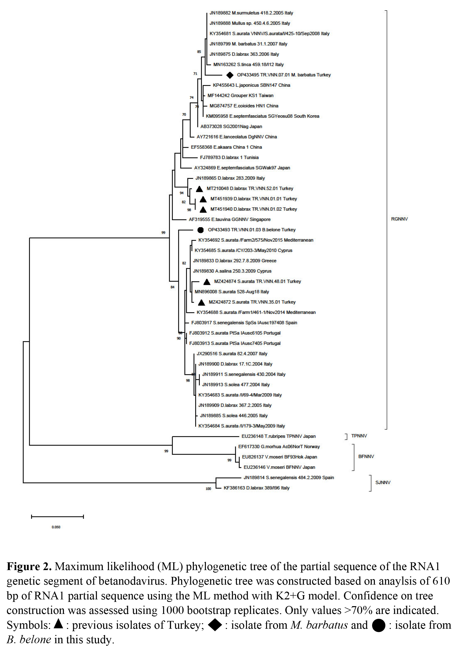
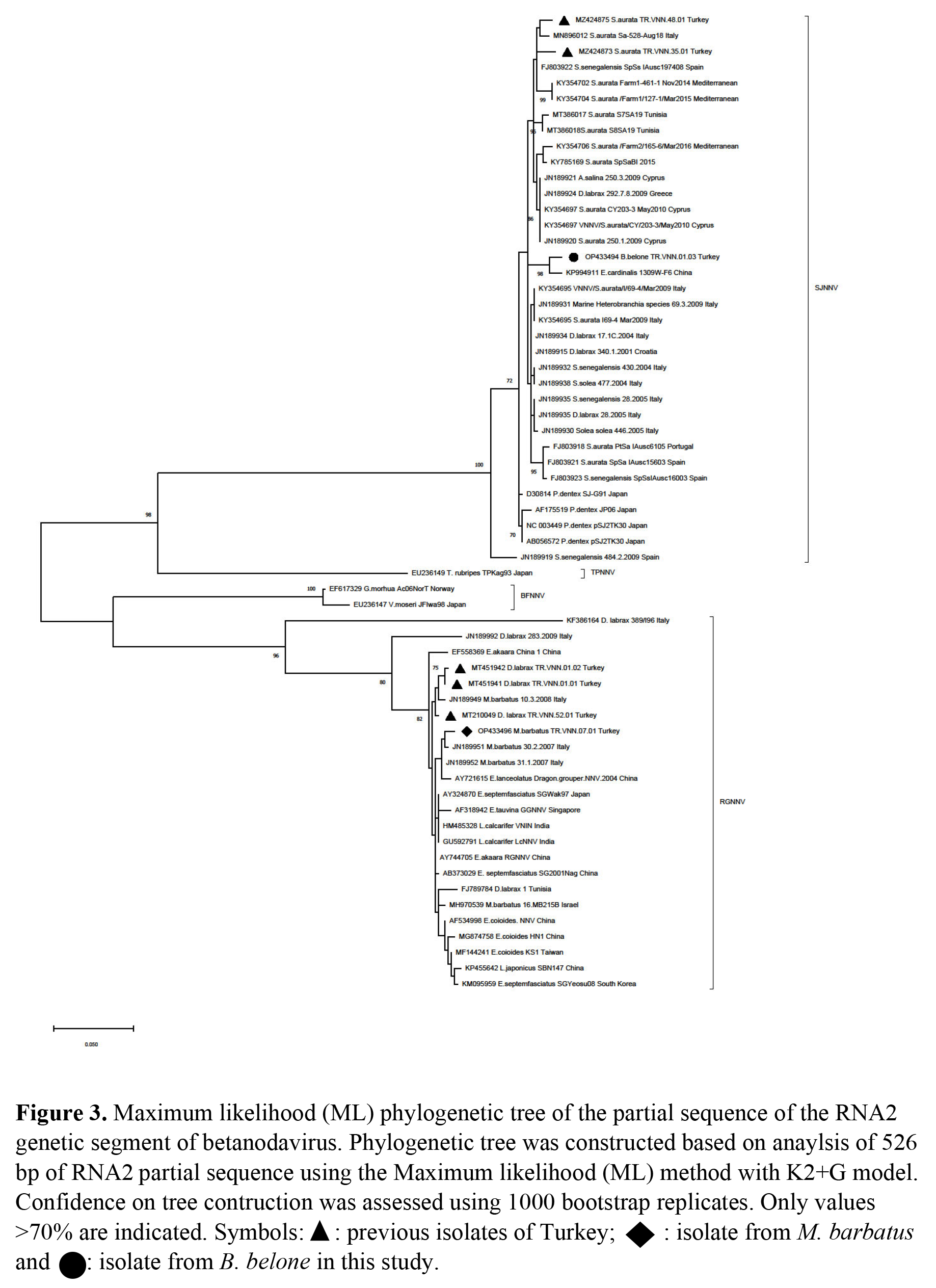
Partial sequencing of TR.VNN.01.03 and TR.VNN.07.01 were performed based on both RNA1 and RNA2 segments. The RNA1 segment of TR.VNN.01.03 and TR.VNN.07.01 were sequenced as 630 and 942 nt, respectively. The RNA2 segment of TR.VNN.01.03 and TR.VNN.07.01 were sequenced as 594 and 526 nt, respectively. Obtained sequences were uploaded to GenBank, and OP433493, OP433495 (RNA1), and OP433494, OP433496 (RNA2) accession numbers were obtained for TR.VNN.01.03 and TR.VNN.07.01, respectively. Isolates of this study were compared with the sequences in GenBank. The RGNNV was determined to be genotype of both isolates according to the RNA1 segment (Figure 2). However, TR.VNN.01.03 was a SJNNV, and TR.VNN.07.01 was a RGNNV genotype according to RNA2 segment (Figure 3). Thus, the TR.VNN.01.03 isolate was reassortant RGNNV/SJNNV, whereas the TR.VNN.07.01 isolate was RGNNV genotype. Interestingly, each of the isolates were detected in different location (Iskenderun and Antalya bays), from different fish species (garfish and red mullet fish), and belonged to the different genotypes (RGNNV/SJNNV and RGNNV). A 610 bp product of the RNA1 segment and 526 bp of RNA2 segment were used to perform further comparisons with other isolates in phylogenetic analysis.

In the phylogenetic analysis of RNA1 segment of TR.VNN.07.01, nt similarity was 98.22% to Italy isolates, while their aa proximity was 100%. TR.VNN.07.01 was found to be 94-95% similar to Turkish isolates previously detected in the Mediterranean (TR.VNN.01.01, TR.VNN.01.02) (Kaplan and Karaoğlu 2021), but aa proximity was 99.47%. The RNA2 segment of TR.VNN.07.01 isolate was 99.22% and 99.02% similar to Italy and China isolates, respectively. The aa proximity was 100% similar to many isolates from both the Mediterranean and the China, India, and Japan coasts (Appendix 1). NNVs previously isolated from red mullet fish worldwide (Berzak et al. 2019; Bitchava et al. 2019; Ciulli et al. 2007; Panzarin et al. 2012) were also examined only in proximity-similarity among themselves. However, because the gene regions where previous researcher’s studied were different, an evaluation could be made on a very short gene region isolated in İtaly, Greece and Israel. According to RNA1 (464 bp) and RNA2 (309 bp) nt similarity, the closest isolate was 31.1.2007 isolated in Italy with 98.46% (aa; 100%) and 99.01% (aa; 100%), respectively (data not shown).

Phylogenetic analysis of RNA1 segment of TR.VNN.01.03 revealed the closest relatedness was Portugal isolates with 97.85 % nt similarity. Furthermore, there was 97.87% similarity in aa with many isolates isolated from the Mediterranean Sea by Turkey, Italy and Portugal. According to the TR.VNN.01.03 isolate’s RNA2 segment, the closest isolate was KP994911 isolated from E. cardinalis in China with 98.46% similarity, the proximity of aa was 98.85% with the same isolate, and the similarity was 97.1% with the majority of Mediterranean isolates used in the phylogenetic study (Appendix-1). Around the world, only in the Mediterranean Sea was NNV reported in one garfish (Ciulli et al. 2007). However, since there were no records in the studies conducted in GenBank, we could not make a comparison with the NNV isolated from the garfish we found in the present study.

Discussion

Study of wild fish in VNN endemic regions in Turkey resulted in finding the virus presence in garfish and red mullet. While the RGNNV/SJNNV reassortant strain was detected in garfish, the RGNNV genotype was detected in red mullet. Both NNV positive fish were asymptomatic and viral loads (>35 Ct) were low in garfish, as expected; but the viral loads in red mullets were very high (22-25 Ct). This suggests that red mullet may play a more significant role in the transmission of NNV. A study of farmed and wild thicklip grey mullets supported the alternative theory. Researchers detected NNV genome in mullets after confirmation of outbreaks in sea bass (Zrnčić et al. 2022). However, much more data and validation are necessary to prove this hypothesis, farm to wild or wild to farm.

VNN is endemic in the Mediterranean basin, and the disease has occurred many times in both farm and wild fish (Berzak et al. 2019; Costa and Thompson 2016; Munday, Kwang, and Moody 2002; OIE 2017; Toffan et al. 2017). RGNNV is the most common genotype in this region (Costa and Thompson 2016), but the SJNNV genotype was also present in the Iberian Peninsula in 2009 (Panzarin et al. 2012). Reassortant RGNNV/SJNNV and SJNNV/RGNNV strains have only been reported in the Mediterranean Sea (He and Teng 2015). NNV has been detected clinically and subclinically in sea bass in Turkey; however, it has only been detected subclinically in sea bream thus far. The first NNV was first detected in Turkey in clinically infected sea bass on the Mediterranean coast in 2011 (Özkan Özyer et al. 2014). In ongoing studies, NNV has been detected frequently in sea bass and the RGNNV genotype was identified (Kalaycı et al. 2016; Kaplan et al. 2021; Kaplan and Karaoğlu 2021). The RGNNV/SJNNV genotype was also isolated from asymptomatic sea bream only in the Aegean Sea (Kaplan, Pekmez, Cagirgan, et al. 2022).

NNV research on wildlife along the Turkish Mediterranean coast has not been documented. So, in terms of taking epidemiological pictures, this research is essential. Here, multiple sampling was performed at five different points in different fish species, and as a result, both RGNNV (red mullet) and RGNNV/SJNNV (garfish) genotypes were isolated. The RGNNV/SJNNV genotype was detected for the first time on the Mediterranean coast of Turkey. Despite the fact that the RGNNV/SJNNV genotype was found in garfish (Ciulli et al. 2007), it was not possible for molecular comparisons to be performed since the records for this genotype were not accessible in GenBank. However, sampling had been performed many times in farms in the region where the garfish were collected (Kaplan and Karaoğlu 2021) and only the RGNNV genotype (TR.VNN.01.01 and TR.VNN.01.02) was identified. Two viruses isolated from the same location, TR.VNN.01.03 and TR.VNN.01.01, show a difference of 5% in the RNA1 segment, suggesting that either different viruses circulate in this region or mutations may have occurred in these segments. However, to clarify this, it is necessary to conduct whole genome studies of the viruses and subsequently molecular evolution studies. Here, the detection of the RGNNV/SJNNV genotype was surprising. In molecular analysis, TR.VNN.01.03 isolated from garfish was closer to the Portuguese reassortants according to the RNA1 segment, while it was closer to the Chinese isolates than the Mediterranean isolates according to the RNA2 segment. As previously reported in the literature, betanodavirus can transmit between different species independently of the host (Costa and Thompson 2016). More importantly, it suggests that different genotypes can circulate across different geographic regions. As a result, there is a constant flow to the Mediterranean Sea through the Red Sea, and the species exotic to the Mediterranean Sea come here from the Indian Ocean through the Red Sea (Al-Sulaimani et al. 2022; Mehanna and Osman 2022; Tomanić et al. 2022). This fact supports the idea that exotic virus species can be transferred through exotic fish by different geographical regions. Additionally, the RGNNV genotype suggests that high diversity among genetic clusters may play a critical role in virus evolution (Sethi et al. 2018). Epistasis and mutation resulting from reassortants within RGNNV may also shape the evolutionary process and diversity of RGNNV (Bitchava et al. 2019). Considering the genome characteristics of betanodavirus, both the segmented structure and especially the RNA1 segment being more prone to mutation, combined with the migration of viruses or exotic fish species, may lead to even more negative consequences. In addition, global warming causes significant changes in aquatic environmental parameters, which may affect the epidemiology of aquatic animal diseases as in other animal diseases (Bandín and Souto 2020). Worse still, this may exacerbate the disease severity in aquaculture worldwide (Bitchava et al. 2019). Rapid increases in viral load due to high numbers and temperature changes in wild asymptomatic species infected with NNV, particularly in Asian and Mediterranean countries, may cause an increase in deaths (Bandín and Souto 2020; Souto, Olveira, and Bandín 2015). In addition, increasing ocean temperatures may increase the number of reported cases of wild species and cause the decline of some endangered species (Bandín and Souto 2020). VNN outbreaks occur mostly in warm conditions (Doan et al. 2016). It is also possible for wild fish species to transmit the virus to farms or vice versa (Bitchava et al. 2019; Zrnčić et al. 2022).

In the molecular analysis of the TR.VNN.07.01 isolated from red mullet, the fact that it is similar to the RGNNV isolates previously isolated from the Mediterranean according to both RNA1 and RNA2 segments, shows that similar virus circulation in red mullets continues in a wide area in this region. Considering that red mullet fish inhabit deeper regions, both the high positivity rate (25/50) and the high viral loads found show that red mullets can play a crucial role in the epidemiology of betanodavirus. With this study, NNV isolation from red mullets has been performed for the first time on the Mediterranean coast of Turkey. The high positivity rate poses a risk in this region. Considering aquaculture in Turkey, further studies on betanodavirus in red mullets in both Mediterranean and Aegean seas can contribute to sustainable cultural fisheries.

NNV has been detected in more than 120 wild aquatic species globally (Bandín and Souto 2020). Although most are asymptomatic, NNV-related mortality has been reported in milk fish (Chanos chanos) (Sethi et al. 2018), European sea bass (Vendramin et al. 2013), mullet (Liza aurata and L. saliens) (Zorriehzahra et al. 2016) and wild grouper (E. costae and E. marginatus) (Kara et al. 2014; Nishioka et al. 2016; Valencia et al. 2019; Vendramin et al. 2013). In previous studies in the Mediterranean, NNV were detected in asymptomatic (Berzak et al. 2019; Bovo et al. 2011; Ciulli et al. 2007; Panzarin et al. 2012) or clinically infected (Haddad-Boubaker et al. 2014; Lopez-Jimena et al. 2010; Maltese et al. 2005; Terlizzi, Tedesco, and Patarnello 2012; Vendramin et al. 2013; Valencia et al. 2019) wild fish many times. Although RGNNV was identified frequently in wild populations in the Mediterranean (Bandín and Souto 2020), RGNNV/SJNNV reassortant was isolated from wild Mediterranean horse mackerel (Trachurus mediterraneus) and greater amberjack (Seriola dumerili) in Greece (Bitchava et al. 2019), thicklip grey mullet (Chelon labrosus) in Spain (Haddad-Boubaker et al. 2014). In Asia, the RGNNV/SJNNV was isolated from whitefin trevally (Carangoides equula) and Japanese jack mackerel (T. japonicus) species (Gomez et al. 2004; Liu et al. 2014). In this study, an RGNNV/SJNNV reassortant was identified in garfish in the Mediterranean. With new research, the number of fish species involved in the spread of the betanodavirus is increasing day by day. The increase in risk factors constantly reminds us of the increase in biosecurity measures on farms and their environments. However, in the future, multivalent vaccines may be necessary, rather than monovalent vaccines to prevent outbreaks. On the other hand, increasing fish species in betanodavirus epidemiology poses a threat to aquaculture, which shows the importance of screening studies on wildlife or farms. Asymptomatic infected fish are eaten by predator (hunter‒predator) fish and get contaminated with infected stools of these fish. Another possible transmission of the disease is the feeding of broodstocks with raw fish (Mori et al. 2005). For example, after detecting NNV in wildlife, it could be detected in a farm in a short time (Berzak et al. 2019). Therefore, research on wild fish is critical in sustaining the future of aquaculture industries. The close viral sequences between betanodavirus isolates from wild fish and those isolated during clinical disease outbreaks in cultured fish in the same region in previous years show permanent circulation and interaction and transmission between wildlife and farm fish (Vendramin et al. 2013). Horizontal transmission among subclinical fish after migration or commercial importation for cultural fishery production can play a significant role in new outbreaks (Valencia et al. 2019). Information on the health status of specific wild fish populations is also crucial for aquaculture (Moreno et al. 2014). Considering the origins of NNV isolates detected thus far in Turkey, although most were asymptomatic, high aquaculture production capacity in Turkey necessitates early warning systems. To date, studies on NNV screening in Aegean, Mediterranean, or Black Sea regions have been at the farm level (Kaplan et al. 2021; Kaplan and Karaoğlu 2021; Kaplan, Pekmez, Çagirgan, et al. 2022). Additionally, NNV studies were conducted on mollusks in North Aegean and Marmara seas, but no virus was detected (Kaplan, Pekmez, Çagirgan, et al. 2022). However, a screening study involving wild fish in the Mediterranean Sea, where the virus is endemic, most abundant, and circulation is highest has yet to be conducted. This study involved the collection of data on the epidemiology of betanodavirus for the first time in wildlife of this region.

In a conclusion, after evaluating the genetic similarities between isolates in this study and other isolates, the isolates were similar to those found in Italy and the far-east rather than previous Turkish isolates. The first task at this point is to perform NNV screening in wild fish in a much wider area, i.e. in order to obtain whole genome sequences of possible isolates, and follow-up with a phylogenetic study. In the context of sustainable aquaculture production, it will be vital to reveal whole genome sequences of all isolates and to determine the prevalence or differences to be considered for potential vaccines.


Acknowledgments

This study was supported and officially permissed by the Ministry of Agriculture and Forestry of the Republic of Türkiye.

Submitted: February 09, 2023 CEST

Accepted: April 30, 2023 CEST

References

Ahlquist, Paul, Amine O. Noueiry, Wai-Ming Lee, David B. Kushner, and Billy T. Dye. 2003. “Host Factors in Positive-Strand Rna Virus Genome Replication.” Journal of Virology 77 (15): 8181–86. https:/​/​doi.org/​10.1128/​jvi.77.15.8181-8186.2003.
Google ScholarPubMed CentralPubMed
Al-Sulaimani, Suad, Nicholas Vincent Titelbaum, Ricardo El Ward, Tharwat El Zahran, Sana Chalhoub, and Ziad Kazzi. 2022. “Case Report of Tetrodotoxin Poisoning from Lagocephalus Sceleratus in Lebanon.” International Journal of Environmental Research and Public Health 19 (22): 14648. https:/​/​doi.org/​10.3390/​ijerph192214648.
Google ScholarPubMed CentralPubMed
Altschul, Stephen F., Warren Gish, Webb Miller, Eugene W. Myers, and David J. Lipman. 1990. “Basic Local Alignment Search Tool.” Journal of Molecular Biology 215 (3): 403–10. https:/​/​doi.org/​10.1016/​s0022-2836(05)80360-2.
Google Scholar
Baeck, G.W., Dennis Kaw Gomez, K.S. Oh, J.H. Kim, C.H. Choresca, and S.C. Park. 2007. “Detection of Piscine Nodaviruses from Apparently Healthy Wild Marine Fish in Korea.” Bulletin of the European Association of Fish Pathologists 27:116–22.
Google Scholar
Bandín, Isabel, and Sandra Souto. 2020. “Betanodavirus and VER Disease: A 30-Year Research Review.” Pathogens 9 (2): 106. https:/​/​doi.org/​10.3390/​pathogens9020106.
Google ScholarPubMed CentralPubMed
Barker, D.E., AM MacKinnon, L Boston, M.D.B. Burt, D.K. Cone, D.J. Speare, S Griffiths, M Cook, R Ritchie, and G Olivier. 2002. “First Report of Piscine Nodavirus Infecting Wild Winter Flounder Pleuronectes Americanus in Passamaquoddy Bay, New Brunswick, Canada.” Diseases of Aquatic Organisms 49 (2): 99–105. https:/​/​doi.org/​10.3354/​dao049099.
Google Scholar
Berzak, R, A Scheinin, N Davidovich, Y Regev, R Diga, D Tchernov, and D Morick. 2019. “Prevalence of Nervous Necrosis Virus (NNV) and Streptococcus Species in Wild Marine Fish and Crustaceans from the Levantine Basin, Mediterranean Sea.” Diseases of Aquatic Organisms 133 (1): 7–17. https:/​/​doi.org/​10.3354/​dao03339.
Google Scholar
Bitchava, Konstantina, Taxiarchis Chassalevris, Eirini Lampou, Fotini Athanassopoulou, Vangelis Economou, and Chrysostomos I. Dovas. 2019. “Occurrence and Molecular Characterization of Betanodaviruses in Fish and Invertebrates of the Greek Territorial Waters.” Journal of Fish Diseases 42 (12): 1773–83. https:/​/​doi.org/​10.1111/​jfd.13098.
Google Scholar
Bovo, G, A Gustinelli, F Quaglio, F Gobbo, Valentina Panzarin, A Fusaro, F Mutinelli, M Caffara, and ML Fioravanti. 2011. “Viral Encephalopathy and Retinopathy Outbreak in Freshwater Fish Farmed in Italy.” Diseases of Aquatic Organisms 96 (1): 45–54. https:/​/​doi.org/​10.3354/​dao02367.
Google Scholar
Breuil, G., J.R. Bonami, J.F. Pepin, and Y. Pichot. 1991. “Viral Infection (Picorna-like Virus) Associated with Mass Mortalities in Hatchery-Reared Sea-Bass (Dicentrarchus Labrax) Larvae and Juveniles.” Aquaculture 97 (2–3): 109–16. https:/​/​doi.org/​10.1016/​0044-8486(91)90258-9.
Google Scholar
Ciulli, S., E. Galletti, M. Grodzki, A. Alessi, M. Battilani, and S. Prosperi. 2007. “Isolation and Genetic Characterization of Betanodavirus from Wild Marine Fish from the Adriatic Sea.” Veterinary Research Communications 31 (S1): 221–24. https:/​/​doi.org/​10.1007/​s11259-007-0010-y.
Google Scholar
Ciulli, S., M. Grodzki, G. Bignami, P. Serratore, and S. Prosperi. 2010. “Molecular Detection and Genetic Analysis of Betanodaviruses in Bivalve Mollusks.” Journal of Biotechnology 150 (November):4–4. https:/​/​doi.org/​10.1016/​j.jbiotec.2010.08.026.
Google Scholar
Costa, Janina Z., and Kim D. Thompson. 2016. “Understanding the Interaction between Betanodavirus and Its Host for the Development of Prophylactic Measures for Viral Encephalopathy and Retinopathy.” Fish & Shellfish Immunology 53 (June):35–49. https:/​/​doi.org/​10.1016/​j.fsi.2016.03.033.
Google Scholar
Doan, Q K, M Vandeputte, B Chatain, T Morin, and F Allal. 2016. “Viral Encephalopathy and Retinopathy in Aquaculture: A Review.” Journal of Fish Diseases 40 (5): 717–42. https:/​/​doi.org/​10.1111/​jfd.12541.
Google Scholar
Fenner, Beau J., Winnie Goh, and Jimmy Kwang. 2006. “Sequestration and Protection of Double-Stranded RNA by the Betanodavirus B2 Protein.” Journal of Virology 80 (14): 6822–33. https:/​/​doi.org/​10.1128/​jvi.00079-06.
Google ScholarPubMed CentralPubMed
Fernández Sánchez, José L., Alain Le Breton, Edgar Brun, Niccolò Vendramin, Georgios Spiliopoulos, Dolors Furones, and Bernardo Basurco. 2022. “Assessing the Economic Impact of Diseases in Mediterranean Grow-out Farms Culturing European Sea Bass.” Aquaculture 547 (January):737530. https:/​/​doi.org/​10.1016/​j.aquaculture.2021.737530.
Google Scholar
Gallagher, T. M., and R. R. Rueckert. 1988. “Assembly-Dependent Maturation Cleavage in Provirions of a Small Icosahedral Insect Ribovirus.” Journal of Virology 62 (9): 3399–3406. https:/​/​doi.org/​10.1128/​jvi.62.9.3399-3406.1988.
Google ScholarPubMed CentralPubMed
Gomez, Dennis Kaw, Gun Wook Baeck, Ji Hyung Kim, Casiano H. Jr. Choresca, and Se Chang Park. 2008a. “Molecular Detection of Betanodaviruses from Apparently Healthy Wild Marine Invertebrates.” Journal of Invertebrate Pathology 97 (3): 197–202. https:/​/​doi.org/​10.1016/​j.jip.2007.10.012.
Google Scholar
Gomez, Dennis Kaw, Gun Wook Baeck, Ji Hyung Kim, Casiano Hermopia Jr. Choresca, and Se Chang Park. 2008b. “Molecular Detection of Betanodavirus in Wild Marine Fish Populations in Korea.” Journal of Veterinary Diagnostic Investigation 20 (1): 38–44. https:/​/​doi.org/​10.1177/​104063870802000107.
Google Scholar
Gomez, Dennis Kaw, S. Matsuoka, K. Mori, Y. Okinaka, S.C. Park, and T. Nakai. 2009. “Genetic Analysis and Pathogenicity of Betanodavirus Isolated from Wild Redspotted Grouper Epinephelus Akaara with Clinical Signs.” Archives of Virology 154:343–46. https:/​/​doi.org/​10.1007/​s00705-.
Google Scholar
Gomez, Dennis Kaw, J Sato, K Mushiake, T Isshiki, Y Okinaka, and T Nakai. 2004. “PCR-Based Detection of Betanodaviruses from Cultured and Wild Marine Fish with No Clinical Signs.” Journal of Fish Diseases 27 (10): 603–8. https:/​/​doi.org/​10.1111/​j.1365-2761.2004.00577.x.
Google Scholar
Haddad-Boubaker, S., Wassim Boughdir, Soufien Sghaier, Jamila Ben Souissi, Aida Megdich, Raouf Dhaouadi, Abdelkader Amara, Valentina Panzarin, and Emna Fakhfakh. 2014. “Outbreak of Viral Nervous Necrosis in Endangered Fish Species Epinephelus Costae and E. Marginatus in Northern Tunisian Coasts.” Fish Pathology 49 (2): 53–56. https:/​/​doi.org/​10.3147/​jsfp.49.53.
Google Scholar
Hata, Naomi, Yasushi Okinaka, Tokinori Iwamoto, Yasuhiko Kawato, Koh-Ichiro Mori, and Toshihiro Nakai. 2010. “Identification of RNA Regions That Determine Temperature Sensitivities in Betanodaviruses.” Archives of Virology 155 (10): 1597–1606. https:/​/​doi.org/​10.1007/​s00705-010-0736-7.
Google Scholar
He, Mei, and Chun-Bo Teng. 2015. “Divergence and Codon Usage Bias of Betanodavirus, a Neurotropic Pathogen in Fish.” Molecular Phylogenetics and Evolution 83 (February):137–42. https:/​/​doi.org/​10.1016/​j.ympev.2014.11.016.
Google Scholar
ICTV. 2021. “International Committee on Taxonomy of Viruses.” 2021. https:/​/​talk.ictvonline.org/​taxonomy/​.
Ito, Y, Y Okinaka, K.I. Mori, T Sugaya, T Nishioka, M Oka, and T Nakai. 2008. “Variable Region of Betanodavirus RNA2 Is Sufficient to Determine Host Specificity.” Diseases of Aquatic Organisms 79 (3): 199–205. https:/​/​doi.org/​10.3354/​dao01906.
Google Scholar
Iwamoto, Tokinori, Kazuyuki Mise, Atsushi Takeda, Yasushi Okinaka, Koh-Ichiro Mori, Misao Arimoto, Tetsuro Okuno, and Toshihiro Nakai. 2005. “Characterization of Striped Jack Nervous Necrosis Virus Subgenomic RNA3 and Biological Activities of Its Encoded Protein B2.” Journal of General Virology 86 (10): 2807–16. https:/​/​doi.org/​10.1099/​vir.0.80902-0.
Google Scholar
Iwamoto, Tokinori, Yasushi Okinaka, Kazuyuki Mise, Koh-Ichiro Mori, Misao Arimoto, Tetsuro Okuno, and Toshihiro Nakai. 2004. “Identification of Host-Specificity Determinants in Betanodaviruses by Using Reassortants between Striped Jack Nervous Necrosis Virus and Sevenband Grouper Nervous Necrosis Virus.” Journal of Virology 78 (3): 1256–62. https:/​/​doi.org/​10.1128/​jvi.78.3.1256-1262.2004.
Google ScholarPubMed CentralPubMed
Johnson, Kyle L., B.Duane Price, and L.Andrew Ball. 2003. “Recovery of Infectivity from cDNA Clones of Nodamura Virus and Identification of Small Nonstructural Proteins.” Virology 305 (2): 436–51. https:/​/​doi.org/​10.1006/​viro.2002.1769.
Google Scholar
Kalaycı, G., B. Özkan, K. Pekmez, and Murat Kaplan. 2016. “Levrek ve Çipura Kuluçkahanelerinde Viral Nervöz Nekrozis Hastalığının Durumu (Poster Sunum).” XII. Veterinary Medicines Microbiology Congress with International Attendance, Kapadokya/Nevşehir, 30 August – 02 September 2016.
Google Scholar
Kaplan, Murat, and Mehmet Taner Karaoğlu. 2021. “Investigation of Betanodavirus in Sea Bass (Dicentrarchus Labrax) at All Production Stages in All Hatcheries and on Selected Farms in Turkey.” Archives of Virology 166 (12): 3343–56. https:/​/​doi.org/​10.1007/​s00705-021-05254-0.
Google Scholar
Kaplan, Murat, K Pekmez, B Özkan, A.A. Çağırgan, and G Kalaycı. 2021. “Detection of RGNNV Genotype Betanodavirus in the Black Sea and Monitoring Studies.” Diseases of Aquatic Organisms 144 (April):117–21. https:/​/​doi.org/​10.3354/​dao03583.
Google Scholar
Kaplan, Murat, Kemal Pekmez, Abdurrahman Anil Cagirgan, Fatih Arslan, Buket Özkan, and Gülnur Kalaycı. 2022. “The First Detection of Betanodavirus Reassortant Genotype (RGNNV/SJNNV) Isolated from Gilthead Sea Bream (Sparus Aurata) in the Turkish Coastlines: The Importance of Screening and Monitoring Studies for Identifying the Source of the Infection.” Journal of Fish Diseases 45 (6): 783–93. https:/​/​doi.org/​10.1111/​jfd.13603.
Google Scholar
Kaplan, Murat, Kemal Pekmez, Abdurrahman Anıl Çagirgan, Buket Özkan, Fatih Arslan, Bülent Kafa, and Gülnur KALAYCI. 2022. “Türkiye’de Çift Kabuklu Yumuşakçalarda Betanodavirus Varlığının Araştırılması.” Acta Aquatica Turcica 18 (3): 415–25. https:/​/​doi.org/​10.22392/​actaquatr.1088276.
Google Scholar
Kara, H M, L Chaoui, F Derbal, R Zaidi, C de Boisséson, M Baud, and L Bigarré. 2014. “Betanodavirus‐associated Mortalities of Adult Wild Groupers E Pinephelus Marginatus (Lowe) and E Pinephelus Costae (Steindachner) in Algeria.” Journal of Fish Diseases 37 (3): 273–78. https:/​/​doi.org/​10.1111/​jfd.12020.
Google Scholar
Kim, Y. C., W. J. Kwon, M. S. Kim, K. I. Kim, J. G. Min, and H. D. Jeong. 2017. “High Prevalence of Betanodavirus Barfin Flounder Nervous Necrosis Virus as Well as Red-Spotted Grouper Nervous Necrosis Virus Genotype in Shellfish.” Journal of Fish Diseases 41 (2): 233–46. https:/​/​doi.org/​10.1111/​jfd.12702.
Google Scholar
Kumar, Sudhir, Glen Stecher, Michael Li, Christina Knyaz, and Koichiro Tamura. 2018. “MEGA X: Molecular Evolutionary Genetics Analysis across Computing Platforms.” Molecular Biology and Evolution 35 (6): 1547–49. https:/​/​doi.org/​10.1093/​molbev/​msy096.
Google ScholarPubMed CentralPubMed
Liu, X. D., J. N. Huang, S. P. Weng, X. Q. Hu, W. J. Chen, Z. D. Qin, X. X. Dong, et al. 2014. “Infections of Nervous Necrosis Virus in Wild and Cage-Reared Marine Fish from South China Sea with Unexpected Wide Host Ranges.” Journal of Fish Diseases 38 (6): 533–40. https:/​/​doi.org/​10.1111/​jfd.12265.
Google Scholar
Lopez-Jimena, B., N. Cherif, E. Garcia-Rosado, C. Infante, I. Cano, D. Castro, S. Hammami, J.J. Borrego, and M.C. Alonso. 2010. “A Combined RT-PCR and Dot-Blot Hybridization Method Reveals the Coexistence of SJNNV and RGNNV Betanodavirus Genotypes in Wild Meagre (Argyrosomus Regius).” Journal of Applied Microbiology 109 (4): 1361–69. https:/​/​doi.org/​10.1111/​j.1365-2672.2010.04759.x.
Google Scholar
Maltese, C., P. Antonetti, R. Quartesan, S. Ormelli, F. Borghesan, A. Manfrin, L. Selli, et al. 2005. “Isolation of Viral Encephalopathy and Retinopathy Virus (VERV) from Wild Marine Fish Species in the Mediterranean Sea.” In Proceeding of the Twelfth International Conference Diseases of Fish and Shellfish, European Association of Fish Pathologists, 11–16 September 2005; Copenhagen. Edited by The European Association of Fish Pathologists, 8–19. Aberdeen: Albion Press.
Google Scholar
Mehanna, Sahar F., and Yassein A.A. Osman. 2022. “First Record of the Lessepsian Sammara Squirrelfish, Neoniphon Sammara (Forsskål, 1775), in the Egyptian Mediterranean Waters.” Mediterranean Marine Science 23 (3): 664–67. https:/​/​doi.org/​10.12681/​mms.28204.
Google Scholar
Moreno, Patricia, José G. Olveira, Alejandro Labella, Juan Manuel Cutrín, Jorge C. Baro, Juan Jose Borrego, and Carlos P. Dopazo. 2014. “Surveillance of Viruses in Wild Fish Populations in Areas around the Gulf of Cadiz (South Atlantic Iberian Peninsula).” Applied and Environmental Microbiology 80 (20): 6560–71. https:/​/​doi.org/​10.1128/​aem.02090-14.
Google ScholarPubMed CentralPubMed
Mori, K., T. Sugaya, Gomez Nishiocat, Fujinamy DK, Oka Y, Arimoto M, Okinaka Y. M, and T. Nakai. 2005. “Detection of Betanodaviruses from Feed Fish Used in Marine Aquaculture. Abstract O-142.” In 12th International Conference Diseases of Fish and Shellfish, European Association of Fish Pathologists, 11–16. Copenhagen (Denmark).
Google Scholar
Munday, B. L., J. Kwang, and N. Moody. 2002. “Betanodavirus Infections of Teleost Fish: A Review.” Journal of Fish Diseases 25 (3): 127–42. https:/​/​doi.org/​10.1046/​j.1365-2761.2002.00350.x.
Google Scholar
Nagai, Takahiro, and Toyohiko Nishizawa. 1999. “Sequence of the Non-Structural Protein Gene Encoded by RNA1 of Striped Jack Nervous Necrosis Virus.” Journal of General Virology 80 (11): 3019–22. https:/​/​doi.org/​10.1099/​0022-1317-80-11-3019.
Google Scholar
Nishioka, Toyohiro, Takuma Sugaya, Yasuhiko Kawato, Koh-ichiro Mori, and Toshihiro Nakai. 2016. “Pathogenicity of Striped Jack Nervous Necrosis Virus (SJNNV) Isolated from Asymptomatic Wild Japanese Jack Mackerel Trachurus Japonicus.” Fish Pathology 51 (4): 176–83. https:/​/​doi.org/​10.3147/​jsfp.51.176.
Google Scholar
Nishizawa, T., K.-i. Mori, M. Furuhashi, T. Nakai, I. Furusawa, and K. Muroga. 1995. “Comparison of the Coat Protein Genes of Five Fish Nodaviruses, the Causative Agents of Viral Nervous Necrosis in Marine Fish.” Journal of General Virology 76 (7): 1563–69. https:/​/​doi.org/​10.1099/​0022-1317-76-7-1563.
Google Scholar
OIE. 2017. “Manual of Diagnostic Tests for Aquatic Animals.” In Section 2.3. Diseases of Fish. Chapter 2.3.12 Viral Encephalopathy and Retinopathy. http:/​/​www.oie.int/​fileadmin/​Home/​eng/​Health_standards/​aahm/​current/​chapitre_viral_encephalopathy_retinopathy.pdf.
Google Scholar
Özkan Özyer, B., G. Kalaycı, Ş. İnçoğlu, K. Pekmez, and Y. Küçükali. 2014. “Türkiye’de Kültürü Yapılan Levreklerden Betanodavirus’un Ilk Izolasyonu.” Bornova Veteriner Bilimler Dergisi 36 (50): 13–17.
Google Scholar
Panzarin, Valentina, Elisabetta Cappellozza, Marzia Mancin, Adelaide Milani, Anna Toffan, Calogero Terregino, and Giovanni Cattoli. 2014. “In Vitro Study of the Replication Capacity of the RGNNV and the SJNNV Betanodavirus Genotypes and Their Natural Reassortants in Response to Temperature.” Veterinary Research 45 (1): 56. https:/​/​doi.org/​10.1186/​1297-9716-45-56.
Google ScholarPubMed CentralPubMed
Panzarin, Valentina, Alice Fusaro, Isabella Monne, Elisabetta Cappellozza, Pierpaolo Patarnello, Giuseppe Bovo, Ilaria Capua, Edward C. Holmes, and Giovanni Cattoli. 2012. “Molecular Epidemiology and Evolutionary Dynamics of Betanodavirus in Southern Europe.” Infection, Genetics and Evolution 12 (1): 63–70. https:/​/​doi.org/​10.1016/​j.meegid.2011.10.007.
Google Scholar
Panzarin, Valentina, P. Patarnello, A. Mori, E. Rampazzo, E. Cappellozza, G. Bovo, and G. Cattoli. 2010. “Development and Validation of a Real-Time TaqMan PCR Assay for the Detection of Betanodavirus in Clinical Specimens.” Archives of Virology 155 (8): 1193–1203. https:/​/​doi.org/​10.1007/​s00705-010-0701-5.
Google Scholar
Panzarin, Valentina, Anna Toffan, Miriam Abbadi, Alessandra Buratin, Marzia Mancin, Stine Braaen, Christel Moræus Olsen, Luca Bargelloni, Espen Rimstad, and Giovanni Cattoli. 2016. “Molecular Basis for Antigenic Diversity of Genus Betanodavirus.” PloS One 11 (7): e0158814. https:/​/​doi.org/​10.1371/​journal.pone.0158814.
Google ScholarPubMed CentralPubMed
Sethi, S.N., K. Vinod, N. Rudramurthy, M.R. Kokane, and P. Pattnaik. 2018. “Detection of Betanodavirus in Wild Caught Fry Milk Fish, Chanos Chanos, (Lacepeds 1803).” Indian Journal of Geo-Marine Sciences 47 (8): 1620–24.
Google Scholar
Souto, Sandra, Jose G. Olveira, and Isabel Bandín. 2015. “Influence of Temperature on Betanodavirus Infection in Senegalese Sole (Solea Senegalensis).” Veterinary Microbiology 179 (3–4): 162–67. https:/​/​doi.org/​10.1016/​j.vetmic.2015.07.004.
Google Scholar
Terlizzi, Antonio, Perla Tedesco, and Pierpaolo Patarnello. 2012. “Spread of Pathogens from Marine Cage Aquaculture - A Potential Threat for Wild Fish Assemblages Under Protection Regimes?” Edited by E.D. Carvalho, G.S. David, and R.J. Silva. Health and Environment in Aquaculture, April, 403–14. https:/​/​doi.org/​10.5772/​30826.
Google Scholar
Thiéry, R., K.L. Johnson, T. Nakai, A. Schneemann, J.R. Bonami, and D.Y. Lightner. 2011. “Family Nodaviridae.” In Virus Taxonomy Ninth Report of the International Committee on Taxonomy of Viruses, King A.M.Q., Adams M.J., Carstens E.B. and Lefkowitz E.J, 1061–67. Elsevier Academic Press.
Google Scholar
Toffan, Anna, Francesco Pascoli, Tobia Pretto, Valentina Panzarin, Miriam Abbadi, Alessandra Buratin, Rosita Quartesan, Daniel Gijón, and Francesc Padrós. 2017. “Viral Nervous Necrosis in Gilthead Sea Bream (Sparus Aurata) Caused by Reassortant Betanodavirus RGNNV/SJNNV: An Emerging Threat for Mediterranean Aquaculture.” Scientific Reports 7 (1): 1–12. https:/​/​doi.org/​10.1038/​srep46755.
Google ScholarPubMed CentralPubMed
Toffolo, Vania, Enrico Negrisolo, Chiara Maltese, Giuseppe Bovo, Paola Belvedere, Lorenzo Colombo, and Luisa Dalla Valle. 2007. “Phylogeny of Betanodaviruses and Molecular Evolution of Their RNA Polymerase and Coat Proteins.” Molecular Phylogenetics and Evolution 43 (1): 298–308. https:/​/​doi.org/​10.1016/​j.ympev.2006.08.003.
Google Scholar
Tomanić, Jovana, Ilija Ćetković, Predrag Simonović, Zdravko Ikica, Aleksandar Joksimović, and Ana Pešić. 2022. “New Species of Fish and Crustaceans in Montenegrin Waters (South Adriatic Sea).” Acta Adriatica 63 (1): 109–22. https:/​/​doi.org/​10.32582/​aa.63.1.11.
Google Scholar
Valencia, J.M., A. Grau, T. Pretto, J. Pons, J.A. Jurado-Rivera, J.A. Castro, A. To an, and G. Catanese. 2019. “Viral Encephalopathy and Retinopathy (VER) Disease in Epinephelus Marginatus from the Balearic Islands Marine Protected Areas.” Diseases of Aquatic Organisms 135 (1): 49–58. https:/​/​doi.org/​10.3354/​dao03378.
Google Scholar
Vendramin, Niccolò, Pierpaolo Patarnello, Anna Toffan, Valentina Panzarin, Elisabetta Cappellozza, Perla Tedesco, Antonio Terlizzi, Calogero Terregino, and Giovanni Cattoli. 2013. “Viral Encephalopathy and Retinopathy in Groupers (Epinephelusspp.) in Southern Italy: A Threat for Wild Endangered Species?” BMC Veterinary Research 9 (1): 20. https:/​/​doi.org/​10.1186/​1746-6148-9-20.
Google ScholarPubMed CentralPubMed
Volpe, E., A. Gustinelli, M. Caffara, F. Errani, F. Quaglio, M.L. Fioravanti, and S. Ciulli. 2020. “Viral Nervous Necrosis Outbreaks Caused by the RGNNV/SJNNV Reassortant Betanodavirus in Gilthead Sea Bream (Sparus Aurata) and European Sea Bass (Dicentrarchus Labrax).” Aquaculture 523 (June):735155. https:/​/​doi.org/​10.1016/​j.aquaculture.2020.735155.
Google Scholar
Zorriehzahra, M.E.J., M. Ghasemi, M. Ghiasi, S.Haghighi Karsidani, G. Bovo, A. Nazari, M. Adel, V. Arizza, and K. Dhama. 2016. “RETRACTED: Isolation and Confirmation of Viral Nervous Necrosis (VNN) Disease in Golden Grey Mullet (Liza Aurata) and Leaping Mullet (Liza Saliens) in the Iranian Waters of the Caspian Sea.” Veterinary Microbiology 190 (July):27–37. https:/​/​doi.org/​10.1016/​j.vetmic.2016.04.023.
Google Scholar
Zrnčić, Snježana, Dragan Brnić, Valentina Panzarin, Miriam Abbadi, Ivana Lojkić, Ivana Giovanna Zupičić, and Dražen Oraić. 2022. “Transmission Pathways of the VNN Introduced in Croatian Marine Aquaculture.” Pathogens 11 (4): 418. https:/​/​doi.org/​10.3390/​pathogens11040418.
Google ScholarPubMed CentralPubMed

This website uses cookies

We use cookies to enhance your experience and support COUNTER Metrics for transparent reporting of readership statistics. Cookie data is not sold to third parties or used for marketing purposes.

Powered by Scholastica, the modern academic journal management system