


Introduction
Bacterial septicemia is a common fulminant disease in Pangasius bocourti aquaculture. The mortality rate is high, with sporadic episodes of death occurring in the early stages of disease outbreaks, followed by fulminant deaths. At present, the prevention and control of a disease outbreak are difficult. If the pathogen can be found and accurately isolated and identified from the early diseased fish and the transmission route can be blocked, the spread of the disease can be effectively controlled, and the loss of aquaculture can be reduced.
Basa (P. bocourti) is a type of catfish in the Pangasiidae family (Orban et al. 2008) and is one of the important economic fish species in the Mekong basin in Southeast Asia (Meidong et al. 2018). P. bocourti is one of the major commercial freshwater fish species in China owing to its various advantages such as rapid growth, resistance to low oxygen levels, and polytrophy (Thammapat, Raviyan, and Siriamornpun 2010). The demand for P. bocourti is increasing in China owing to its delicious white meat, which offers a good opportunity for China to develop P. bocourti aquaculture. High demand for this fish has led to intensive farming practices. With intensive farming, fish are subjected to stressful conditions that weaken their immune systems, leading to increased susceptibility to pathogens (Cordero et al. 2016). Bacterial infections are the most common cause of disease in aquaculture. Motile Aeromonas spp., especially A. hydrophila, affect various freshwater fish species (Monwadee et al. 2016; Najiah and Laith 2014), which can severely impact the survival rate of cultured P. bocourti. Therefore, developing effective strategies for the prevention and control of bacterial diseases in P. bocourti is necessary.
A. hydrophila is a Gram-negative aerobic and facultative anaerobic, oxidase-positive motile bacterium that dwells in aquatic environments and the gastrointestinal tract of healthy fish (Khor et al. 2018a). Significant mortality owing to A. hydrophila infection has been reported in aquaculture in China (Mzula et al. 2019; Zhu et al. 2019). A. hydrophila is a well-known pathogen of many species of cultured and wild fish, including carp (Cyprinus carpio), channel catfish (Ictalurus punctatus), striped bass (Morone saxatilis), goldfish (Carassius auratus), largemouth bass (Micropterus salmoides), and tilapia (Oreochromis spp.), and is often associated with disease outbreaks secondary to stress or infection with a primary pathogen (Anusha et al. 2014; Baumgartner, Ford, and Hanson 2017; Darwish, Bebak, and Schrader 2012; Nicholson et al. 2020; Ran et al. 2018; Zhou et al. 2021). The bacterium causes diverse pathological conditions such as dermal ulceration, rotting of the tails, fin hemorrhage, septicemia, red sores, exophthalmia, erythrodermatitis, and scale protrusion (Pękala-Safińska 2018). Chronic infections may lead to ulceration, inflammation, and dermal lesions with focal hemorrhages. During acute septicaemia, the liver and kidney are the common target organs. Therefore, A. hydrophila is an important pathogen in aquaculture.
A few studies have demonstrated the isolation and identification of A. hydrophila from P. bocourti, followed by the antimicrobial susceptibility test. In this study, we isolated a pathogenic bacterial strain from Basa catfish and verified its pathogenicity. The drug resistance phenotype of the strain was analyzed by detecting drug sensitivity via antimicrobial susceptibility testing. The findings of this study provide a theoretical foundation for the prevention and treatment of diseases in cultured Basa catfish.
Materials and methods
Experimental fish
Basa catfish with bacterial septicemia were collected from aquaculture farms in Lingao County, Hainan Province, China. The average body weight and length of the fish were 352.8 ± 28.7 g. Healthy fish (weight, 134.6 ± 12.7 g) were used in the challenge test for verifying bacterial pathogenicity. They were reared in tanks (80 cm × 50 cm × 50 cm), and the stocking density was 45 fish in 80 L of freshwater. During the experimental period of 1 week, the temperature ranged from 26°C to 28°C, and the level of dissolved oxygen was approximately 8 mg/L. The fish were normally fed, and water was replaced once a day during the experiment.
Isolation of pathogenic bacteria
The experimental fish were initially examined via light microscopy to determine the presence of parasites or fungi on their body surface and gills. Subsequently, the fish were dissected under aseptic conditions. The kidneys of the fish were homogenized with phosphate-buffered solution (PBS) and cultured on LB agar plates at 28°C for 24–48 h, and morphological features of the formed colonies were analyzed. Colonies with the same color and shape and the largest area on the plate were considered the dominant bacteria for purification. A single colony was selected and cultured on LB agar plates at least thrice to obtain a pure culture. In addition, a single colony was selected for Gram staining and morphological analysis.
Identification of the isolates
The purified isolates were cultured and amplified in the LB medium, and DNA was extracted using a DNA extraction kit. Subsequently, PCR was performed with the universal 16S rRNA primers 27F (5′-AGAGTTTGAICAIGGCTCAG-3′) and 1492R (5′-TACGGYTACCTTGTTACGACTT-3′), and PCR products were detected via agarose gel electrophoresis and sequenced. To identify the isolates, sequences were identified via comparative analysis using the GenBank database (Flemming, Rawlings, and Chenia 2007).
Re-infection tests
A total of 90 Basa catfish were cultured in tanks for 1 week for their acclimatization to experimental facilities and conditions. Subsequently, they were divided into two groups (45 fish in each group): experimental and negative control groups. The cultured colonies were washed using sterilized PBS to prepare a bacterial suspension, which was diluted to 8.0 × 10⁸ CFU/mL for the challenge test.
Fish in the experimental group were intraperitoneally injected with 0.1 mL of the bacterial suspension, whereas those in the negative control group were injected with an equal amount of sterilized PBS. The re-infection test lasted 7 days. The vitality and mortality of fish in each group were observed every day. If the experimental group had dead fish, they were removed for bacterial re-isolation.
Identification of a pathogenic bacterial strain
The purified isolates were used to prepare a bacterial suspension with sterilized PBS, and bacterial DNA was extracted using the TIANamp Bacteria DNA Kit (TIANGEN). The corresponding sequences of total DNA were amplified using the universal 16S rRNA primers, and amplified products were separated via 1.0% agarose gel electrophoresis and sequenced by Sangon Biotech (Polz and Cavanaugh 1998). The BLAST tool in the NCBI database was used to analyze the obtained gene sequences. Sequences with high sequence identity were selected to construct a phylogenetic tree.
Antimicrobial susceptibility test
Susceptibility to antimicrobial agents was determined using the Kirby–Bauer disc diffusion method as described by the Clinical and Laboratory Standards Institute (Hudzicki 2009). An LB agar plate was selected as the medium for susceptibility testing. The concentration of bacteria was adjusted to approximately 8 × 10⁸ CFU/mL. All plates were maintained in an incubator for 24 h at 28°C. Based on the CLSI recommendations, bacterial isolates were classified as highly sensitive (S), resistant (R), and intermediate (I) (Miao et al. 2018). The 10 antimicrobial agents tested in this study and their content are mentioned in Table 1. The antibiotics were purchased from Hangzhou Tianhe Microbial Reagent Co., Ltd.
The antibacterial activity was evaluated by calculating the percentage of relative inhibition zone diameter (%RIZD) as follows:
% RIZD=IZD sample−IZD resistant breakpoints IZD resistant breakpoints×100
where IZD is inhibition zone diameter (mm). All values were expressed as mean ± SD.
In the abovementioned equation, IZD is the inhibition zone diameter (mm). All values were expressed as mean ± SD.
Statistical analysis
The IZD and %RIZD data were analyzed via one-way analysis of variance (ANOVA), and P-values of <0.05 were considered significant (Rojas et al. 2006). The IBM SPSS Statistics software (version 22) was used for statistical analysis.
Results
Isolation of pathogenic bacteria
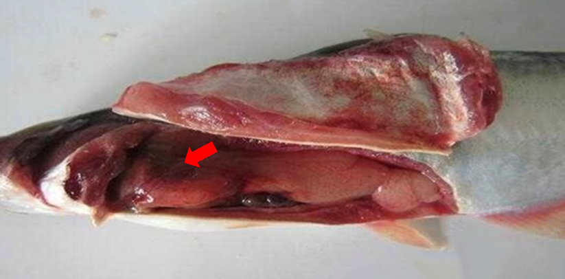
The fish with bacterial septicemia swam slowly, with decreased vitality and reduced food intake, and their body surface was congestive to varying degrees. The diseased and dead fish were dissected, and watery blood was observed in their abdominal cavity. In addition, their gills had swelling and were faint yellow. A small amount of food and minimal congestion were observed in the intestine, and the kidney was severely congested (Figure 1). Bacteria were isolated from the kidney of diseased and dead fish. After 48 h of culture, colonies with the same color and shape and the largest proportion of the total area of the culture medium were purified as the dominant bacteria. A pure bacterial strain was obtained and designated as FXZ01. The results of 16S rDNA gene sequencing showed that the dominant bacterial strain was A. hydrophila.
Re-infection test and pathogenicity of the isolated strain
The re-infection experiment revealed that FXZ01 had a mortality of 100% after 7 days, and fish in the negative control had no abnormalities (Figure 2). In the experimental group, P. bocourti began to get sick within 3 days. In the initial stage, the sick fish swam slowly and had a reduced food intake. Minimal hyperemia was observed around the orbits, at the base of the fins, and on both sides of the body. As the time of infection advanced, the diseased fish had spotted ulcerative muscle on both sides of the body, with bleeding, erosion, a red and swollen anus, and an enlarged abdomen. These features were consistent with the natural symptoms of bacterial diseases. The death rate of fish increased over time, and 15, 24, and 39 fish died on days 3, 4, and 5, respectively (Figure 2). On day 7, all diseased fish in the experimental group died, which ended the experiment. Evident bleeding symptoms were observed on the surface of the diseased fish. The dead fish were dissected, and it was found that the kidney was swollen, the liver was white, and the intestinal tract was bleeding slightly into the body cavity. After the isolation and purification of bacteria from dead fish and re-infection test, the pathogenic strain was identified as A. hydrophila. The symptoms of the disease caused by A. hydrophila are consistent with those of bacterial septicemia observed in naturally infected fish. In the control group, the feeding and activity of the fish were normal, and no disease or death was observed.

General features of FXZ01 and molecular identification
After the FXZ01 strain was cultured on an LB agar plate at 28°C for 24 h, it formed a round, flesh-colored, slightly elevated colony with smooth edges, indicating adequate growth and development. Additionally, FXZ01 could grow in TCBS agar (thiosulfate citrate bile salts sucrose agar), which is a selective medium. The colonies formed on TCBS agar plates were round and light yellow, with a diameter of approximately 1.0–3.0 mm. Based on the results of Gram staining, FXZ01 was identified as a Gram-negative bacterial strain. Microscopic observation of FXZ01 showed short, rod-shaped bacteria arranged in single, with blunt ends. These morphological features are consistent with those of A. hydrophila.
Furthermore, the sequence of the 16S rDNA gene of FXZ01 was 1175 bp in length. Using the BLAST tool in the NCBI database, FXZ01 was found to be highly homologous to Aeromonas. From the NCBI database, 18 strains with sequences most similar to those of FXZ01 and other Aeromonas species were selected to reconstruct a neighbor-joining phylogenetic tree (Figure 3). FXZ01 was found to have 97.3% homology with A. hydrophila MT605959.1.

Based on the comparative analysis of the gene sequence of FXZ01, general features, and molecular identification, the FXZ01 strain was eventually identified as A. hydrophila. The sequence was uploaded to NCBI, and the accession number in GenBank is OK326946.
Drug sensitivity test
The sensitivity of FXZ01 to 10 antibiotics was determined using the Kirby–Bauer method. The results of the drug sensitivity test showed that FXZ01 was susceptible to eight antibiotics (Table 2), including amikacin, cefazolin, ciprofloxacin, norfloxacin, erythromycin, chloramphenicol, gentamicin, and sulfamethoxazole, and resistant to two antibiotics, namely, penicillin and ampicillin. The %RIZD of the eight susceptible antibiotics is shown in Figure 4. Sulfamethoxazole (a sulfonamide antibiotic) had the largest IZD of 35.37 mm at 1.25 μg/disc, with 53.77% RIZD, whereas gentamicin (an aminoglycoside antibiotic) had the shortest IZD of 15.03 mm at 10 μg/disc, with 25.28% RIZD. Erythromycin (a macrolide antibiotic) had the highest antibacterial activity, with 135.13% RIZD and a zone of 30.57 mm at 15 μg/disc, whereas gentamycin had the lowest antibacterial activity. Overall, erythromycin was found to be the most effective antibacterial agent against A. hydrophila.

Discussion
Isolation and identification of pathogens is the primary aspect of research on pathogenic mechanisms and disease control. The 16S rDNA gene is an ideal target for gene classification owing to its highly conserved molecular structure, ubiquitous distribution, and the presence of a large amount of information and is widely used in bacterial identification (Srinivasan et al. 2015). To date, various integrated methods have been used for identifying pathogens, such as the use of the combination of morphological analysis and 16S rRNA sequencing to comprehensively assess the types of pathogens. Nahar et al. isolated a pathogen from the eye lesions, kidney, and liver of Pangasianodon hypophthalmus with disease and identified it as A. hydrophila by analyzing physiological and biochemical characteristics and using the 16S rDNA gene sequence of the pathogen (Nahar et al. 2016). Yuan et al. identified Vibrio harveyi as the pathogen of enteritis in Scophthalmus maximus using the 16S rDNA gene sequence (Yuan et al. 2021). Shen et al. identified Vibrio harveyi as the pathogen causing skin ulcers through physiological and biochemical characterization and amplification of the 16S rDNA gene sequence of the isolate (Shen et al. 2017). In the present study, we collected diseased P. bocourti with bacterial septicemia, isolated a pathogenic bacterial strain designated as FXZ01 from the infected fish, and performed a re-infection test to confirm the pathogenicity of the strain. The 16S rDNA gene sequence of the pathogen was 97.3% similar to that of A. hydrophila in the NCBI database. Additionally, the phylogenetic tree showed that the pathogen and A. hydrophila were clustered together. Based on the morphological characteristics of the pathogen and the 16S rDNA gene sequence, FXZ01 was determined to be A. hydrophila.
The opportunistic pathogen A. hydrophila is widely distributed in various natural water bodies and is the primary pathogen of various aquatic animals (Suarez et al. 2012). It can cause systemic sepsis or local infection in various animals in aquaculture and cause death (Janda and Abbott 2010). A. hydrophila can infect Siniperca chuatsi, ornamental fish, Nile tilapia, Carassius auratus red var., Pseudosciaena crocea, and Ictalurus punctatus, causing hemorrhage on the body surface and in internal organs, resulting in death (Anjur et al. 2021; Baumgartner, Ford, and Hanson 2017; Chen et al. 2018; Farag et al. 2021; Mu et al. 2010; Xiong et al. 2021). In this study, the FXZ01 strain was isolated from the kidney tissue of diseased P. bocourti. The re-infection test showed that the lethal dose reached 100% within 7 days, indicating that the strain was pathogenic to the cultured P. bocourti to a certain extent.
With the rapid development of aquaculture, the use of antibiotics has become an efficient and inexpensive measure for the prevention and control of bacterial diseases in aquaculture (Assefa and Abunna 2018). However, with the widespread use of antimicrobial drugs, their drawbacks are becoming serious. In particular, bacterial resistance has emerged as a global public health concern and a limiting factor for the rapid development of aquaculture (Rodríguez-González, Zanin, and Menasalvas-Ruiz 2019). A. hydrophila have a high drug resistance rate and broad drug resistance spectrum (Khor et al. 2018b). The A. hydrophila strain isolated from Mugil cephalus was found to be 84% resistant to ampicillin (Ramadan et al. 2018), that strain isolated from diseased Nile tilapia (Oreochromis niloticus) was found to be resistant to ampicillin and amoxicillin (Tartor et al. 2021), and that isolated from freshwater fish was found to be sensitive to erythromycin but resistant to penicillin G (Hayati et al. 2018). In this study, the A. hydrophila strain FXZ01 had different susceptibility to different antibacterial drugs. Erythromycin (a macrolide antibiotic) had the optimal antibacterial activity (RIZD, 135.13%). FXZ01 was resistant to two antibiotics, with a drug resistance rate of 20.00%. However, both drugs were β-lactams, suggesting that FXZ01 is resistant to most β-lactams. The resistance of A. hydrophila to β-lactams is a common phenomenon (Mbiada et al. 2015; Najiah and Laith 2014). Therefore, before using antibacterial drugs, drug susceptibility tests should be performed to select drugs with optimal effects. Rational use of antibacterial drugs is necessary in aquaculture.
Overall, this study showed that A. hydrophila is a pathogenic bacterium that causes bacterial septicemia in P. bocourti and has strong infectivity. The pathogen is resistant to β-lactams, and erythromycin is the most effective antimicrobial agent. The findings of this study provide a reference for the rapid prevention and control of bacterial diseases caused by A. hydrophila in cultured fish.
Conclusion
A pathogenic bacterial strain was isolated from diseased P. bocourti with bacterial septicemia and designated as FXZ01. The strain was identified to be Gram-negative. Microscopic observation of FXZ01 showed short, rod-shaped bacteria arranged in single, with blunt ends. These morphological features are consistent with those of A. hydrophila. To the best of our knowledge, this study is the first to report the presence of pathogenic A. hydrophila in the intestines of P. bocourti in China. The FXZ01 strain was found to be resistant to penicillin and ampicillin. Erythromycin (a macrolide antibiotic) had the optimal antibacterial activity and can be used for the prevention and treatment of disease caused by A. hydrophila in P. bocourti.
Acknowledgments
We extend our gratitude to Guangxi Natural Science Foundation Program (2021GXNSFAA075008); The Central Project Guide local science and technology for development (ZY21195020); the National Key R&D Program of China (2018YFD0901406); the National Natural Science Foundation of China (31873042); Guangxi Beibu Gulf Key Laboratory of Marine Biodiversity Conservation Major Cultivation Project (2021ZB02); Guangxi Beibu Gulf Key Laboratory of Marine Biodiversity Conservation Major Science and Technology Special Project (2022ZA01) and Beibu Gulf University Marine Science Guangxi First-Class Subject Construction Special Project (DRB001, DRB002).
Corresponding author
Dahui Yu
Email: pearlydh@163.com