
Peru is a significant contributor to the global production of rainbow trout, ranking fifth worldwide with a production volume of 54,187,00 tons in 2021 (FAO 2022). However, the emergence of fish diseases, particularly those of uncertain aetiology, poses a significant challenge for this industry. Red Mark Syndrome (RMS) is a prime example of such a disease, affecting rainbow trout as a non-lethal skin condition caused by a Midichloria-like organism (MLO) in both traditional and recirculating aquaculture (Metselaar et al. 2022; Orioles, Saccà, et al. 2022). The widespread syndrome affects rainbow trout populations in at least three continents, including South America, specifically Chile (Sandoval et al. 2016). For a decade, there has been unofficial speculation about the existence of RMS in Peru, yet no official reports have been documented in the scientific literature.
Typical gross RMS-related lesions present as bright red skin spots that can be focal or multifocal, circular, flat, or protruding, often with scattered petechial haemorrhages or scale loss (Oidtmann et al. 2013). Characteristic histologic features include deep chronic lymphohistiocytic and granulomatous dermatitis associated with panniculitis and myositis (Galeotti et al., 2021). The development and healing of these lesions essentially depend on water temperature (Orioles, Galeotti, et al. 2022). While RMS does not cause significant mortality, it has a high morbidity rate and has been deemed to have a considerable economic impact due to the downgrading of commercial products and management costs (Verner-Jeffreys and Taylor 2015; Metselaar et al. 2022). RMS diagnosis heavily relies on clinical features and histopathology (Oidtmann et al. 2013; Galeotti et al., 2021), although several molecular techniques to detect MLO-DNA are available (Lloyd et al. 2011; Cafiso et al. 2016; Orioles, Bulfoni, et al. 2022).
The outbreak under investigation occurred on an extensive rainbow trout farm in a one-hectare area in the Lagunillas lagoon, Santa Lucia district, Lampa province, Puno, Peru (Figure 1). The farmer reported recurrent disease outbreaks every year during the dry season (May-November). Rainbow trout were farmed in floating cages with a rearing density of 15 Kg/m3 and fed a commercial diet. Two cages were affected, with 50-60% of the fish in each showing external signs of red spots and raised, skin lesions on fish. At the time of the outbreak, the water temperature was approximately 14 +/- 1°C, with an oxygen level of five ppm and a salinity of one g/L. Embryonated eggs were supplied from the USA. Furthermore, no mortality was recorded during the outbreak.
._other_two_farming_sites_(.png)
Five fish exhibiting clinical signs (as shown in Figure 2) were selected and euthanized using an overdose of benzocaine (20% benzocaine [BZ-20], Veterquimica, Chile). Immediately following euthanasia, postmortem examinations were performed on the fish, and samples of internal organs and skin and muscle lesions were collected and fixed in 10% buffered formaldehyde for subsequent standard histological assessment with hematoxylin and eosin (HE). Moreover, scrapings from the gills, skin, kidney, liver, and spleen of each diseased fish and healthy fish were obtained for microscopic observation and samples for standard bacterial isolation were collected in aseptic conditions from each individual’s external skin, kidney, liver, and spleen.
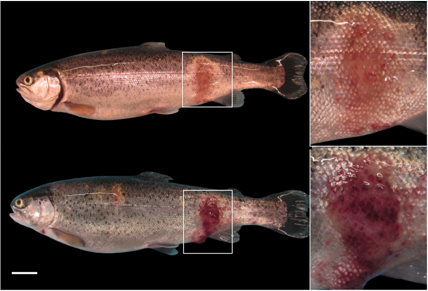
_and_severe_skin_lesions_(on_bottom)_as_describe.png)
Gross skin lesions were characterized by bright pink or red spots, often displaying scattered petechial haemorrhages on the flanks, dorsal and ventral areas of various sizes ranging from 2 to 10 cm in diameter, with frequent scale loss, and in rare cases, small central ulcers (Figure 2). No relevant parasites were detected by direct microscopic examination of wet mounts of skin and gills.
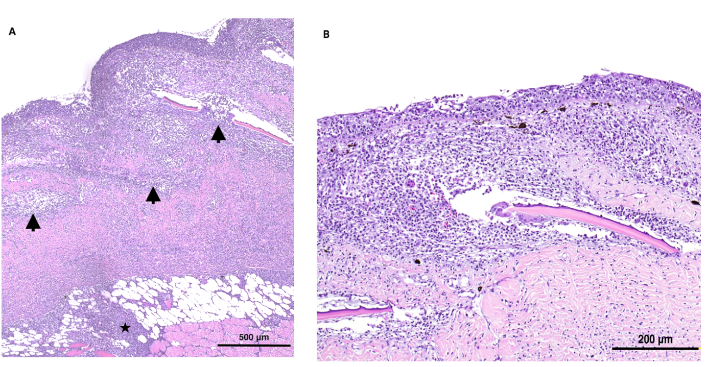
Microscopically, skin samples showed severe full-thickness dermatitis with marked lympho-histiocytic infiltration involving all skin layers, including the hypodermis, and muscular tissue (Figure 3a). Scale resorption was observed in moderate and severe cases concomitant with osteoclasts and oedema-induced pocket distention (Figure 3b). In advanced cases, scales were usually absent, having been completely resorbed by osteoclasts, and erosion and ulceration of the epidermis were observed. Collagen fibers of the dermis were disrupted and infiltrated by lymphocytes. In some cases, the infiltrate extended from the subcutis into the muscle, sometimes accompanied by myonecrosis.
A histological examination of the remaining organs did not reveal significant abnormalities. Microscopic examination of smears from the gills, skin, kidney, liver, and spleen of affected rainbow trout found no detectable bacteria. The clinical signs resolved spontaneously in most fish approximately 21-28 days after the first observation.
The affected fish displayed clinical signs, gross and microscopic lesion features compatible with RMS, and according to the criteria proposed by current literature (Verner-Jeffreys et al. 2008; Oidtmann et al. 2013; Metselaar et al. 2022).
_and_moderate_(3b)_typical_rms_skin_lesions_._figur.png)
The current study presents the first documented red mark syndrome (RMS) report in Peruvian salmonid aquaculture, representing noteworthy outbreaks. In the case described here the authors believe there were enough evidences to establish the presumptive diagnosis of RMS. These were essentially the distinctive clinical and histopathological features and the lack of significant results from microbiology. While PCR techniques for MLO DNA are not considered the definitive diagnostic gold standard, their inclusion in diagnostics is highly recommended in the diagnostic process, as MLO seems to be strongly associated with this syndrome.
RMS in Perù has been suspected for years, but never officially reported. The report of this disease in a new location should raise significant concerns within the aquaculture industry. The documentation of RMS in Peruvian aquaculture highlights the urgent need for effective monitoring and control measures to prevent the further spread of the disease, safeguard the industry’s economic viability, and ensure the sustainable management of aquatic resources. Laguna Lagunillas, situated at an altitude of 4,174 meters above sea level, in the district of Santa Lucia, Province of Lampa, and within the Lake Titicaca Basin, spans over a surface area of 65.71 Km2, with an average depth of 15.2 m and a maximum depth of 53.4 m (Torres 2021). Trout culture is developed in a system of floating cages with approximately 70 formal producers (RNIA 2023). Orestias luteus and Oncorhynchus mykiss are the main species populating this lagoon (IMARPE and PELT 2014).
The Cabanillas River feeds the lagoon, eventually converging with other tributaries to form the Coata River, which empties its waters into the Bay of Chucuito, an integral part of the vast Titicaca Lake. The interconnectedness of these water bodies underscores the possibility of disseminating various pathogens among fish farms. Moreover, the clustering of cages within the Lagunillas Lagoon presents a significant risk factor for the spread of disease-causing agents. Transferring fingerlings from one farm to another and exchanging equipment and materials for fish management can facilitate the transmission of pathogens to previously unaffected farms. This threat is further compounded by fingerlings often originating from the same source. As a result, the risk of disease dissemination is heightened in this open system, warranting adequate and proactive disease management measures to mitigate the potential impact of pathogenic outbreaks. Furthermore, the same fish farmer in Lagunillas Lagoon referred to the presence of the same clinical signs in subsequent production lots the following year, testifying to the stable presence of the disease in this river basin.
RMS responds to antibiotics (Schmidt, Henriksen, and Olesen 2021), but these are not commonly used due to the long withdrawal period and cost related to the medicated feed. In this case, the signs spontaneously and completely receded in 3-4 weeks from onset noticed by farmers without the use of any treatment; RMS is reported to resolve in a much longer period in experimental settings (Schmidt, Henriksen, and Olesen 2021), unless water temperature rises above 15.5°-16°C (Orioles, Galeotti, et al. 2022). We believe the water temperature, which was raised after the onset of skin lesions, can, in this case, justify the apparent quicker healing of the skin lesions.
During the same dry season, two separate farm sites from distinct districts (Lima and Junin) were observed having outbreaks of similar signs (Figure 1). As often suspected in literature, the authors believe that the route of entry of RMS may have been through the importation of embryonated rainbow trout eggs (Verner-Jeffreys et al. 2008; Galeotti et al. 2021; Metselaar et al. 2022). It is important to highlight that Peruvian salmonid production depends upon more than 90% on imported eggs from Spain, Denmark, United Kingdom, the USA, and Chile (PNIPA 2020), where disease has already been reported. In addition, there is a possibility of illegally introducing eggs and contaminated equipment or material from other-positive countries with RMS.
Given the current situation in the Peruvian trout farming sector, where biosecurity measures and good aquaculture practices are lacking, specialized diagnostic laboratories are not commonly utilized to determine the definitive diagnosis of fish diseases. There is little recurrence or assistance from veterinarians due to distances from fish farmers concerning diagnostic laboratories, as well as the illegal trade of fish and eggs, it is clear that urgent action is required to address the potential spread of RMS in Peruvian aquaculture.
To mitigate this issue, it is imperative to implement biosecurity procedures and sanitation measures, such as disinfection and quarantine protocols. Additionally, further research is necessary to identify specific features of MLO, including the mode of transmission and epidemiology, which can assist in developing effective control measures to prevent the further dissemination of the disease. These measures are critical to safeguard the economic viability of the aquaculture industry in Peru, and ensure the sustainable management of aquatic resources.