Loading [Contrib]/a11y/accessibility-menu.js

This website uses cookies

We use cookies to enhance your experience and support COUNTER Metrics for transparent reporting of readership statistics. Cookie data is not sold to third parties or used for marketing purposes.

Skip to main content
null
Bulletin of the EAFP
  • Menu
  • Articles
    • Case study
    • Method
    • Note
    • Opinion
    • Research article
    • Review
    • Workshop Report
    • All
  • For Authors
  • Editorial Board
  • About
  • Issues
  • search
  • Facebook (opens in a new tab)
  • LinkedIn (opens in a new tab)
  • RSS feed (opens a modal with a link to feed)

RSS Feed

Enter the URL below into your favorite RSS reader.

https://eafpbulletin.scholasticahq.com/feed
P-ISSN 0108-0288
E-ISSN 3005-4648
Research article
Vol. 44, Issue 3, 2025September 18, 2025 CEST

First report on bacterial isolation from marbled flounder (Pseudopleuronectes yokohamae) associated with skin ulcers

Shuai Wang, Hui Liu, Zheng Zhang, Xilin An, Xiaoyan Wei, Yanyan Shi, Yan Chen, Xuejie Li, Wei Wang,
Pseudopleuronectes yokohamaeIsolated strainSusceptibilityAntibioticsVibrio
Copyright Logoccby-4.0 • https://doi.org/10.48045/001c.144590
Photo by Brian Yurasits on Unsplash
Bulletin of the EAFP
Wang, Shuai, Hui Liu, Zheng Zhang, Xilin An, Xiaoyan Wei, Yanyan Shi, Yan Chen, Xuejie Li, and Wei Wang. 2025. “First Report on Bacterial Isolation from Marbled Flounder (Pseudopleuronectes Yokohamae) Associated with Skin Ulcers.” Bulletin of the European Association of Fish Pathologists 44 (3). https:/​/​doi.org/​10.48045/​001c.144590.
Download all (6)
  • Figure 1. Gross lesions of diseased marbled flounder infected with Bacteria 1.
    Download
  • Figure 2. Observation of liver, kidney, and spleen tissue.
    Download
  • Figure 3. Observation of tissue sections of liver, gills and skin.
    Download
  • Figure 4. The phylogenetic tree following blasting the gyrB gene sequence of the isolated strain with other homologous sequences available in GenBank using a Neighbour-Joining method.
    Download
  • Figure 5. Survival rates of P. yokohamae after infection with different bacterial strains.
    Download
  • Supplementary materials
    Download

Sorry, something went wrong. Please try again.

If this problem reoccurs, please contact Scholastica Support

Error message:

undefined

View more stats

Abstract

Skin ulceration syndrome (SUS) is a significant disease affecting the marbled flounder (Pseudopleuronectes yokohamae) in aquaculture. This study aimed to identify the pathogen responsible for SUS outbreaks. Three bacterial species were isolated from infected flounder, with isolate Isolated strain 1 confirmed as pathogenic through artificial infection experiments. Isolated strain 1 is highly pathogenic, significantly reducing the survival rate of P. yokohamae. The strains were identified using morphological, physiological, biochemical analyses, 16S rRNA, and gyrB gene sequencing. Isolated strain 1 was found to be a Gram-negative bacillus, closely related to Vibrio splendidus. Phylogenetic analysis showed that the 16S rRNA and gyrB gene sequences of Isolated strain 1 clustered with V. splendidus, with high bootstrap confidence levels (99.09% and 100%, respectively). Consequently, V. splendidus was identified as the causative agent of SUS in P. yokohamae. Antibiotic susceptibility tests revealed that Isolated strain 1 was sensitive to norfloxacin, ciprofloxacin, enrofloxacin, ceftazidime, doxycycline and florfenicol, but resistant to tetracycline, gentamicin, erythromycin and amoxicillin. These findings provide a foundation for developing disease prevention and treatment strategies in marbled flounder aquaculture.

1. Introduction

The marbled flounder (Pseudopleuronectes yokohamae) is widely distributed in China, Japan, and North Korea (C. Zhang et al. 2016) and serves as a key aquaculture species in northern China, especially in Shandong and Dalian (Cui et al. 2019). Due to its favorable traits—such as low-temperature tolerance, strong adaptability, and high nutritional value—it has been artificially bred since the 1960s (Cho et al. 2021). However, recent declines caused by disease, overfishing, and environmental stressors have led to significant economic losses (Z. Zhang et al. 2022), highlighting the need to investigate disease control strategies.

Skin ulceration syndrome (SUS) is a major disease threat in aquaculture, particularly under high-density farming conditions where physical injuries increase susceptibility (Yang et al. 2023). Similar symptoms have been reported in other flatfish and species such as Cynoglossus semilaevis (Zuo et al. 2023), Epinephelinae, and Pseudopleuronectes americanus (Bodammer 2000), with ulcers often leading to high mortality and reduced market value (Xu et al. 2015). Treatment approaches vary, with antibiotic immersion being commonly used.

Among the pathogens, Vibrio spp. are widespread marine bacteria with many known fish and shellfish pathogens, including V. harveyi, V. parahaemolyticus, V. alginolyticus, V. vulnificus, and V. splendidus (Lin et al. 2010; Ghasemieshkaftaki et al. 2023). Some, such as V. parahaemolyticus, cause AHPND in shrimp (Fadel et al. 2025), while others infect a range of hosts including Oreochromis niloticus (Elgendy et al. 2022) and Scorpaena porcus (Eissa et al. 2015). V. splendidus, in particular, is highly genotypically diverse and has been implicated in disease outbreaks in turbot (Thomson et al. 2005), cod (Gadus morhua) (Reid et al. 2009), and bivalves (Dubert, Barja, and Romalde 2017), as well as ectoparasitic hosts (Jensen et al. 2003; Gulla et al. 2015). It is also linked to SUS outbreaks in sea cucumbers (C. Li et al. 2012), underscoring its broad host range and economic impact.

Due to the host- and environment-specific characteristics of V. splendidus, further study of its pathogenic traits is crucial for disease control. In this study, we isolated and identified the causative agent of SUS in P. yokohamae using morphological analysis, biochemical profiling, 16S rRNA sequencing, antibiotic susceptibility testing, and bacterial growth experiments (Liao et al. 2024), aiming to provide a scientific basis for the prevention and treatment of SUS in cultured marbled flounder.

2. Materials and Methods

2.1. Animals

Diseased P. yokohamae were collected from Xinchangxing Market in Shahekou District, Dalian city, Liaoning Province, China, and used for the isolation and detection of pathogenic bacteria. Prior to bacterial isolation, all diseased individuals underwent microscopic examination and PCR detection for common parasitic infections and bacterial pathogens to rule out co-infections. Only fish confirmed to be free of co-infections were included in the study. Healthy P. yokohamae specimens, exhibiting intact body surfaces, normal pigmentation, and good vitality, with an average weight of 150±20 g, were selected for the artificial infection experiment. Before the experiment, the P. yokohamae were acclimated at the Key Laboratory of Northern Mariculture of the Ministry of Agriculture at Dalian Ocean University. The breeding tanks were filled with naturally filtered seawater obtained through sedimentation and sand filtration. The water temperature was maintained at 15 ± 1°C, the salinity was 30, and the pH was 7.4 ± 0.1. During the temporary breeding period, sandworms were fed twice a day, each time at 3±1% of body weight to ensure their adaptation to the new environment. Feeding was stopped 12 hours before the artificial recall experiment.

2.2. Pathogenic and histopathological properties

Clinical and postmortem examinations of naturally infected Pseudopleuronectes yokohamae were conducted to identify gross lesions and internal abnormalities, following the standard procedures described by Jiang (X. Jiang et al. 2022). To exclude co-infections, all diseased fish underwent microscopic and PCR screening for common pathogens. Nine infected and three healthy laboratory-acclimated fish were selected. After rinsing with sterile saline, fish were dissected aseptically, and gill, liver, and intestinal tissues were fixed in Davidson’s solution. Samples were processed using standard histological techniques, stained with H&E, and examined under a light microscope (Olympus, Japan). Images were acquired with Leica LAS X 3.1.1 software.

2.3. Phenotypic observation of pathogenic bacterial isolates from fish specimens

Bacteria were isolated from the skin ulcers, liver, and spleen of diseased fish using Tryptic Soy Broth (TSB; Hopebio, China), and then streaked onto 2216E agar (Hopebio, China) for incubation at 16 °C for 12–16 h. Single colonies were subcultured on 2216E agar at least three times to obtain pure strains, which were subsequently transferred to thiosulfate-citrate-bile salt-sucrose (TCBS) agar for colony morphology observation. Identification of Vibrio species followed Bergey’s Manual (9th Edition) (Mahbub, Paul, and Ahmed 2011). Gram staining was performed during the late logarithmic phase according to Cruickshank (Cruickshank et al. 1975), and bacterial morphology was observed under a microscope to differentiate Gram-positive (purple) and Gram-negative (red) cells.

2.4. Determination of biochemical characteristics

Purely cultured colonies were selected to prepare a bacterial suspension under sterile conditions, and ID 20E (BioMérieux, France) Enterobacteriaceae bacteria identification test strips were utilized to identify the biochemical characteristics of bacteria (Varettas, Mukerjee, and Schmidt 1995) following the manufacturer’s protocol. In brief, 100 μL of bacterial suspension was added to the biochemical reaction cup, and mineral oil was added to create an anaerobic environment. Afterward, the test strips were covered. After incubation at 36°C for 18–24 h, the identification reagent was added dropwise according to the manufacturer’s instructions. To identify Vibrio species, a series of biochemical tests were performed via API 20E strips, each containing 20 microtubes for specific tests. These tests collectively provided valuable information on the metabolic characteristics of the bacterial isolate, aiding in its identification (Aly et al. 2023).

2.5. Molecular Strategies for Bacterial Characterization

Genomic DNA was extracted from bacterial cultures using the Bacterial Genomic DNA Extraction Kit (TIANGEN, China), following the manufacturer’s protocol. DNA concentration and quality were assessed via agarose gel electrophoresis and NanoDrop spectrophotometry. The 16S rRNA gene was amplified using universal primers 27F and 1492R (H. Jiang et al. 2006) on a ProFlex™ PCR System (Thermo Fisher, USA). To improve species-level resolution, the gyrB gene was also amplified using primers gyrBF and gyrBR (Yamamoto and Harayama 1995; Wang et al. 2007). Primer sequences and PCR conditions are listed in Table 1. PCR products were separated by 1% agarose gel electrophoresis using a DL2000 DNA Marker (Takara, Japan), visualized under UV light, and purified for sequencing (Sangon Biotech, Shanghai, China). Consensus sequences were aligned with NCBI database references, and phylogenetic trees were constructed using the neighbor-joining method based on 16S rRNA and gyrB gene sequences.

Table 1.Oligonucleotide primers and their cycling conditions used in the current study
Amplification (35 cycles)
Target gene Sequences Amplified product Primary denaturation Secondary denaturation Annealing Extension Final extension
Bacterial 16SrRNA 27F: AGAGTTTGATCCTGGCTCAG
1492R: TACGGCTACCTTGTTACGACTT
1500bp 94°C
5 min
94°C
30 s
56°C
30 s
72°C
1 min
72°C
10 min
Bacterial gyrB gyrBF: AGCAAGTTCGGCAACGG 1200bp
gyrBR: AGCATCTTCTCGAAGCG

2.6. Antimicrobial resistance analyses

Antimicrobial susceptibility was evaluated using the Kirby–Bauer disk diffusion method, following the Clinical and Laboratory Standards Institute (CLSI) M100 guidelines (CLSI 2020). Marine bacterial isolates were cultured on Mueller–Hinton agar (MHA; Hopebio, China) supplemented with 1.5% NaCl. Bacterial suspensions were adjusted to a turbidity equivalent to 0.5 McFarland standard and evenly inoculated onto the surface of MHA plates. Antibiotic discs were applied to the agar surface with sterile forceps after drying for 3–5 minutes. The antibiotics tested included norfloxacin, ciprofloxacin, enrofloxacin, ceftazidime, tetracycline, doxycycline, oxytetracycline, kanamycin, gentamicin, erythromycin, amoxicillin, sulfadiazine-trimethoprim, and florfenicol. Plates were incubated at 28 °C for 22–24 hours, after which the diameters of the inhibition zones were measured. All assays were performed in duplicate. Results were interpreted according to the CLSI VET03 guidelines (CLSI 2020).

2.7. Bacterial growth experiment

All strains (Isolated strain 1, Isolated strain 2, and Isolated strain 3) were cultured at 15°C overnight. Three hundred microliters of each bacterial culture was subsequently extracted and diluted to a ratio of 1:100. The mixture was sequentially inoculated into 30 mL of fresh 2216E Liquid Medium (Hopebio, China) and then placed in an incubator at 15 C with shaking at 200 rpm. The optical density at 600 nm was determined hourly for 10 h via a spectrophotometer (Xie et al. 2020). Sterile 2216E broth was used as a blank control, and all OD₆₀₀ measurements were performed using the same calibrated spectrophotometer. Data from triplicate cultures were averaged, and the entire experiment was independently repeated three times.

2.8. Artificial infection experiment

The tested strains were cultured in 2216E liquid medium (Hopebio, China) at 28 °C for 12–16 h and resuspended in sterile saline (0.9% NaCl) to prepare bacterial suspensions. Fish were randomly assigned to three experimental and three control groups, each with three replicates (15 fish per 0.5 m³ bucket) (Zuo et al. 2023; Liao et al. 2024). Experimental groups were intraperitoneally injected with 0.2 mL of bacterial suspension (1×10⁷ CFU/mL) (Ghasemieshkaftaki et al. 2023), while control groups received an equal volume of saline, PBS, or heat-killed bacteria. Fish were maintained at 15 ± 1 °C for 14 days, fed as described in section 2.1, with adjusted feeding as needed. Mortality, clinical signs, and pathological features were recorded (Ghasemieshkaftaki et al. 2023). Moribund and dead P. yokohamae were dissected to examine lesions, and tissues and ascitic fluid were collected for pathogen re-identification via 16S rRNA and gyrB gene PCR and sequencing.

2.9. Ethics statement

The present study was carried out following the recommendation of the ARRIVE guidelines for animal research. The Animal Research and Ethics Committee of Dalian Ocean University approved this study. Anatomical experiments were performed under anesthesia using ethyl 3-aminobenzoate mesylate (MS-222. Sigma‒Aldrich. USA) to minimize fish discomfort.

3. Results

3.1. Clinical examination

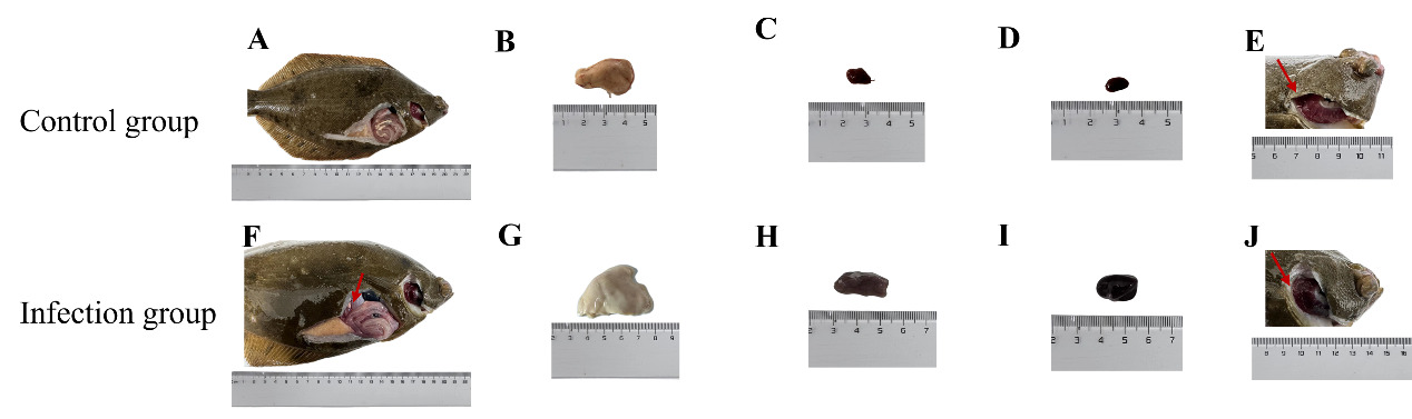
Clinical signs of SUS included anorexia, reduced resistance, abnormal swimming, and lethargy, with ulcer formation and tail bleeding in severe cases (Figure 1). Necropsy revealed pale livers, swollen discolored kidneys and spleens, gill congestion and edema, mucus in gill filaments, thin gut walls, and ascites (Figure 2). Histopathology showed hepatic vacuolation and hyperemia (Figure 3D), gill cell swelling with blood cell infiltration (Figure 3E), and thinning of the mucous layer with increased immune cells and scale loss (Figure 3F).

Figure 1
Figure 1.Gross lesions of diseased marbled flounder infected with Bacteria 1.

(B, F) No apparent external lesions were observed on the abdominal region of either healthy or diseased fish. (C, G) Black arrows indicate ulcerative lesions on the body surface of infected individuals, characterized by localized tissue erosion. (D, H) The caudal fins of diseased fish exhibited signs of fin rot, with frayed edges and visible hemorrhaging at the margins.

Figure 2
Figure 2.Observation of liver, kidney, and spleen tissue.

(B, G) The liver of infected fish appeared pale or whitish in color, indicating potential hepatocellular damage or lipid degeneration. (C, H) The kidneys exhibited a color shift from normal reddish hue to dark black, suggesting renal congestion or necrosis. (D, I) The spleen was notably enlarged and darkened, consistent with splenic hyperplasia or hemorrhage. (E, J) The gill filaments turned dark red, possibly due to hyperemia or hemorrhagic response.

Figure 3
Figure 3.Observation of tissue sections of liver, gills and skin.

(A, D) Liver sections: black arrows indicate hepatic vacuolization and dilated sinusoids. (B, E) Gill sections: the gill filaments are markedly shortened and thickened; black arrows denote vacuole formation and hemorrhagic spots on the filaments. (C, F) Skin sections: the mucous layer on the epidermal surface is diminished, with increased infiltration of immune cells and exfoliation of scales. (G–I) Semi-quantitative histopathological scores of lesions in the liver (G), gill (H), and skin (I) tissues. Scoring was based on predefined histopathological criteria under light microscopy. Data are expressed as mean ± standard deviation (SD). Statistical significance was determined using an independent t-test. *p < 0.05; **p < 0.01.

3.2. Culture conditions and morphological characteristics

The isolates were designated as Isolated strains 1–3. On 2216E medium at 15°C for 24 h, Isolated strain 1 formed circular, orange colonies, while strains 2 and 3 were white. On TCBS agar, strain 1 produced yellow, swarming colonies (2–3 mm), strain 2 formed green colonies (1–2 mm), and strain 3 showed no growth. Microscopically, all isolates were Gram-negative, short, motile rods, either straight or curved (Supplementary Figure 1).

3.3. Biochemical identification

Three bacterial isolates were identified using the API 20E system (bioMérieux, France) based on their biochemical profiles. Isolate 1 tested positive for several sugar fermentations (GLU, MAN, SAC, ARA), LDC, ODC, and IND, with an API code of 6144600, indicating Vibrio spp. Isolate 2 was only positive for citrate (CIT), with an API code of 0200000, suggesting Pseudoalteromonas spp. Isolate 3 showed positive reactions for GLU, LDC, H₂S, and GEL, with an API code of 1006100, identifying it as Shewanella spp. These API codes enable species-level presumptive identification based on metabolic traits (Table 2).

Table 2.The physiological and biochemical characteristics
Biochemical project Result
Isolated strain 1 Isolated strain 2 Isolated strain 3
β-Galactosidase - - -
Arginine dihydrolase - - -
Lysine decarboxylase + - +
Ornithine decarboxylase + - -
Citric acid utilization - + -
H2S generation - - +
Urease - - -
Tryptophan deaminase - - -
Indole is produced + - -
3-Hydroxybutanone produces Acetylmethylmethanol - - -
Gelatinase + - +
Glucose + - +
Mannitol + - -
Inositol - - -
Sorbitol - - -
L-rhamnose monohydrate - - -
Sucrose + - -
Melibiose - - -
Amygdalin - - -
Arabinose + - -

Note: +: Positive; -: Negative

3.4. Phylogenetic analyses

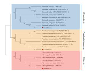
PCR amplification of the 16S rRNA gene from all three isolates yielded ~1,500 bp amplicons. BLAST analysis showed >99% identity with Vibrio splendidus (99.09%), Pseudoalteromonas distincta (99.93%), and Shewanella baltica (99.86%) for Isolates 1–3, respectively. Phylogenetic analysis confirmed clustering with reference strains (Supplementary Figure 2). To improve species resolution, gyrB was also analyzed, revealing 99.49%, 99.31%, and 100% identity with the corresponding species. The phylogenetic tree based on gyrB supported these identifications (Figure 4). Sequences were submitted to GenBank (16S rRNA: PP330039–PP330041; gyrB: PQ189395–PQ189397).

Figure 4
Figure 4.The phylogenetic tree following blasting the gyrB gene sequence of the isolated strain with other homologous sequences available in GenBank using a Neighbour-Joining method.

3.5. Antimicrobial resistance analyses

Antimicrobial susceptibility testing of Isolated strains 1, 2, and 3 against 13 antibiotics showed distinct resistance profiles (Table 3). Isolated strain 1 was sensitive to norfloxacin, ciprofloxacin, enrofloxacin, ceftazidime, doxycycline, and florfenicol, but resistant to tetracycline, gentamicin, erythromycin, and amoxicillin. Isolated strain 2 exhibited sensitivity to norfloxacin, ciprofloxacin, enrofloxacin, tetracycline, doxycycline, kanamycin, and gentamicin, but was resistant to ceftazidime, oxytetracycline, erythromycin, amoxicillin, and florfenicol. In contrast, Isolated strain 3 was sensitive to florfenicol, ceftazidime, doxycycline, gentamicin, and trimethoprim, but resistant to ciprofloxacin, ceftazidime, erythromycin, and amoxicillin. The interpretation of antimicrobial resistance was based on the CLSI VET03 guidelines (Supplementary Table 1).

Table 3.Drug sensitivity test of 3 strain
Drugs Concentration μg/disc Isolated strain 1 Isolated strain 2 Isolated strain 3
Diameter of inhibition/mm Sensitivity Diameter of inhibition/mm Sensitivity Diameter of inhibition/mm Sensitivity
Florfenicol 10 22±0.5 S 19±0.6 S 9±0.3 R
Ciprofloxacin 5 24±0.3 S 22±0.3 S 17±0.4 I
Enrofloxacin 5 26±0.4 S 26±0.7 S 28±0.2 S
Ceftazidime 30 28±0.3 S 0 R 12±0.3 R
Tetracycline 30 8±0.5 R 22±0.5 S 22±0.6 S
Doxycycline 30 30±0.6 S 19±0.3 S 24±0.2 S
Oxytetracycline 30 17±0.4 I 4±0.4 R 16±0.3 I
Kanamycin 30 16±0.2 I 21±0.3 S 17±0.4 I
Gentamicin 10 0 R 24±0.5 S 21±0.6 S
Erythromycin 15 13±0.6 R 0 R 0 R
Amoxicillin 10 15±0.4 R 6±0.2 R 14±0.7 R
Sulfadiazine-Trimethoprim 25 14±0.2 I 13±0.4 I 22±0.1 S
Florfenicol 30 39±0.5 S 13±0.3 R 20±0.6 S

Note: (Sensitivity determination criteria) R. means drug resistance; I. means medium sensitivity; S. means high sensitivity.

3.6. Bacterial growth patterns of pathogenic bacteria

The strains of Isolated strain 1, 2, and 3 displayed similar representative growth patterns. Nonetheless, the proliferative rate of Isolated strain 1 was significantly greater than that of Isolated strain 2 and Isolated strain 3 (Supplementary Figure 3).

3.7. Artificial recall experiment

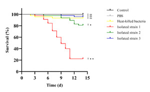
Artificial infection assays showed that only fish injected with Isolated strain 1 developed typical SUS symptoms within 48 hours, while strains 2 and 3 caused only mild behavioral changes. No symptoms appeared in the PBS, heat-killed, or control groups. Kaplan–Meier analysis revealed that strain 1 caused high mortality (~80% by day 12), strain 2 caused moderate mortality (~20%), and the other groups had no significant deaths. Survival in the strain 1 group was significantly lower than all others (p < 0.01), and strain 2 showed a mild but significant effect (p < 0.05) (Figure 5). Strain 1 also matched the dominant bacteria re-isolated from diseased fish, and 16S rRNA and gyrB gene sequencing confirmed it as the causative agent of SUS in P. yokohamae.

Figure 5
Figure 5.Survival rates of P. yokohamae after infection with different bacterial strains.

The survival rate of P. yokohamae was monitored for 14 days after infection with the three strains. The black curve is the control group, the gray curve is PBS, the yellow curve is heat-killed bacteria, the red curve is Isolated strain 1, the green curve is Isolated strain 2, and the blue curve is Isolated strain 3. Statistical differences between groups were assessed using the Kaplan-Meier survival test. Significant differences are indicated by asterisks (*p < 0.05; **p < 0.01).

4. Discussion

Pseudopleuronectes yokohamae is widely distributed in East Asia, including the Yellow Sea, southern Hokkaido, and Korean coastal regions (Ji, Kim, and Kim 2016), and holds significant commercial value (Huh et al. 2012). Despite its importance, the etiology of skin ulcer syndrome (SUS) remains unclear. In the present outbreak, fish showed clinical and pathological signs such as liver pallor, gill edema, intestinal thinning, and ascites under moderate temperature and salinity conditions. Histopathological lesions included vacuolar degeneration in the liver, epithelial swelling in gills, and immune cell infiltration.

Biochemical and molecular identification revealed that isolates belonged to Vibrio splendidus, Pseudoalteromonas distincta, and Shewanella baltica. The use of gyrB gene sequencing, with higher taxonomic resolution than 16S rRNA (Urdaci et al. 1998; Romalde et al. 1999; Cepeda and Santos 2000; Liu et al. 2021; Kong et al. 2022), clarified species identity.

Among the isolates, V. splendidus exhibited strong pathogenicity. This bacterium is metabolically versatile—capable of fermenting sugars, producing indole (S. Zhang et al. 2017), utilizing L-glutamate (Y. Li, Shi, and Zhang 2023), and secreting metalloproteases like Vsm (Binesse et al. 2008)—all contributing to tissue damage and immune evasion (Mey, Craig, and Payne 2012; Rivera-Posada et al. 2011; J. Li et al. 2020). Its infection patterns are consistent across multiple hosts, including turbot (Gatesoupe, Lambert, and Nicolas 1999), New Zealand brill (Diggles et al. 2000), carp, and trout, with mechanisms involving adhesion, biofilm formation, and toxin secretion (Roux and Blokesch 2018; Letchumanan, Chan, and Lee 2014). However, marbled flounder may be more susceptible due to slower epithelial repair and immune response under environmental stress (Shi et al. 2023).

Environmental factors such as water temperature, salinity, and organic load significantly affect V. splendidus pathogenicity. Infections are often associated with temperatures above 15 °C (Thomson et al. 2005; Austin and Austin 2016), and salinity (2–8%) and dissolved organic carbon enhance biofilm and virulence expression (Eiler, Johansson, and Bertilsson 2006; Oyanedel et al. 2020). High-density culture, poor water quality, and skin damage from handling or parasitism increase infection risk (Spanggaard et al. 2000; Kokou et al. 2020; Sobhana et al. 2009). Rapid bacterial growth observed in this study (peaking within 10–12 h) suggests that early intervention post-injury is critical for disease control.

Antibiotic sensitivity tests showed that quinolones (enrofloxacin, ciprofloxacin) and amides (florfenicol) were effective against all isolates, while resistance to erythromycin and amoxicillin was widespread. Doxycycline showed good activity, whereas sensitivity to ceftazidime and sulfadiazine-trimethoprim varied. These findings highlight the need for treatment based on specific susceptibility profiles (Zanetti et al. 2001). Monitoring resistance patterns and determining MIC values in future studies will help optimize therapeutic regimens.

Given concerns over antibiotic resistance, alternative strategies such as probiotics and improved husbandry should be integrated into disease management (Hai 2015; Chauhan and Singh 2019). However, this study has limitations: it captures only a snapshot in time and may not reflect long-term pathogen dynamics or geographic variation (Ke Huei et al. 2017) Liang et al., 2022; Lion and Metz 2018). Therefore, multi-year studies across broader environments are necessary to generalize these findings.

5. Conclusions

In summary, Vibrio splendidus infection causes skin ulcers and varying degrees of lesions in the kidneys, intestines, gills, and liver. However, the severity of the disease under laboratory conditions was significantly lower than that in natural artificial breeding environments. Moreover, in the natural environment, the probability of disease in P. yokohamae is significantly lower than that in cultured environments. The disease may be triggered by V. splendidus infection and exacerbated by factors such as overcrowding, handling, environmental fluctuations, biofouling, and predator interactions, all of which collectively induce stress in fish.

Submitted: January 17, 2025 CEST

Accepted: September 18, 2025 CEST

References

Aly, Salah M., Mohamed A. Elatta, Noha I. ElBanna, Mustafa A. El-Shiekh, Mahmoud Mabrok, Mahmoud S. Kelany, and Mohamed Fathi. 2023. “Comprehensive Analysis of Vibrio Alginolyticus: Environmental Risk Factors in the Cultured Gilthead Seabream (Sparus Aurata) under Seasonal Fluctuations and Water Parameter Alterations.” Journal of Fish Diseases, jfd.13860.
Google Scholar
Austin, Brian, and Dawn A. Austin. 2016. Bacterial Fish Pathogens. Cham: Springer International Publishing.
Google Scholar
Binesse, Johan, Claude Delsert, Denis Saulnier, Marie-Christine Champomier-Vergès, Monique Zagorec, Hélène Munier-Lehmann, Didier Mazel, and Frédérique Le Roux. 2008. “Metalloprotease vsm Is the Major Determinant of Toxicity for Extracellular Products of Vibrio Splendidus.” Applied and Environmental Microbiology 74 (23): 7108–17.
Google Scholar
Bodammer, J. E. 2000. “Some New Observations on the Cytopathology of Fin Erosion Disease in Winter Flounder Pseudopleuronectes Americanus.” Diseases of Aquatic Organisms 40 (1): 51–65.
Google Scholar
Cepeda, C., and Y. Santos. 2000. “Rapid and Low-Level Toxic PCR-Based Method for Routine Identification of Flavobacterium Psychrophilum.” International Microbiology: The Official Journal of the Spanish Society for Microbiology 3 (4): 235–38.
Google Scholar
Chauhan, Arun, and Rahul Singh. 2019. “Probiotics in Aquaculture: A Promising Emerging Alternative Approach.” Symbiosis 77 (2): 99–113.
Google Scholar
Cho, J. h., S. Lee, B. j. Lee, S. w. Hur, K. w. Kim, M. h. Son, and D. j. Yoo. 2021. “A Preliminary Study of Dietary Protein Requirement of Juvenile Marbled Flounder (Pseudopleuronectes Yokohamae).” Animal Nutrition (Zhongguo Xu Mu Shou Yi Xue Hui) 7 (2).
Google Scholar
CLSI. 2020. Methods for Antimicrobial Broth Dilution and Disk Diffusion Susceptibility Testing of Bacteria Isolated From Aquatic Animals. 2nd ed. Clinical & Laboratory Standards Institute. https:/​/​clsi.org/​standards/​products/​veterinary-medicine/​documents/​vet03/​.
Google Scholar
Cruickshank, R., J. P. Duguid, B. P. Marmion, and R. H. A. Swain. 1975. Medical Microbiology: The Pratice of Medical Microbiology.
Google Scholar
Cui, Qianjin, Lihua Qiu, Xu Yang, Shengnan Shang, Boxue Yang, Mingkang Chen, Xia Liu, et al. 2019. “Transcriptome Profiling of the Low-Salinity Stress Responses in the Gills of the Juvenile Pseudopleuronectes Yokohamae.” Comparative Biochemistry and Physiology. Part D, Genomics & Proteomics 32:100612.
Google Scholar
Diggles, B.K., J. Carson, P.M. Hine, Hickman Hickman, and M.J. Tait. 2000. “Vibrio Species Associated with Mortalities in Hatchery-Reared Turbot (Colistium Nudipinnis) and Brill (C. Guntheri) in New Zealand.” Aquaculture 183 (1–2): 1–12.
Google Scholar
Dubert, Javier, Juan L. Barja, and Jesús L. Romalde. 2017. “New Insights into Pathogenic Vibrios Affecting Bivalves in Hatcheries: Present and Future Prospects.” Frontiers in Microbiology 8:762.
Google Scholar
Eiler, Alexander, Mona Johansson, and Stefan Bertilsson. 2006. “Environmental Influences on Vibrio Populations in Northern Temperate and Boreal Coastal Waters (Baltic and Skagerrak Seas).” Applied and Environmental Microbiology 72 (9): 6004–11.
Google Scholar
Eissa, A., M. Abdelsalam, A. Abumhara, A. Kammon, Fateim T. Gammoudi, K. Naser, T. Borhan, and A. Asheg. 2015. “First Record of Vibrio Vulnificus/Anisakis Pegreffi Concurrent Infection in Black Scorpionfish (Scorpaena Porcus)from the South Mediterranean Basin.” Research Journal of Pharmaceutical, Biological and Chemical Sciences 6 (3): 1537–48.
Google Scholar
Elgendy, Mamdouh Y., Mohamed Abdelsalam, Amany M. Kenawy, and Shimaa E. Ali. 2022. “Vibriosis Outbreaks in Farmed Nile Tilapia (Oreochromis ⁠niloticus) Caused by Vibrio ⁠mimicus and V. ⁠cholerae.” Aquaculture International 30 (5): 2661–77.
Google Scholar
Fadel, Amr, Amal Khafage, Mohamed Abdelsalam, and Mohamed M. Abdel-Rahim. 2025. “Comparative Evaluation of Three Herbal Extracts on Growth Performance, Immune Response, and Resistance against Vibrio Parahaemolyticus in Litopenaeus Vannamei.” BMC Veterinary Research 21 (1): 166.
Google Scholar
Gatesoupe, F. J., C. Lambert, and J. L. Nicolas. 1999. “Pathogenicity of Vibrio Splendidus Strains Associated with Turbot Larvae, Scophthalmus Maximus.” Journal of Applied Microbiology 87 (5): 757–63.
Google Scholar
Ghasemieshkaftaki, Maryam, Ignacio Vasquez, Aria Eshraghi, Anthony Kurt Gamperl, and Javier Santander. 2023. “Comparative Genomic Analysis of a Novel Vibrio Sp. Isolated from an Ulcer Disease Event in Atlantic Salmon (Salmo Salar).” Microorganisms 11 (7): 1736.
Google Scholar
Gulla, Snorre, Henning Sørum, Øyvind Vågnes, and Duncan J. Colquhoun. 2015. “Phylogenetic Analysis and Serotyping of Vibrio Splendidus-Related Bacteria Isolated from Salmon Farm Cleaner Fish.” Diseases of Aquatic Organisms 117 (2): 121–31.
Google Scholar
Hai, N. V. 2015. “The Use of Probiotics in Aquaculture.” Journal of Applied Microbiology 119 (4): 917–35.
Google Scholar
Huh, Sung-Hoi, Ki-Mun Nam, Joo-Myun Park, Jae-Mook Jeong, and Gun-Wook Baeck. 2012. “Feeding Habits of the Marbled Flounder, Peluronectes Yokohamae in the Coastal Waters off Tongyeong, Korea.” Korean Journal of Ichthyology 24 (2): 77–83.
Google Scholar
Jensen, Sigmund, Ole B. Samuelsen, Kari Andersen, Lise Torkildsen, Christophe Lambert, Gwénaëlle Choquet, Christine Paillard, and Øivind Bergh. 2003. “Characterization of Strains of Vibrio Splendidus and V. Tapetis Isolated from Corkwing Wrasse Symphodus Melops Suffering Vibriosis.” Diseases of Aquatic Organisms 53 (1): 25–31.
Google Scholar
Ji, Hwan-Sung, Jin-Koo Kim, and Byung-Jik Kim. 2016. “Molecular Phylogeny of the Families Pleuronectidae and Poecilopsettidae (PISCES, Pleuronectiformes) from Korea, with a Proposal for a New Classification.” Ocean Science Journal 51 (2): 299–304.
Google Scholar
Jiang, Hongchen, Hailiang Dong, Gengxin Zhang, Bingsong Yu, Leah R. Chapman, and Matthew W. Fields. 2006. “Microbial Diversity in Water and Sediment of Lake Chaka, an Athalassohaline Lake in Northwestern China.” Applied and Environmental Microbiology 72 (6): 3832–45.
Google Scholar
Jiang, Xinyu, Xiaoyu Wang, Lei Li, Chen Niu, Chao Pei, Lei Zhu, and Xianghui Kong. 2022. “Identification of Shewanella Putrefaciens as a Novel Pathogen of the Largemouth Bass (Micropterus Salmoides) and Histopathological Analysis of Diseased Fish.” Frontiers in Cellular and Infection Microbiology 12.
Google Scholar
Ke Huei, Mien, Prachumwat Anuphap, Yu Chun-Ping, Yang Yi-Ting, Promsri Sutitcha, Liu Kuan-Fu, Lo Chu-Fang, et al. 2017. “Comparative Genomics of Vibrio Campbellii Strains and Core Species of the Vibrio Harveyi Clade.” Scientific Reports 7:41394.
Google Scholar
Kokou, Fotini, Goor Sasson, Itzhak Mizrahi, and Avner Cnaani. 2020. “Antibiotic Effect and Microbiome Persistence Vary along the European Seabass Gut.” Scientific Reports 10 (1): 10003.
Google Scholar
Kong, Delong, Qingqing Li, Yanzheng Zhou, Yan Wang, Xu Jiang, Zhiye Wang, and Zhiyong Ruan. 2022. “Pseudomonas Tumuqii Sp. Nov., Isolated from Greenhouse Soil.” Archives of Microbiology 204 (5): 249.
Google Scholar
Letchumanan, Vengadesh, Kok-Gan Chan, and Learn-Han Lee. 2014. “Vibrio Parahaemolyticus: A Review on the Pathogenesis, Prevalence, and Advance Molecular Identification Techniques.” Frontiers in Microbiology 5.
Google Scholar
Li, Chenghua, Weida Feng, Lihua Qiu, Changge Xia, Xiurong Su, Chunhua Jin, Tingting Zhou, Yuan Zeng, and Taiwu Li. 2012. “Characterization of Skin Ulceration Syndrome Associated microRNAs in Sea Cucumber Apostichopus Japonicus by Deep Sequencing.” Fish & Shellfish Immunology 33 (2): 436–41.
Google Scholar
Li, Jia, Richard W. McLaughlin, Mo Chen, Ying Li Liu, Hia Xia Xie, Xiao Ling Wan, Jun Ying Zhou, and Jin Song Zheng. 2020. “First Case of Shewanella Indica Isolated from a Bryde’s Whale (Balaenoptera Edeni) Stranded in the Northern Beibu Gulf, China.” Antonie Van Leeuwenhoek 113 (9): 1385–91.
Google Scholar
Li, Ya, Weibo Shi, and Weiwei Zhang. 2023. “Vibrio Splendidus AJ01 Promotes Pathogenicity via L-Glutamic Acid.” Microorganisms 11 (9): 2333.
Google Scholar
Liao, Wenyu, Dongdong Wei, Mingzhu Liu, Lin Huang, Bingzheng Li, Yunyi Wei, Shuyu Han, Shuaishuai Huang, Qing Yu, and Pengfei Li. 2024. “Phenotypic Characteristics and Immune Response of Procypris Merus Following Challenge with Aquatic Isolate of Klebsiella Pneumoniae.” Journal of Fish Diseases 47 (1): e13875.
Google Scholar
Lin, Baochuan, Zheng Wang, Anthony P. Malanoski, Elizabeth An O’Grady, Charles F. Wimpee, Varaporn Vuddhakul, Nelson Alves, Fabiano L. Thompson, Bruno Gomez-Gil, and Gary J. Vora. 2010. “Comparative Genomic Analyses Identify the Vibrio Harveyi Genome Sequenced Strains BAA-1116 and HY01 as Vibrio Campbellii.” Environmental Microbiology Reports 2 (1): 81–89.
Google Scholar
Lion, Sébastien, and Johan A. J. Metz. 2018. “Beyond R0 Maximisation: On Pathogen Evolution and Environmental Dimensions.” Trends in Ecology & Evolution 33 (6): 458–73.
Google Scholar
Liu, Yang, Tao Pei, Shuoxing Yi, Juan Du, Xianjiao Zhang, Xiaoqin Deng, Qing Yao, Ming-Rong Deng, and Honghui Zhu. 2021. “Phylogenomic Analysis Substantiates the gyrB Gene as a Powerful Molecular Marker to Efficiently Differentiate the Most Closely Related Genera Myxococcus, Corallococcus, and Pyxidicoccus.” Frontiers in Microbiology 12.
Google Scholar
Mahbub, Khandaker Rayhan, Komal Prasad Paul, and Monzur Morshed Ahmed. 2011. “Prevalence of Vibrio Spp and Antibiogram of Isolates from Shrimp Rearing Ponds in Bangladesh.” Journal of Advanced Scientific Research 2 (04): 74–80.
Google Scholar
Mey, Alexandra R., Stephanie A. Craig, and Shelley M. Payne. 2012. “Effects of Amino Acid Supplementation on Porin Expression and ToxR Levels in Vibrio Cholerae.” Infection and Immunity 80 (2): 518–28.
Google Scholar
Oyanedel, Daniel, Yannick Labreuche, Maxime Bruto, Hajar Amraoui, Etienne Robino, Philippe Haffner, Tristan Rubio, Guillaume M. Charrière, Frédérique Le Roux, and Delphine Destoumieux-Garzón. 2020. “O-Antigen Structure: A Trade-off between Virulence to Oysters and Resistance to Grazers.” Environmental Microbiology 22 (10): 4264–78.
Google Scholar
Reid, Helen I., James W. Treasurer, Berit Adam, and T. Harry Birkbeck. 2009. “Analysis of Bacterial Populations in the Gut of Developing Cod Larvae and Identification of Vibrio Logei, Vibrio Anguillarum and Vibrio Splendidus as Pathogens of Cod Larvae.” Aquaculture 288 (1–2): 36–43. https:/​/​doi.org/​10.1016/​j.aquaculture.2008.11.022.
Google Scholar
Rivera-Posada, J. A., M. Pratchett, A. Cano-Gomez, J. D. Arango-Gomez, and L. Owens. 2011. “Refined Identification of Vibrio Bacterial Flora from Acanthasther Planci Based on Biochemical Profiling and Analysis of Housekeeping Genes.” Diseases of Aquatic Organisms 96 (2): 113–23.
Google Scholar
Romalde, J. L., B. Magariños, C. Villar, J. L. Barja, and A. E. Toranzo. 1999. “Genetic Analysis of Turbot Pathogenic Streptococcus Parauberis Strains by Ribotyping and Random Amplified Polymorphic DNA.” FEMS Microbiology Letters 179 (2): 297–304.
Google Scholar
Roux, Frédérique Le, and Melanie Blokesch. 2018. “Eco-Evolutionary Dynamics Linked to Horizontal Gene Transfer in Vibrios.” Annual Review of Microbiology 72 (Volume 72, 2018): 89–110.
Google Scholar
Shi, Yue, Changyu Liao, Fa Dai, Yiwei Zhang, Chenghua Li, and Weikang Liang. 2023. “Vibrio Splendidus Fur Regulates Virulence Gene Expression, Swarming Motility, and Biofilm Formation, Affecting Its Pathogenicity in Apostichopus Japonicus.” Frontiers in Veterinary Science 10.
Google Scholar
Sobhana, K. S., K. C. George, G. Venkat Ravi, Gijo Ittoop, and R. Paulraj. 2009. “Development of a Cell Culture System from Gill Explants of the Grouper, Epinephelus Malabaricus (Bloch and Shneider).” Asian Fisheries Science 22 (2): 541–47.
Google Scholar
Spanggaard, B., I. Huber, J. Nielsen, T. Nielsen, K. F. Appel, and L. Gram. 2000. “The Microflora of Rainbow Trout Intestine: A Comparison of Traditional and Molecular Identification.” Aquaculture 182 (1): 1–15.
Google Scholar
Thomson, R., H. L. Macpherson, A. Riaza, and T. H. Birkbeck. 2005. “Vibrio Splendidus Biotype 1 as a Cause of Mortalities in Hatchery-Reared Larval Turbot, Scophthalmus Maximus (L.).” Journal of Applied Microbiology 99 (2): 243–50.
Google Scholar
Urdaci, M. C., C. Chakroun, D. Faure, and J. F. Bernardet. 1998. “Development of a Polymerase Chain Reaction Assay for Identification and Detection of the Fish Pathogen Flavobacterium Psychrophilum.” Research in Microbiology 149 (7): 519–30.
Google Scholar
Varettas, K., C. Mukerjee, and M. Schmidt. 1995. “A Comparative Study of the BBL Crystal Enteric/Nonfermenter Identification System and the Biomerieux API20E and API20NE Identification Systems after Overnight Incubation.” Pathology 27 (4): 358–61. https:/​/​doi.org/​10.1080/​00313029500169303.
Google Scholar
Wang, Li-Ting, Fwu-Ling Lee, Chun-Ju Tai, and Hiroaki Kasai. 2007. “Comparison of gyrB Gene Sequences, 16S rRNA Gene Sequences and DNA–DNA Hybridization in the Bacillus Subtilis Group.” International Journal of Systematic and Evolutionary Microbiology 57 (8): 1846–50.
Google Scholar
Xie, Jiasong, Lingfei Bu, Shan Jin, Xinyi Wang, Qingsong Zhao, Suming Zhou, and Yongjian Xu. 2020. “Outbreak of Vibriosis Caused by Vibrio Harveyi and Vibrio Alginolyticus in Farmed Seahorse Hippocampus Kuda in China.” Aquaculture 523:735168.
Google Scholar
Xu, Xiaoli, Peng Shao, Hemi Li, Xueliang Yao, Yankun Zheng, and Haiyan Bao. 2015. “Isolation and Identification of the Pathogen of Body Surface Ulcer Disease in Glossus Semilaevis.” Journal of Ocean University of China (Natural Science Edition) 45 (11): 29–35.
Google Scholar
Yamamoto, S., and S. Harayama. 1995. “PCR Amplification and Direct Sequencing of gyrB Genes with Universal Primers and Their Application to the Detection and Taxonomic Analysis of Pseudomonas Putida Strains.” Applied and Environmental Microbiology 61 (3): 1104–9.
Google Scholar
Yang, Ying, Wenyue Xu, Xinglin Du, Yucong Ye, Jiangtao Tian, Yiming Li, Qichen Jiang, and Yunlong Zhao. 2023. “Effects of Dietary Melatonin on Growth Performance, Antioxidant Capacity, and Nonspecific Immunity in Crayfish, Cherax Destructor.” Fish & Shellfish Immunology 138:108846.
Google Scholar
Zanetti, S., T. Spanu, A. Deriu, L. Romano, L. A. Sechi, and G. Fadda. 2001. “In Vitro Susceptibility of Vibrio Spp. Isolated from the Environment.” International Journal of Antimicrobial Agents 17 (5): 407–9.
Google Scholar
Zhang, Chi, Weikang Liang, Weiwei Zhang, and Chenghua Li. 2016. “Characterization of a Metalloprotease Involved in Vibrio Splendidus Infection in the Sea Cucumber, Apostichopus Japonicus.” Microbial Pathogenesis 101:96–103.
Google Scholar
Zhang, Shanshan, Weiwei Zhang, Ningning Liu, Tongxiang Song, Huijie Liu, Xuelin Zhao, Wei Xu, and Chenghua Li. 2017. “Indole Reduces the Expression of Virulence Related Genes in Vibrio Splendidus Pathogenic to Sea Cucumber Apostichopus Japonicus.” Microbial Pathogenesis 111:168–73.
Google Scholar
Zhang, Z., W. Wang, Y. Wei, Y. Gu, Y. Wang, X. Li, and W. Wang. 2022. “Cloning, Tissue Distribution of Desert Hedgehog (Dhh) Gene and Expression Profiling during Different Developmental Stages of Pseudopleuronectes Yokohamae.” Gene Expression Patterns 46:119277. https:/​/​doi.org/​10.1016/​j.gep.2022.119277.
Google Scholar
Zuo, Zhihan, Bijiao Shang, Hongrui Liu, Jiacheng Sun, Wenyue Li, Yichen Liu, and Jinsheng Sun. 2023. “Identification and Evaluation of Potential Probiotics against Skin-Ulceration Disease in the Chinese Tongue Sole (Cynoglossus Semilaevis).” Fish & Shellfish Immunology 137:108769.
Google Scholar

Powered by Scholastica, the modern academic journal management system