
1. INTRODUCTION
The gills are one of the most commonly investigated organs in fish toxicology studies, likely superseded only by the liver and kidney. As the only one of those three tissues that has direct exposure to the environment, it could be assumed that the fragile-appearing gills are highly vulnerable to the toxic effects of waterborne substances. Given the abundance and array of contaminants in natural waterways, it is therefore surprising that fish are often (but clearly not always) able to survive, and frequently thrive, despite the existence of such constant chemical threat. Fortunately for fish, this can occur because of the many biochemical, physiological, and cellular defense mechanisms that their gills have developed during eons of adaptive evolution. Adaptive responses to toxicologic exposures may include: increased mucus production and mucous cell metaplasia; pavement cell proliferation; chloride cell proliferation (to maintain osmotic homeostasis); immune cell recruitment; the triggering of oxidative stress response proteins (such as glutathione peroxidase, catalase, and superoxide dismutase) and heat shock proteins; and the activation of molecules tailored to export specific toxicants, such as metals (Kwong 2024). Additional morphologic indicators of toxic substance exposure can include: vascular compromise that leads to lamellar hemorrhage and/or edema; enlargement of pavement cells caused by degenerative hydropic swelling or by metabolic activation (i.e., hypertrophy); pavement cell necrosis/apoptosis; lamellar adhesions (synechia) that may occur as a consequence of pavement cell surface alteration; and an advanced degree of pavement cell proliferation that results in lamellar and/or filament fusion (Speare and Ferguson 2006; Mallatt 1985). However, most of these microscopic changes are not specific for exposure to any particular category of toxic substance, or even gill toxicity in general, and alternative etiologies such as physical trauma, water quality issues, overcrowding in experimental systems, or infections caused by various pathogens can produce visually similar gill alterations (Mallatt 1985; Rodger et al. 2011; Smith et al. 2000). Further complicating the histopathologic diagnosis of gill toxicity is the fact that peri-mortem or postmortem artifacts, attributable to fish handling and euthanasia practices, or tissue collection and histologic processing procedures, can be mistaken for pathologic findings (Mitchell et al. 2023; Wolf et al. 2015). Such diagnostic issues are not restricted to investigations of gill toxicity, however, as indicated by the outcomes of several previous systematic reviews of histopathology results reliability involving other organ systems investigated in fish toxicology studies (Wolf and Maack 2017; Wolf and Wheeler 2018; Wolf 2021).
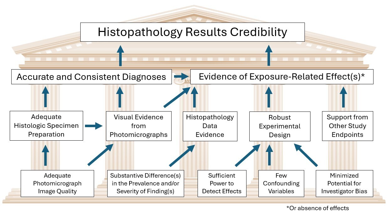
There are two essential components of histopathology results credibility in published fish toxicology papers (Figure 1). First, it is imperative that the reported pathologic findings are fundamentally accurate and have been diagnosed consistently. Although consistency can only be confirmed by a re-examination of the original histologic slides, diagnostic accuracy can be assessed to a limited degree via examination of the published photomicrographic figure images. Diagnostic accuracy in turn requires that the quality of tissue preparation is at least adequate for interpreting potential toxicologic findings, and that the pathologist examining the slides has appropriate diagnostic expertise and species-specific experience. The second component of toxicologic histopathology results credibility is the provision of sufficient evidence that one or more pathologic findings were (or were not) caused by the toxic exposure, as opposed to background (i.e., spontaneous) occurrence unrelated to exposure. This component requires the authors to effectively demonstrate that the prevalence and/or severity of pathologic findings were increased (or decreased) substantively in substance-exposed fish relative to non-exposed individuals. For the purpose of this review, the prevalence of a particular gill finding is defined as the number of fish affected by that finding, divided by the number of fish for which the gills were examined (e.g., in a given comparison group). Meanwhile, severity is defined as the visual extent to which a particular histopathologic finding was observed in the gills of an individual fish. Additional elements that contribute to confidence in reliability of the histopathology results include: a robust experimental design with sufficient power to detect effects (e.g., adequate number of animals per exposure group); effective mitigation of potential confounding variables; the incorporation of procedures used to minimize both sampling and observer bias (e.g., randomized sampling, observer blinding, and/or pathology peer review); and evidentiary support from other concurrent experimental endpoints and/or the scientific literature.

The 100 articles that comprise the current review were originally assembled to provide material for a keynote presentation given at the European Association of Fish Pathologists meeting that took place on 31 August 2025 in advance of the 22nd International Conference on Diseases of Fish and Shellfish Conference in Heraklion, Greece. Similar to that presentation, the primary objective of the current paper is to describe an investigation into the reliability of gill histopathology results in fish toxicology studies. Additional goals are to increase awareness of commonly observed issues associated with the histopathology endpoint in ecotoxicological investigations, demonstrate the extent of such problems, identify specific areas of concern, and provide recommendations for improvement. It is especially important that investigators fully appreciate the potential consequences that may ensue following the publication of inaccurate results from toxicological bioassays, which can include irretrievable expenditure of funds and animal life, misguided future research efforts, inappropriate regulatory agency decisions, and loss of confidence in the scientific process.
2. MATERIALS AND METHODS
2.1. Paper Selection
To obtain material for this review, two popular free-for-use scientific internet search engines, Google Scholar (https://scholar.google.com) and Science Direct (https://www.sciencedirect.com) were queried on 30 June, 2025, and 7 July, 2025, respectively, using the following four search terms in single combination: “fish”, “gill”, “toxicity”, and “histology”. No additional search engine filters or advanced settings were used. Initially, the first 50 papers from each search engine that met three specific inclusion criteria were downloaded as PDF files. These inclusion criteria were as follows: first, each peer-reviewed paper needed to describe an original hypothesis-based cause-and-effect type study that involved the intentional exposure of fish to one or more potentially toxic substances, or alternatively, the capture of fish from contaminated and non-contaminated surface waters to investigate the potential effects of contamination on gill morphology. In practice, this first criterion was used primarily to exclude review papers from the current exercise. Second, each paper needed to include histopathologic evaluation of the gills as an experimental endpoint. Third, if exposure-related effects (i.e., positive results) were reported, the paper needed to contain photomicrographic image examples of relevant findings. One paper that reported histopathologic gill effects but failed to provide image examples did not meet this last criterion and was consequently excluded from the current review. Additionally, the Google Scholar and Science Direct queries produced three papers that were common to both search engines; as a result, three additional papers (i.e., the next three to appear following the original 50) were downloaded from Science Direct. Each downloaded paper was obtained in its entirety, including supplemental data as available. Each paper was read completely, although special attention was paid to the overall experimental design, and aspects of the methods, results, discussion, figure images, and tables that pertained specifically to the histopathology endpoint. Specific types of information were obtained from each of the 100 papers and entered into a spreadsheet; a detailed accounting of the information obtained is presented in Table 1. Reasons for selecting those particular types of information should become apparent later in this review when they re-emerge as discussion points.
2.2. Assessment Criteria
Criteria used to assess the credibility of gill histopathology results in each paper were based on methodology described previously for similar histopathology results reviews (Wolf and Maack 2017; Wolf and Wheeler 2018; Wolf 2021). As in those earlier reviews, the histopathology results in each paper were scored according to the following scale: Grade 5 = highly credible, Grade 4 = credible, Grade 3 = equivocal credibility, Grade 2 = low (dubious) credibility, and Grade 1 = no credibility. For convenience, a summary of Histopathology Credibility (HC) scoring criteria is included as Supporting Information Table 2. In broad terms, HC scores were based on: the robustness of the experimental design for detecting potential histopathologic effects; the approach used for data collection and reporting (to be discussed subsequently); diagnostic accuracy; and the reliability of presented evidence linking toxicologic exposure to the reported histopathologic effects. Information not used as credibility scoring criteria included: the type(s) of investigated test substance; the identities of the authors or their affiliated institutions; the journal in which the paper was published; non-fundamental (i.e. preferential) differences in diagnostic terminology; the credibility of reported histopathologic findings in organs other than the gill; and the results from non-histopathology endpoints. Because key methodological details required to assess the reliability of quantitative (morphometric) histopathology data are seldom provided in published papers, the credibility of such data, as available, was not evaluated during the course of the current review; however, the results of such analyses were considered when determining whether sufficient evidence of exposure-related effects existed. Papers in which the credibility of the gill histopathology results seemed to straddle the imagined line between two adjacent HC scores (e.g., Grade 2 versus 3, or Grade 3 versus 4) were assigned the higher score by default.
Concerning the topic of diagnostic accuracy, a novel set of thirteen Pathology Assessment Factors (PAF) was formulated ad hoc to specifically evaluate diagnostic issues in photomicrographs and figure legends (Table 2). These thirteen PAF (indicated by the letters “A” through “M”) were created to improve objectivity and transparency in the credibility scoring process, and also to highlight specific problems following review of the histopathology results produced by the PAF assessment. The first six PAF concerned the accuracy of the reported histopathology findings, the next four concerned the strength of evidence that exposure-related effect(s) had occurred, and the last three were related to general histopathology results credibility. Each paper was assigned (tagged with) as many PAF as were deemed applicable based on a review of provided photomicrographic gill images and their accompanying figure legends. One exception to this rule involved papers in which all of the included gill images depicted specimens that were considered to be of non-diagnostic quality (PAF = “B”); for such papers, the assignment of additional PAF was not considered relevant. For papers that had a mixture of diagnostic quality and non-diagnostic specimens, assignment of additional PAF (as appropriate) was based on the former. Specimens considered non-diagnostic were those in which issues involving sample collection and/or preparation (e.g., moderate to severe autolysis, traumatic excision resulting in specimen fragmentation, poor orientation during embedding, and/or inadequate tissue processing or microtomy procedures) resulted in artifactual distortion of the tissue to a degree that would seriously impact the ability of a well-trained fish pathologist to reliably obtain useful diagnostic information from that sample. As much as possible, issues of specimen quality were differentiated from problems involving suboptimal image quality; in practice, distinguishing specimen quality from image quality issues was typically not difficult. Results of PAF assessment will be discussed subsequently.
For each of the 100 papers, recorded information, PAF assignments, and HC scores are presented in Table 1. Each paper was assigned an identification number (1-100) based on the order in which the article was encountered in each search engine. Thus, Papers 1-50 originated from the Google Scholar search, while Papers 51-100 were found via Science Direct. The literature references that correspond to those identification numbers are available in the Supporting Information, while in the body of this review, each paper is referred to only by its identification number. Median values and other basic arithmetic calculations were performed using Microsoft Excel; no further statistical analyses were conducted.
3. RESULTS
3.1. General Search Results
Publication years for the 100 selected papers ranged from 1988 to 2025, with 2021 as the median year. The 50 papers generated by the Google Scholar search were of a distinctly earlier vintage (median year = 2012) than the 50 produced by the Science Direct search (median year = 2024). The overall number of journals represented was 48. The number of different substances/conditions tested among the 100 papers was 81, and examples included algal toxins, atmospheric particulates, contaminated sediments, contaminated surface waters, fish therapeutics, food additives, fuels, human pharmaceuticals, manufactured products and byproducts, metals, microplastics, mycotoxins, nanomaterials, pesticides, phytotoxins, pH, salinity, and water quality components. The overall number of fish species tested was 46 (all teleosts), and the two most frequently utilized species were Nile tilapia Oreochromis niloticus (16 papers) and zebrafish Danio rerio (12 papers). These metrics suggest that the two online literature searches produced a diverse assortment of relevant articles for review.
3.2. Experimental Designs of Reviewed Papers
The following results pertain specifically to fish that were evaluated histopathologically. The median duration of the longest experimental exposure reported in each of the reviewed papers was 14 days (range = 0.17-120 days). The number of papers in which the longest exposure was 4 or fewer days was 33. When reviewing papers, noting the exposure duration of the histopathology phase can be particularly important, because certain types of findings are more likely to legitimately occur as treatment effects in short-term trials than they are in long-term studies, and vice versa. For example, it would be highly unusual to observe chronic changes such as fibrosis or severe epithelial proliferation as a consequence of toxicologic exposure in a two-day study, while acute changes such as fresh hemorrhage, edema, or individual cell necrosis are less likely to be test-article-related in studies that terminate following weeks or months of exposure.
The number of papers that were designed with lethality as an intentional endpoint (e.g., LC50 experiments) was 15. The majority of those lethal experiments involved comparatively short-term (e.g., 4-day) exposures. Two additional experiments were not designed with the stated purpose of generating lethality or LC50 values, but still reported some degree of unintended mortality among fish that were examined histopathologically. In one further study (Paper 39), fish that were investigated histopathologically were exposed to test substance concentrations as high as 70% of the LC50; however, the occurrence of mortalities at that concentration, or their absence, was not reported. In yet another study (Paper 94), the highest tested concentration for the histopathology phase was 80% of the LC50, and somewhat surprisingly, the authors reported specifically that no mortality had occurred at that test level. Among the papers in which any degree of lethality occurred, there was no reported evidence to suggest that respiratory compromise was the primary cause of death. Interestingly, only 1/100 papers described clinical evidence of visible respiratory compromise (e.g., labored breathing, piping at the surface) in substance exposed fish (Paper 53); however, there were no reported mortalities in that particular study.
Among the 100 papers, the number of fish per exposure group used for histopathologic assessment of the gills ranged from 1 to 75, with a median of 10 fish per group. Often, the number of fish per group used for histopathology was far fewer than the number that originally began the experiment. However, for many papers it was challenging to determine precisely how many fish per group were examined microscopically, because this quantity was not clearly indicated in the text, tables, or figure legends. In fact, for 4 papers, the number of fish per group used for this endpoint could not ultimately be determined. The number of papers in which 5 or fewer fish per exposure group were used for histopathology was 26. This result is mentioned because experience suggests that the statistical power of such experiments to detect exposure-related effects may be limited (Vollmer 2011).
Histopathological findings in the gills were reported according to sex for 5 of the 100 reviewed papers. Those 5 papers conducted either single-sex experiments (2 papers), utilized both sexes and reported results according to each sex individually (1 paper), or used a 1:1 ratio of both sexes in each group (1 paper). For the remaining 95 studies, either the sex of the fish was unknown (or at least not reported), or the sexes were known but histopathologic gill findings from the two sexes were pooled.
The number of papers in which studies used replicates was 59, and for those papers, the number of replicates used per study generally ranged from 2 to 4 (one paper reported 12 replicates and another possibly indicated 20 replicates, although this could not be confirmed). Among some papers there was confusion regarding the exact meaning and usage of the term “replicate”. Traditionally, this term is used to indicate a tank (i.e., test container or vessel) in which the exposure concentration and test conditions are kept essentially identical to those of one or more concurrently maintained tanks. Replication is frequently employed in fish toxicity studies to ensure that differences between control and treated tanks are actually the result of treatment rather than other uncontrolled factors, because the tank may be used as the statistical unit for comparison in some cases, and also to ensure adequate sampling if one or more tanks encounters a problem that causes it to be disqualified from the study. However, a review of the text indicated that the term replicate was used creatively in some papers to indicate various types of alternative units, such as one of multiple animals in a group, one of multiple samplings obtained over time, one in a series of repeated experiments, or one of in a series of repeated analyses within a single experiment. Other papers simply stated that the study had been conducted “in triplicate”, without further explanation as to whether this meant that there were three replicate tanks, three repeated experiments, or three samplings performed within a given experiment. In order to establish whether differences between control and treated tanks were the result of treatment versus uncontrolled factors, study data can be analyzed to determine if the degree of variation among tanks was significantly different from the amount of variation observed within tanks. However, not one of the 59 papers that used replicates reported performing any type of statistical analysis to detect potential replicate-dependent effects.
In 8 of 100 papers, the authors reported having performed blinded (masked) examination of the histologic slides. The remaining 92 papers did not mention whether blinding was used.
3.3. Approaches Used for Data Collection and Reporting
Although technically a component of the experimental design, this topic has special considerations that merit its own subheading. Among the 100 reviewed papers, the most common approach for data collection and reporting was to provide no semi-quantitative or quantitative histopathology data (45 papers). Those 45 papers instead furnished morphologic descriptions of microscopic findings in varying degrees of detail, with observations concerning the comparative abundance of histopathologic findings among the different exposure groups. There was no evidence in any of those papers that such descriptions were derived from semi-quantitative or quantitative scoring data, nor were there indications that the fish were evaluated on an individual animal basis. For the purpose of this review, semi-quantitative assessment was defined as the assignment of ordinal scores or grades (e.g., minimal mild, moderate, or severe), each of which represented the relative degree to which the gills were affected by a particular histopathologic finding. This is in contrast to quantitative histopathology data, which are based on measurements obtained from photomicrographs, typically. The number of papers that provided morphologic descriptions and quantitative data in lieu of semi-quantitative data was 13.
Among the 42 papers that reported semi-quantitative histopathology data, various tactics for semi-quantitative scoring were employed, including the group-wise severity approach (21 papers), the index system approach (14 papers), mean severity scoring (3 papers), scoring that generated both prevalence and severity data (2 papers), and the reporting of group-wise prevalence or group-wise occurrence data (1 paper each). In the most frequently used approach, referred to here as “group-wise severity”, scoring most often employed a 0-4 grade scheme with the absence of effect represented by a dash, and progressively increasing severity of effect indicated by one, two, or three plus signs, respectively (Table 3A). There was no evidence provided in any of those 21 papers to document or even suggest that fish were scored individually. Of the 21 papers that used the group-wise severity approach, 19 reported zero pathologic findings in control fish, one paper did not have a control group (that study involved the use of one substance to potentially mitigate toxicity induced by another), and another paper did not report histopathologic results pertaining to their control fish. Reasons for noting the absence of findings in control fish will be discussed subsequently.
The second most commonly used semi-quantitative scoring approach involved index systems that were based on methodology proposed primarily by Bernet et al. 1999 or Poleksić and Tutundžić, 1994. The canonical procedure common to such systems is to record a severity score for each type of pathologic finding in each examined organ or tissue from each fish. The severity score for each finding is then multiplied by a weighted “importance factor” (IF), that typically ranges from 1-3, and is intended to represent the perceived health impact and/or potential reversibility of each finding type. The resulting values created by this mathematical operation are then added together to create a whole-organ score for each fish. Inter-group differences in mean index scores have been used as evidence of exposure-related histopathologic effects; however, scientific and logical flaws associated with the index system approach have been reported previously (Wolf 2018), and for convenience these issues will be summarized briefly in the Discussion section.
The four additional approaches to collecting and reporting histopathology data appeared in only 7/100 papers; therefore, they will not be discussed in detail here. Instead, a preferred method commonly used in pre-clinical toxicology studies will be described in the Recommendations for Improvement section of the Discussion.
3.4. Histopathology Endpoint Outcomes
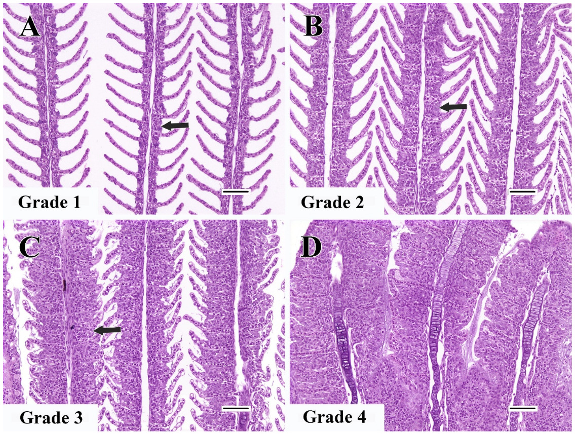
Each of the 100 reviewed papers reported histopathologic effects in the gills associated with substance exposure (i.e., 100% positive study outcomes). The number of papers in which zero pathologic changes were reported in the gills of control fish was 81, which equated to approximately 1,090 fish. This included 59 papers in which the controls were specifically reported to be free of pathologic findings, and 22 papers that did not happen to state whether the controls had findings. However, a total of 24 papers failed to report at least one pathologic finding in the gills that was evident in the published photomicrographs (PAF = “L”), and often these undiagnosed findings were evident in control fish. The most common unreported finding in controls was lamellar epithelial hyperplasia. Lamellar epithelial hyperplasia is characterized by proliferation of pavement cells primarily, which may be accompanied by lesser degrees of mucous cell metaplasia, chloride cell hyperplasia, and/or inflammatory cell infiltration. This cellular proliferation can occur anywhere along the length of the (secondary) lamellae, but often begins proximally at the base; in minimally to moderately affected gills this causes partial filling of interlamellar sulci, while in severe cases it results in lamellar fusion (Wolf et al. 2015). Although lamellar epithelial hyperplasia can occur focally or multifocally, experience suggests such patterns tend to be more characteristic of parasitic or bacterial infections, while general irritation of the gills or chemical exposure are often associated with diffuse or segmental changes that may be more prominent toward the apical ends of filaments. For reference, Figure 2 of the current review provides an example of lamellar epithelial hyperplasia severity grading. Examples of undiagnosed lamellar epithelial hyperplasia in control fish were observed in Papers 23, 30, 31, 42, 51, 54, 55, 56, 58, 62, 66, 68, 70, 72, 75, 76, 81, 82, 83, 91, and 94. In a few instances, the severity of undiagnosed epithelial hyperplasia in controls could be graded as moderate (Grade 3) or even severe (Grade 4), and in some of those papers, the magnitude of hyperplasia actually appeared greater in control fish as compared to the exposed fish.
._example_of_visual_severity_grading_criteria_for_the_diagnosis_of_lamellar_epithelial.png)
Only one paper reported antemortem or post-mortem observations of gill pathogens. Paper 24 indicated that a low prevalence of parasites was found in the gills of both control and substance-exposed fish, but stated that the presence of parasites did not appear to impact the study results. One additional paper (Paper 33), reported that the gills of experimental fish were free of observed parasitic or bacterial pathogens prior to testing. Three further papers described prophylactic dips with potassium permanganate for external pathogens prior to experimentation, but did not establish if such organisms were present prior to dipping (this can be important, because the presence of pre-existing pathogens can cause inflammation and/or proliferative changes that may persist into the experimental period), or if the dipping was successful at eliminating pathogens. Overall, 95 papers did not in any way address the possible existence of gill pathogens in the tested fish. Ironically, unrecognized gill pathogens can be seen in figure images from at least two papers: in Figure 1C1 of Paper 66, there is evidence of undiagnosed epitheliocystis (i.e., infection with chlamydia or chlamydia-like organisms), while in Figure 5F of Paper 45, the authors failed to diagnose an embedded Ichthyophthirius multifilis ciliate protozoan, and instead mistakenly diagnosed the presence of this parasite as “necrosis”.
3.5. Diagnostic Accuracy and PAF Assignment
Percentages of papers that received each of the 13 PAF are listed in Table2, and a detailed accounting of the PAF assigned to each paper can be found in Table 1. The following description of results is focused on the most commonly observed and consequential PAF.
The number of papers in which at least one histopathologic finding was considered to be accurately diagnosed was 67 (PAF = “A”). Conversely, 33 papers contained zero accurately diagnosed findings. Among those 33 papers were 12 papers in which all of the provided photomicrographs depicted histologic gill specimens that were deemed to be of non-diagnostic quality (PAF = “B”; Papers 9, 20, 21, 22, 25, 47, 57, 63, 77, 80, 89, and 99). For each of the remaining 21 papers that had zero accurately diagnosed findings, gill tissue in at least one image was of satisfactory quality, but none of the indicated diagnoses were evident, or at least correctly identified. In Paper 3, for example, reported findings of gill epithelium desquamation, lamellar telangiectasis, secondary lamellar hemorrhage, and lamellar hypertrophy and hyperplasia were not appreciable to any degree (PAF = “C”), despite figures that portrayed reasonably good tissue and image quality.
After “A”, the second most commonly recorded PAF was “C” (53 papers). In papers tagged with this PAF, at least one indicated finding in an image was not evident to any degree, and this included the mischaracterization of normal anatomic structures as pathologic findings. Clear examples of the latter included the misidentification of: filament cartilage as “swollen mucocytes” (Paper 83) or “cartilaginous tissue edema” (Paper 64); mineralized filament cartilage as “vascular swelling” (Paper 74); goblet mucous cells as “cellular necrosis” (Paper 5) or “lamellar aneurysms” (Paper 32); normal capillary lumina (lacunae) as “epithelial lifting” (Paper 59); blood within the central venous sinus of a filament as “hemorrhage”; and a normal-appearing pseudobranch (an entirely separate gill-like anatomic structure) as a gill that had “severe (lamellar) fusion with hyperemia and capillary vasodilation” (Paper 71). In some papers, reported effects were not only inapparent, they were not characteristic responses of gills to injury induced by toxicologic substance exposure (or any other pathogenic causes, for that matter); examples of this included: “hypertrophy of mucous cells” (Paper 15); “atrophy of mucous cells” (Paper 27); “abnormally elongated lamellae” (Paper 45); and “epithelial congestion” (Paper 96).
The third most commonly recorded PAF was “E” (53 papers), in which at least one purported pathologic finding appeared instead to be a post-mortem artifact. This is perhaps not surprising, because collection and histologic processing of the gills can be challenging, and artifacts related to specimen collection, preservation, and histologic processing are all too common (George et al. 2016). Artifacts observed frequently in figure images included changes consistent with post-mortem autolysis, fragmentation of filaments or lamellae caused by traumatic gill excision or poor microtomy technique, “epithelial lifting” (artifactual separation between the lamellar epithelium and underlying lamellar capillaries, which in some cases can be attributed to suboptimal fixation), and plane-of-section artifacts. Plane-of-section artifacts are observed in histologic sections in which tissue structures are oriented in uncharacteristic angles or have been cut tangentially. A prototypical example of this is provided by Paper 35, in which slightly twisted lamellae were misdiagnosed as lamellar hypertrophy and lamellar fusion. Further examples of various types of collection/processing artifacts that were misinterpreted as pathologic changes are readily apparent in Papers 17, 19, 26, and 42.
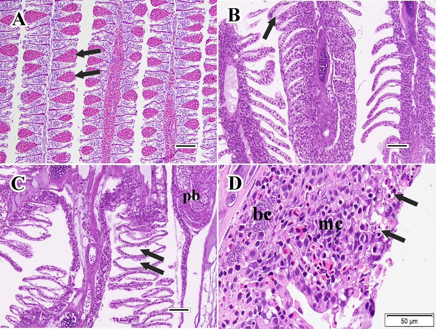
A PAF that pertained to evidence of exposure-related effect rather than diagnostic accuracy was “H” (30 papers). In papers tagged with this PAF, the nature and visual extent of at least one indicated finding appeared essentially the same in images of exposed versus unexposed fish. Although it is recognized that photomicrographic images can be selected so as to portray experimental results that may not fully represent the true study outcome, the authors’ ability to demonstrate the existence of clear morphologic differences between negative control and substance-exposed animals can add some measure of evidentiary weight to the validity of reported exposure-related effects. Conversely, the inability of investigators to provide that type of visual histopathologic confirmation may be even more informative in terms of assessing study credibility. Examples in which the gills of substance-exposed fish appeared essentially identical to those of controls included Papers 30, 55, and 70. Other PAF related to “H” included “I” (23 papers) and “J” (23 papers). A PAF of “I” was recorded when at least one indicated finding was considered to be a common background lesion in untreated controls, and there was insufficient data available to suggest that the prevalence or severity of that finding was greater in substance-exposed fish versus controls. Common types of background findings in this category include telangiectasis (lamellar aneurysms; Figure 3A) and the previously mentioned epithelial lifting artifact (Figure 3C), which can resemble gill edema (Figure 3B). Although telangiectasis can be a potential pathologic effect in substance-exposed fish, especially during the acute exposure phase, it can also be observed in control fish, in some cases as a consequence of procedures performed immediately prior to or during sacrifice (Mitchell et al. 2023; Figure 3A). Consequently, the relationship between telangiectasis and substance exposure should be interpreted cautiously, especially when telangiectasis is not accompanied by resolution via capillary thrombosis, which at least suggests that the lesions likely occurred prior to the peri-mortem period (Wolf et al. 2015). In papers tagged with “J”, at least one incorrect result interpretation was based on examination of non-comparable areas or angles of gill in one exposure group versus another. Although many types of pathologic changes may occur diffusely along the gill filaments and arches, other findings may be localized to certain areas. For example, it is not uncommon to observe greater degrees of inflammation, mucous cell metaplasia, and/or epithelial hyperplasia near the distal filament tips (the last of which is often referred to as “clubbing”) as opposed to the more proximal filament segments or the gill arches, and the presence or magnitude of findings (and artifacts) may also depend on whether a given filament was sectioned along its central axis versus tangentially; therefore, it is imperative that all inter-group comparisons are “apples to apples” in terms of precise microanatomic location (Wolf et al. 2015). Figure 4 of Paper 100 provides just one of many examples in which visual differences were misinterpreted as treatment effects as a consequence of comparing different gill areas and section planes in one exposure group relative to another.
_in_the_gills_of_an_untre.jpeg)
3.6. Histopathology Credibility (HC) Scores
The number of papers that received each of the five credibility scores was as follows: Grade 1 (no credibility) = 33; Grade 2 (low credibility) = 34; Grade 3 (equivocal credibility) = 22; Grade 4 (credible) = 10; and Grade 5 (highly credible) = 1.
Papers scored as Grade 1 (no credibility) could be subdivided into two categories. In the first category, specimens in all of the photomicrographic figure images were considered to be of non-diagnostic quality (PAF = “B”). In the second category, at least one of the images depicted a diagnostic quality specimen, but none of the reported histopathologic diagnoses in that paper appeared to be accurate. Comparatively few Grade 1 papers (4/33 = 12%) included histopathology data derived from semi-quantitative scoring as a component of the reported results.
In papers that received a Grade 2 HC score (low credibility), at least one of the images in the paper depicted a diagnostic quality specimen, and at least one of the reported histopathologic diagnoses in that paper appeared to be accurate. However, it was often the case that such papers also reported multiple incorrect diagnoses, based on a review of the supplied figure images. Importantly, Grade 2 papers provided little or no credible evidence of an exposure-related effect, either in terms of images that illustrated a higher degree of pathogenic changes in substance-exposed fish relative to controls, or histopathology data that demonstrated convincing evidence of such effect. In fact, only 18/34 (53%) of Grade 2 papers reported semi-quantitative histopathology data.
Grade 3 papers (equivocal credibility) also tended to fall into one of two categories. Papers in the first category had at least one histopathologic finding that could plausibly have been a genuine treatment effect, but once again, supporting data were either not convincing or were not provided (the number of Grade 3 papers that reported histopathology data was 12/24 = 50%). The provision of persuasive supporting data was considered especially important for studies in which reported exposure-related effects involved types of findings that can be observed commonly in the gills of control fish, such as telangiectasis, lamellar epithelial hyperplasia, or epithelial lifting artifact. The second category for Grade 3 papers included studies in which reported exposure-related effects were based on inconclusive diagnostic interpretations. For example, Paper 75 reported epithelial cell necrosis as an effect of substance exposure, and while the gills of substance-exposed fish did appear slightly fragmented as compared to those of controls, morphologic evidence of necrosis in the figure images was not considered definitive. Definitive characteristics of gill necrosis (Figure 3D of the current review) typically include the presence of apoptotic-looking pavement epithelial cells, epithelial cells with fragmented (karyorrhectic) nuclei, and/or cellular debris within adjacent phagocytic cells; meanwhile, none of those features were clearly evident in the figure photomicrographs of Paper 75. As for Grade 1 and Grade 2 papers, diagnostic errors were also relatively common in Grade 3 papers, and this included the reporting of diagnoses that were patently non-existent, and/or the misinterpretation of normal anatomic structures or tissue preparation artifacts as pathologic findings.
Similar to Grade 3 papers, papers that received Grade 4 scores (credible) had one or more histopathologic findings that could reasonably be interpreted as exposure-related effects. However, those articles additionally provided compelling evidence that pathologic findings occurred to a greater degree in substance-exposed fish as compared to controls, based on review of the provided figure images and/or accompanying histopathology data. Regarding the latter, 8/9 (89%) of Grade 4 papers provided at least some form of histopathology data in addition to morphological descriptions in the text. The single Grade 4 paper that did not furnish semi-quantitative data (Paper 16) was able to make a fairly convincing case by providing a series of superior quality photomicrographic images that were accompanied by detailed morphologic descriptions. Although diagnostic errors were occasionally present in some Grade 4 papers, those tended to be fewer and less blatant relative to papers assigned Grades of 1, 2, or 3.
Attributes of the sole Grade 5 paper (highly credible; Paper 43) that enhanced its credibility included: a 30-day sublethal exposure period; decent histologic and figure image quality; standardization of diagnostic criteria; blinded initial microscopic evaluation; review of diagnoses by a second pathologist as a quality control measure; histopathology data that were recorded on an individual fish basis and reported as mean severity scores; and fundamentally accurate-appearing diagnoses, with the severity of most findings reported as minimal to mild (i.e., severity scoring was not exaggerated, and was consistent with the appearance of the figure images). The included histopathology data indicated that at least some degree of abnormal findings were additionally observed in control fish. Although the reporting of findings in controls is certainly not a requirement for credibility (i.e., it is possible that negative control fish in some studies could be completely free of gill abnormalities), willingness of the authors to report findings in controls tends to promote confidence in the results. Despite being considered highly credible, there was still room for improvement in Paper 43. For example, the number of fish evaluated histopathologically per group was relatively small (i.e., n = 5), and examination of fish for gill pathogens was not reported.
4. DISCUSSION
4.1. General Considerations
The credibility of the gill histopathology results in this current review of 100 fish toxicology papers was lower than originally anticipated, as 67% of the papers were considered to have no or low credibility, while 89% were scored as having no, low, or equivocal credibility. Other published systematic reviews of histopathology results have yielded somewhat less alarming results. For example, a credibility review of histopathology results from 189 environmental endocrine toxicity studies performed as part of a SETAC Pellston Workshop® (Wolf and Maack 2017) reported that 46% of examined papers had equivocal, dubious, or no credibility. However, it is important to note that each of the studies in that review involved exposure to one of six prototypical endocrine active substances: ethinylestradiol (EE2), perchlorate, propiconazole, trenbolone, tributyltin, and vinclozolin. Because the toxicological and/or pharmacological mechanisms of those substances are well established, and characteristic exposure effects have been widely reported, it might be expected that a comparatively higher percentage of such studies would generate reliable histopathology results. In another review of histopathology findings from 117 papers that reported findings of hepatotoxicity in fish (Wolf and Wheeler 2018), 63% of papers were considered to have equivocal, dubious, or no credibility. Meanwhile, a review of histopathology results from 16 papers that investigated Leydig cell hyperplasia in fish (Wolf et al. 2026) found that the results in 62% of the 16 reviewed papers had equivocal, dubious or no credibility; and a review of individual findings from 14 papers that investigated the experimental exposure of fish to diclofenac (Wolf 2021) reported that 85% of the 78 total findings were deemed to have equivocal, dubious, or no credibility. More specific to the current review, Bjørgen et al. (2025) encountered diagnostic misinterpretations, poor quality photomicrographs, and/or a failure to include figure images in 48% of the 21 papers that reported histopathologic effects in the gills of fish maintained in recirculating aquaculture systems. Suspected reasons why gill studies may be particularly prone to producing sub-par histopathology results will be addressed later in this discussion section.
The methodology used to generate material for the current literature review provided a broad selection of tested chemicals, fish species, scientific journals, and publication years. The choice to use simplified search criteria (e.g., a single search string of four terms used in combination, and selection of the first 50 papers from each search engine) was entirely intentional, as each added bit of complexity involves the making of a decision, and each decision point provides an opportunity to introduce bias into the search process. However, reviews of this type inevitably involve compromises, and the current example is no exception. Arguably, a survey of greater than 100 papers could have been conducted, and additional and/or alternative search term combinations could have been used to query additional and/or alternative search engines. Possibly, artificial intelligence software could have been employed to redact any and all information in each paper specifically related to the test chemical or authorship, in order to further lessen the potential for biased assessment during the review process. Factors used for credibility grading could have been weighted mathematically for relative importance (although the wisdom of that is debatable), and credibility assessments could have been performed by one or more additional fish pathologists. However, there is no obvious reason to suspect that the overall outcome would have been fundamentally different, or in any way less biased, had an alternative selection approach been used. Additionally, there is a point at which increased effort yields diminishing returns, and to focus on the methodological limitations of the present review may be to lose sight of the larger issue at hand. Ultimately, whether the percentage of less-than-credible papers was in fact 99% or 79% rather than 89% is of little actionable importance, as any remotely comparable outcome (e.g., even 50%) connotes a serious problem involving the reliability of gill histopathology results reported in fish toxicology papers.
4.2. Quality of Specimen Preparation and Diagnostic Accuracy
As described in the introduction, the first pillar of histopathology results credibility is a demonstrated ability to make fundamentally accurate and consistent diagnoses based on the microscopic examination of tissue sections. It seems fairly clear that this cannot be accomplished unless the sections have been adequately prepared, as even the most skilled pathologist cannot generate valid results based on the evaluation of poor quality specimens. Tissue artifacts caused by substandard tissue collection and histologic preparation techniques can not only mask certain types of genuine findings, resulting in the failure of such diagnoses to be recorded, their presence also increases the odds that pathologists (especially those who have limited experience) will misinterpret such artifacts as pathologic changes. In the current review, 12% of the reviewed papers contained photomicrographs in which all gill specimens were judged to be of non-diagnostic quality, while 20% of papers overall published at least one image of a non-diagnostic quality specimen. Additionally, gill tissue quality appeared suboptimal in a number of other reviewed papers (although the occurrence of suboptimal quality specimens was not specifically recorded during the current review). These results suggest that many research laboratories are less than proficient in the post-mortem collection and histologic processing of fish gill tissue. Potential challenges faced by laboratories include: suboptimal methodology involving one or more procedural steps pertaining to tissue collection and histologic preparation; inadequate or poor quality tissue processing equipment; insufficiently skilled histology technicians; and/or the inability of all involved personnel to recognize when histologic sections do not meet reasonable quality standards. Superior preparation of gill specimens is particularly challenging due to factors that include the fragile nature of the gill lamellae, the tendency of elongated anatomic structures such filaments and lamellae to twist, fold, or clump, and the comparatively rapid decomposition of gill tissue that may occur post-mortem prior to fixation (George et al. 2016). Even when the histologic procedures per se have been performed adequately, a number of potential pitfalls involving prior in-life gill sampling, euthanasia method, interval between death and fixation, and gill excision technique have been demonstrated experimentally to produce artifacts that can readily be confused with pathologic findings (Mitchell et al. 2023; Wolf et al. 2015). Conversely, many laboratories have demonstrated that careful planning and knowledgeable execution can generate excellent quality gill sections, including examples encountered during the current review (e.g., Papers 27, 30, 33, 53, 59, and 94), and elsewhere in the literature (Smith et al. 2018; Mitchell et al. 2023). Nevertheless, it seems logical to conclude that pathologic diagnoses stemming from the evaluation of inadequate quality histologic specimens are inherently untrustworthy, and that the same caveat pertains to toxicological interpretations of such findings.
Results of the current review also demonstrate that the creation of adequate (or even excellent) quality histologic specimens does not necessarily ensure that diagnostic findings will be interpreted accurately. The three most common types of diagnostic errors observed throughout this review involved papers in which at least one indicated finding in an image was not evident to any degree, papers in which at least one purported pathologic finding appeared instead to be a post-mortem artifact, and papers in which at least one reported pathologic finding was fundamentally mischaracterized/misdiagnosed. Of the 88 papers that had specimens that were considered to be of diagnostic quality, 78 (89%) exhibited at least one of these three types of errors, while substantial proportions of those papers exhibited two or all three types. Additionally, one third of all papers did not contain a single accurately reported gill diagnosis based on review of the figure images, annotations, and accompanying legends. Taken together, these results suggest that many of the histopathological evaluations were performed by personnel who did not possess the requisite skill set for the task, which includes a clear understanding of fish gill microanatomy, and sufficient training in the microscopic recognition and characterization of gill disease (whether of natural or chemically-induced etiology). Although a certain degree of subjectivity is inherent in histopathology, it should be reiterated that, in the vast majority of instances, the particular types of diagnostic errors described during this review could not be fairly portrayed as minor disagreements in diagnostic terminology, lesion severity scoring, or the finer points of morphological description, but instead involved fundamentally incorrect interpretations of normal and/or abnormal visual features.
4.3. Evidence of Exposure-related Effect
Although diagnostic accuracy is undeniably a critical component of histopathology results credibility, a further requirement for toxicological challenge studies that report exposure-related effects is that the authors provide reasonably compelling empirical evidence that the prevalence and/or severity of one or more pathologic findings differed substantively in substance-exposed fish when compared to concurrent unexposed or reference site fish. In the context of a journal article, this type of evidence ideally consists of representative photomicrographic figure examples, accompanied by semi-quantitative and/or quantitative histopathology data.
4.3.1. Photomicrographic Figure Images
Although it is easy to understand how figure photomicrographs can be used to assess the accuracy of diagnostic findings, it may be less obvious why the inclusion of images is also important for confirming the validity of exposure-related effects. Admittedly, the only definitive way to determine if reported substance-induced histopathologic effects are genuine is via a re-examination of the original study slides and individual fish data (assuming such materials are available) by one or more experienced fish pathologists. However, the inclusion of photomicrographic figures that demonstrate clear differences in the presence or extent of pathologic findings in substance-exposed fish versus controls can still inspire confidence in the reliability of the histopathology results. Conversely, and perhaps more importantly, figure images that fail to accomplish this evidentiary threshold may bring the credibility of reported results into question. For example, in the present review there were 30 papers in which the nature and visual extent of at least one indicated finding appeared essentially the same in images of substance-exposed versus unexposed fish (PAF = “H”). In such cases, both sets of gills appeared approximately identical to one another, either because they were both largely “normal” (i.e., unremarkable), or because the nature and degree of abnormalities in both sets of gills were comparable. Given that authors naturally tend to select images that best portray the reported effects, an inability to successfully demonstrate such visual differences reduces confidence in the legitimacy of the histopathology results. Although photographic image quality can certainly affect the ability of the reader to assess the ability of reported diagnoses, insufficient image quality per se was not a major issue encountered during the current review. In some papers, images of poor quality simultaneously featured non-diagnostic quality specimens, the latter of which rendered the former issue moot. Typical image quality problems encountered included magnification that was too low (or occasionally too high) to effectively demonstrate the reported gill effects, insufficient image resolution, and poor focus. Other types of image issues, such as lighting and color imbalances, and dirt shadows from uncleaned slides, were considered primarily aesthetic, as they are less likely to interfere with the verification of diagnoses; however, the presence of such quality issues can still suggest inexperience or lack of care taken during photography.
4.3.2. Histopathology Data
While the inclusion of photomicrographic figure images that demonstrate clear effects is helpful for establishing credibility in published reports, further evidence in the form of histopathology data should be provided to support the contention that such images accurately represent the study outcome. Additionally, certain types of effects may occur exclusively as an increased prevalence (versus severity) of findings in substance-exposed fish when compared to controls, and it is not possible to demonstrate increases in prevalence using images alone. In the current review, 45/100 papers did not furnish any type of semi-quantitative or quantitative histopathology data, instead supplying only morphologic descriptions in the text. In the vast majority of cases, that outdated approach to histopathology results reporting, which lacks both precision and specimen accountability, cannot provide adequate evidence for the existence (or absence) of exposure-related effects. When tabulated summaries of individual animal data are not made available, it is difficult for the reader to have confidence that all available tissues from all fish were actually examined, and frequently, it is not even possible to confirm the exact number of animals that were assessed microscopically. As much as possible, it is especially important to ensure that specimens from unexposed control fish were evaluated (sufficiently or at all), as there exists much evidence which suggests that the failure to adequately examine tissues from control or reference site fish is a major problem in fish toxicologic pathology. One noteworthy example described in Wolf et al. 2014 involved a study in which fish exposed to diclofenac were reported to exhibit 19 different types of exposure-related histopathologic findings in the gills, liver, and kidney that were not observed to any degree in the six control fish. However, a subsequent blinded review of the original study slides by a panel of expert fish pathologists (pathology working group) found that most of those same types of findings were also present in the controls, and that the overall prevalence and severity of findings in control and treated fish were in fact comparable. Meanwhile, in the current review, 81/100 papers did not report any histopathologic findings in the gills of control (or reference site) fish. In 22 of those 81 papers, the authors did not specifically state that the gills of control fish had zero findings, however, they also did not report that any were present. In the remaining 59 papers, the authors specifically stated that there were no findings in control fish, or that the gills of controls appeared completely normal. Although it is certainly possible that the control fish in a particular study might not exhibit any pathologic gill findings, experienced fish pathologists recognize that this is highly uncommon, especially for experimental animals sourced from less controlled environments, such as native surface waters or commercial aquaculture facilities. Pre-existing or current pathogenic infections, crowded conditions, and water quality issues characteristic of many recirculating or static containment systems, e.g., may all contribute the presence of background gill lesions in control fish (Bjørgen et al. 2025; Smith et al. 2000). Concerning the 81 studies in this review that did not report findings in controls, the probability that not one of those approximately 1,090 control fish had a single reportable finding in the sampled gills seems infinitesimally small. Further evidence from the present review that control fish are not being scrutinized sufficiently was the frequent failure to report findings in controls that were patently evident in figure images, the most common of which was minimal to severe lamellar epithelial hyperplasia. Rigorous examination of control fish can be essential for distinguishing handling or processing artifacts from pathologic findings, and can help the pathologist to determine if the nature or severity of certain findings in substance-exposed fish is patently different from those same findings in control fish, as opposed to background-type changes that are incidental to the study outcome. Additionally, if findings in controls are not routinely recorded, is not possible to establish a legitimate historical control database, which can be useful for identifying outlier results.
Among the reviewed papers that did furnish histopathology data, the most common approach was to present the results as a tabulated representation of group-wise scores for each finding type (Table 3A). This method, which is generally not used to any extent in mammalian pre-clinical toxicologic pathology, appears to have widespread acceptance in ecotoxicological studies. However, the group-wise severity approach is not highly informative, and actually offers little benefit when compared to merely providing text descriptions of morphologic findings. First, among the reviewed papers that used this approach, it was never stated whether each score represented the percentage of affected animals per group (i.e., lesion prevalence), as opposed to an approximation of the average extent to which gills were affected (i.e., lesion severity), or possibly some combination of those two metrics. Ultimately, the lack of granularity associated with this method, i.e., the inability to assess prevalence and severity independently, can lead to false-positive or false-negative interpretations with regard to the presence or absence of exposure-related effects. Second, the group-wise severity approach provides no evidence to support author claims that all specimens were thoroughly examined. Again, it is noteworthy that zero findings were reported for the negative control fish in 19 of the 20 papers that had control groups and used the group-wise scoring approach. Third, the group-wise scoring method also provides no evidence that findings were recorded on an individual fish basis. Therefore, while the tabulated results convey the appearance of summarized semi-quantitative data, in truth there are no underlying data points; instead such scores represent general perceptions of the pathologist, as opposed to meticulously recorded scientific observations. Another drawback to the absence of underlying individual animal data is that it prevents a second pathologist from subsequently reviewing the original findings in concert with a re-examination of the histologic slides, i.e., pathology peer review, which increasingly has become the gold standard for ensuring quality control in toxicologic pathology studies. Finally, the group-wise severity method lacks both transparency and specimen accountability, which are important for establishing trust in the intentions and technical capabilities of the authors, and confidence in the reported results.
The second most common approach used among the reviewed papers for evaluating results of the histopathology endpoint was the index system method, which like the group-wise severity method, is peculiar to ecotoxicological studies. One clear advantage of this approach as compared to the aforementioned method is that findings are recorded and scored for severity on an individual fish basis. Interestingly, in 11 of the 14 reviewed papers in which the index approach was used, pathologic findings were reported to occur in control fish, which again highlights the value of scoring fish individually. However, the index system approach also has several major drawbacks that make it largely unsuitable for use in toxicological bioassays. The first problem with this approach is that the various types of findings that are combined to create single organ scores often include handling or tissue preparation artifacts, non-lesions, misdiagnoses, and background-type changes, in addition to (or in lieu of) potential exposure-related lesions (Wolf 2018). This is illustrated in Paper 64 for example, in which the scores of 15 different types of histopathologic findings were combined to calculate a single gill score. However, substantive scoring difference compared to control existed for only one of those reported findings (pavement cell hypertrophy). Meanwhile, many of the other reported findings, such as lamellar epithelial lifting, lamellar curling, blood congestion, and marginal channel dilatation, can often be observed as artifacts or incidental occurrences that are unrelated to substance exposure. The index system approach attempts to compensate for the influence of less relevant (superfluous) findings by assigning an “importance factor” (e.g., 1, 2, or 3) to each finding type, which is used as a weighted multiplier. It should be emphasized that these importance factors are arbitrary designations that were not generated by empirical testing, and the importance factor ascribed to a given diagnosis often varies from paper to paper. It should also be understood that each importance factor represents an intuitive notion of the health impact and/or reversibility of a particular histopathologic finding; consequently, the incorporation of importance factors in calculations that are used to determine causality represents an inherent logical flaw in this method, because the importance factors themselves have absolutely no bearing on cause-and-effect relationships (e.g., gunshot wounds can have serious health consequences, but that fact alone provides no information as to who pulled the trigger). The index system approach also relies on various unproven assumptions (e.g., that the health impacts of unrelated types of histopathologic findings are necessarily additive), and statistically invalid mathematical manipulations of ordinal data that are further discussed in Wolf 2018.
A recommended approach for collecting and reporting histopathology data will be described in the following section.
4.4. Recommendations for Improvement
While the following recommendations are particularly relevant for investigations of potential toxicological effects in the gills, many can also be applied to the histopathology endpoint in general, and even to other study endpoints. Admittedly, certain recommendations, such as the inclusion of replicates in the experimental design, will be more applicable to controlled laboratory experiments as opposed to field/survey type studies.
-
Gill histopathology workups should be avoided for studies in which test concentrations are associated with high levels of mortality, whether this is intentional (e.g., LC50 experiments) or inadvertent. This is because the gill tissues of moribund and dead fish are often of suboptimal quality due to autolytic change, and because any observed morphologic changes are more apt to reflect the near death condition of the fish, as opposed to toxicologic effects characteristic of the test substance at or near concentrations that fish are likely to encounter in the environment. One reason for this is that sublethal effects may be produced by mechanisms that are entirely different from those responsible for lethality (Wolf and Segner 2023). Consequently, histopathologic analysis of specimens from high lethality studies may not only represent a waste of funding resources, it can also generate information that is inaccurate and/or of questionable toxicological relevance. For a more in-depth discussion of this topic, see Wolf and Segner 2023.
-
Ideally, investigations of gill toxicity in reproductively mature fish should be conducted either as single sex experiments, or experiments in which the findings are recorded and reported according to fish sex. Although there is currently little evidence in the literature to support the contention that sex-related differences in the nature or degree of chemically-induced gill effects are important (or even exist), that data scarcity may be a product of limited investigation rather than empirical testing (i.e., the absence of evidence is not evidence of absence). There is at least anecdotal evidence that the gills of male and female fish may not respond identically to toxic exposures (unpublished experience of the author), while the mammalian literature contains more concrete examples of gender-related differences in chemically-induced respiratory tract effects (Gochfeld 2017; Price et al. 2011), and fish studies have shown sex-related differences involving other organs such as the liver (Koehler 2004). However, whether or not such evidence exists, the absence of sex-specific responses in the gills cannot be assumed, and until proven otherwise, sex represents a confounding factor for all adult animal toxicological studies that fail to account for gender. In the current review, only 5 papers reported findings according to fish sex. In the one paper in which findings from males and females were reported separately (Paper 60), at least one finding that was reported in males was not described for females; unfortunately, that cannot serve as a legitimate example, since none of the reported gill pathology findings in that study were considered credible based on a review of the figure images.
-
Because of the very real potential for pathogen-induced changes to confound the experimental results, the occurrence and extent of gill pathogens should be assessed for every study. Ideally, this should be first accomplished prior to acclimation, either by examining wet mount preparations of gill clips, and/or via the histologic examination of gills from several sentinel animals sacrificed for that purpose. While the latter option involves the sacrifice of additional animals, it has the benefit of providing a preview of initial background-type changes caused by pathogens or other conditions. Prophylactic treatment alone may not be sufficient to counteract effects of pathogens on the histopathologic results, not only because treatment success can vary, but also because pathogen-induced lesions may persist for a variable time period following pathogen removal. Examination for pathogens should also be performed during the definitive histopathological examinations that follow study termination, to determine if the presence of gill pathogens could have confounded the study results, and such information should be reported in any published results.
-
Whenever possible, replicate containers (typically 2-4 total per group) should be incorporated into experimental designs, in order to account for potential confounding effects associated with inter-tank differences in tank location, lighting, water quality, temperature, stocking density, behavioral issues, etc. This may be especially important for gill investigations, because of the more immediate connection between ambient water conditions and the gill tissue when compared to internal organs. However, including replicates alone is of little benefit unless the resulting data are subsequently statistically evaluated for potential replicate-dependent effects.
-
Investigators should assess whether they have the ability to produce histologic sections of sufficient quality for diagnostic evaluation. This can be done by comparing the appearance of their test specimens against textbook examples of gill rather than photomicrographs encountered in journals, which too often have their own quality issues. If improved training in histological technique cannot be obtained, investigators should either pursue further training, contract that work to an experienced laboratory, or as a last resort, consider omitting histopathology as an endpoint for that study. On the technical side, and especially if gills are considered to be an important target organ for the test substance, superior results are often obtained by excising the entire holobranch at the gill arch and processing the gills separately, as opposed to examining the gill tissue in whole head or whole body sections, because in whole head/body sections the gills are often oriented oblique with respect to the filament axis. Modified Davidson’s, Dietrich’s, or Bouin’s solutions can also provide superior fixation for the gills when compared to initial fixation in formalin (Speare and Ferguson 1989; Wolf et al. 2015).
-
Similar to the prior recommendation, if investigators lack sufficient diagnostic pathology expertise, they should consider other options as described above, and not rely on journal reviewers to identify any interpretive shortcomings in manuscripts submitted for publication. Scientists who wish to supplement their training have a number of resource options that include reference materials and fish histopathology training courses. In addition to excellent text references such as the “Gills and Pseutobranch” chapter in Speare and Ferguson (2006), a recently available open access resource is the fish respiratory system chapter developed as part of the International Harmonization of Nomenclature and Diagnostic Criteria (INHAND) global initiative (Baumgartner et al. 2026). The intent of that 10-plus years initiative was to standardize diagnostic terminology and criteria specifically for toxicology studies, and the respiratory chapter of the fish INHAND publication features dozens of diagnostic terms, morphologic descriptions, photomicrographic image illustrations, and salient references.
-
During the histopathologic evaluation, the severity of each type of non-neoplastic finding should be scored semi-quantitatively for each fish, typically using a 0-3, 0-4, or 0-5 scale. The resulting data points can be recorded in either a spreadsheet or in dedicated pathology data recording software. This method of recording a severity score for each type of histopathological finding on an individual animal basis is consistent with best practice principles espoused by the Society of Toxicologic Pathology and its membership (Morton et al. 2006; Crissman et al. 2004; Adams and Crabbs 2013; Schafer et al. 2018), and should likewise be considered the threshold standard for fish toxicology studies. Whenever possible, findings should be scored as absolute changes rather than relative to control, because the latter method requires a greater degree of subjective judgement and is therefore more prone to bias. However, that recommendation may not be feasible for certain types of findings for which no clear baseline of “normal” exists. For example, because the abundance of chloride cells in the gills of unexposed fish can vary markedly (Perry 1998), it may be that the only practical way to score such changes is to record them as increased or decreased relative to control.
-
Blinding (masking of the slide identities) should be used to minimize the potential for observer bias. For studies in which histopathologic effects characteristic of the test substance are not well-established, the microscopic examination is best conducted as a two-phase process. In the first phase, the slides are examined unblinded, to allow the pathologist the best opportunity to detect potential exposure-related findings and avoid false-negative results (i.e., Type II errors). In the second phase, suspected exposure-related findings, especially those involving subtle or incremental differences between control and substance-exposed fish, are re-reviewed using masked slides to prevent false-positive results (i.e., Type I errors). This blinded re-evaluation approach is a procedure routinely followed by many professional toxicologic pathologists (Crissman et al. 2004; Gibson-Corley et al. 2013; Wolf 2011). While it is not possible to determine the degree to which investigator bias exists in published reports without re-examination of the original histologic slides, certain reported results strongly suggest the presence of bias. For example, bias can be suspected for papers that demonstrate perfect dose-response patterns for exposure-related effects that are actually mis-diagnosed artifacts (e.g., Paper 65), or are based on the evaluation of non-diagnostic quality samples (e.g., Paper 21). However, it should be understood that those two illustrative examples do not represent the extent of this problem; in fact, nearly every reviewed paper that featured multiple exposure concentrations reported the occurrence of increased microscopic gill effects as a function of increased test substance concentration, including the vast majority of those papers that received HC scores of 1 or 2.
-
In any publication that reports histopathologic effects in the gills (or other tissues for that matter), semi-quantitative histopathology data (typically summarized) should be included the paper in addition to morphologic text descriptions. Summarized histopathology data should be presented in a format that allows the prevalence and severity of each finding type to be assessed for each control or substance-exposed group (Table 3B). This approach to data reporting and interpretation allows the existence and magnitude of potential exposure-related effects to be readily determined. For example, in the hypothetical example in Table 3B, the data indicate that the occurrence of gill telangiectasis was unrelated to substance exposure, as neither the prevalence nor severity were increased substantively in exposed fish versus controls. In contrast, lamellar epithelial hyperplasia does appear to be an exposure-related effect, based on the increased severity observed in substance-exposed fish of the 0.1 and 1 mg/L groups. It can also be concluded that lamellar epithelial swelling in those same groups was exposure-related, based on increases in both prevalence and severity. In situations where the results may appear less clear cut, statistical methods such as the purpose-specific approach designed by Green et al. (2014) can be used to further parse the histopathology data. For additional transparency, the individual animal data, and specific morphologic criteria used for lesion severity grading, can be provided as supplemental materials to the main manuscript. As mentioned previously, the prevalence/severity approach is not only consistent with pathology best practices, it is also the manner in which pre-clinical and ecotoxicological histopathology data are routinely submitted for regulatory agency review (for examples, see U.S. EPA, 2009; U.S. EPA, 2015). Additionally, when data are presented in a format similar to Table 3B, this provides support for the assertion that each available tissue type from each animal was examined for each type of reported finding, while confirming the number of animals per group whose tissues were examined microscopically. Meanwhile, reporting data as group-wise severity scores, or as index system values, should be consistently avoided, for the many reasons discussed in Section 4.3.2.
-
Representative high quality image examples of exposure-related gill findings should be included in published reports. This can help assure readers that the quality of the histologic specimens was adequate, that diagnoses were made accurately, and that visible morphologic differences between the gills of control and substance-exposed fish are readily demonstrable.
-
If there is any question as to the validity of recorded diagnoses, or simply as a quality control measure for any histopathological evaluation destined for publication, peer review of the original slides can be conducted by a second pathologist who has the appropriate qualifications, preferably one from an external institution or facility. If that is not feasible, it may be possible for the second pathologist to review a subset of photomicrographs or whole slide scans that illustrate the exposure-relevant findings.
-
For fish toxicology manuscripts that report histopathologic effects, editors should strive to select reviewers who have the appropriate expertise in fish pathology and/or toxicologic pathology. Results of the current review of 100 peer-reviewed papers suggest that many of the scientists who contributed their time and effort serving as reviewers during the peer review process did not possess sufficient fish pathology expertise. In a similar vein, professionals who perform toxicological risk assessments may want to have an experienced pathologist evaluate pathology results encountered during the literature review process, because data assessment tools such as Klimisch scoring (Klimisch et al. 1997) do not adequately address the histopathology endpoint (Wolf and Maack 2017). To facilitate the assessment of that endpoint specifically, a Histopathology Checklist for Fish Toxicology Studies is provided as Supporting Information Table 2. Creation of that table was inspired by the STROBE-VET list (https:/ /meridian-network.org/strobe-vet/ (https:/ /meridian-network.org/strobe-vet/). The intent of the current checklist is to provide guidance for both authors and manuscript reviewers.
It can be conceded that some of the twelve recommendations listed above may be especially challenging to enact in practice (e.g., recommendations 5, 6, 11 and 12). Limited opportunities exist worldwide for improving skills in histological tissue preparation and diagnostic pathology, at least relative to the number and geographical distribution of scientists who would benefit from advanced training in those disciplines. Finding sufficient numbers of capable pathologists to review the seemingly endless stream of fish toxicology manuscripts submitted to a growing array of journals can also be quite difficult. In sharp contrast, however, another three of the twelve recommendations (7, 8, and 9) could be easily incorporated in all fish toxicology studies that have histopathology endpoints, and the inclusion of those specific procedures could potentially have a meaningful positive impact on results reliability. Recording gill findings on an individual fish basis actually adds little time to any thoroughly conducted microscopic examination, as does blinded re-examination of slides to confirm (or invalidate) potential exposure-related effects following the initial slide evaluation. Tabulating the semi-quantitative histopathology data in the recommended format (Table 3B) requires slightly more work than the creation of a simplistic group-wise scoring table (Table 3A), but it takes far less time than the unnecessary and complex calculations associated with the index system approach. Most importantly, faithful adherence to such practices can help to prevent investigators from misinterpreting many types of tissue collection/preparation artifacts, non-lesions, and background findings as exposure-related effects, when it becomes evident to the investigator that such findings are actually present to a comparable degree in the gills of control fish. Additional benefits include improved data accountability, transparency, and precision.
4.5. Quantitative Histopathology Data
It would be tempting to suggest that many of the problems unearthed during the current review could be solved by collecting histopathology data in the form of quantitative gill measurements as opposed to semi-quantitative scoring. Quantitative measurement methods, such as conventional image analysis, stereology, or machine learning-enhanced artificial intelligence (AI) have the potential to be more sensitive, accurate, and consistent than semi-quantitative scoring, and are also generally perceived to be more objective. However, it should be recognized that none of those potential benefits are inherently (i.e., automatically) true, and many of the same issues highlighted during the current review can also impact quantitative studies. For example, it is easy to envision how poor histologic preparation, misidentification of anatomic structures, and/or misdiagnosis of pathologic effects, could likewise prevent a quantitative investigation from generating accurate experimental results. One example of this in the literature is Sweidan et al. 2014, in which histologic specimens of extremely poor quality (based on provided figure images) were used to train machine learning-based algorithms to detect potential histopathologic effects of pollution in fish livers. It can also be argued that the quality and consistency of histologic specimen collection and preparation should be even higher for studies in which measurements are obtained, in order to compensate (at least partially) for inevitable inter-animal differences in animal size, three-dimensional specimen orientation, microanatomic sampling location, histologic staining intensity, and photographic focus, among other possible variables. Such differences typically have less impact on semi-quantitative scoring because of the mental adjustments that are made routinely by well-trained pathologists. Additionally, mechanisms used to minimize sampling and observer bias, such as randomization and blinding, are at least equally imperative for quantitative studies, since many of these require the user to accurately identify microanatomic structures, decide which of those representative structures to measure in a given image field, determine precisely where linear measurements should begin and end, and/or select where outlined areas of interest should be drawn. Because many published reports of quantitative histopathology data do not provide sufficient methodological detail, it can be impossible to determine if such guardrails were used, and therefore, the resulting numerical results often have to be accepted purely on a trust basis. Furthermore, the potential for increased sensitivity using quantitative methods may mean that statistically significant differences do not always equate to toxicologically meaningful results, either because the magnitude of difference was too small to be biologically important, or because the types of measurements obtained were unlikely to be toxicologically relevant. For example, while it is possible to measure the comparative size of mucous cells in the gills of substance-exposed fish versus controls, there is little plausible empirical or conceptual evidence to suggest such size differences, independent of changes in mucous cell number, would be biologically or toxicologically important. Although quantitative data were not evaluated in the current review, 17/100 papers provided such data, and it is noteworthy that this number included 8 papers that were scored as having low or no histopathology results credibility (i.e., HC scores of 2 or 1) based primarily on examination of the figure images.
4.6. Conclusions
Each of the 100 reviewed papers reported that exposure to the test substance(s) produced microscopically-evident effects in the gills, despite indications of questionable or patently incorrect histopathology results in a large majority of those papers, as revealed by the current review. It is possible that the specific terms used to query the Google Scholar and Science Direct search engines (i.e., “fish”, “gill”, “toxicity”, and “histology”) caused an inappropriately high (i.e., non-representative) percentage of positive papers to be selected, because investigators may have been less inclined to report the outcomes of studies that generated no gill effects. However, it is important to recognize that 60 of those 100 papers additionally reported substance-related histopathologic effects in organs other than the gills; therefore, such studies could still have published papers with positive histopathologic outcomes, even if no effects had been observed in the gills. So what can be learned from this? First, the high proportion of papers that had poor or no credibility (67% total), when combined with the previously described evidence of investigator bias (i.e. concentration-responsive effects involving artifactual changes or findings in non-diagnostic quality samples), suggests that there is intense pressure to publish reports that contain as many positive results as possible, in order to generate maximum impact and possibly improve the chances of manuscript acceptance in a preferred journal. Because successful publication in prestigious journals can potentially affect career advancement and future research funding, this is no small incentive. However, as reported previously (Callaham et al. 1998; Mlinarić et al. 2017; Emerson et al. 2010; Dirnagl and Lauritzen 2010), bias toward the publication of scientific papers with positive outcomes is not exclusive to the histopathology endpoint nor to fish toxicology research. Second, it is evident from this investigation that current manuscript peer review processes are not adept at identifying and addressing histopathology endpoint issues in fish gill toxicity studies. The most likely reason for this is that journal editors often fail to select reviewers who have sufficient specific expertise. Experienced pathologists with appropriate levels of targeted expertise are few, and the amount of time they can devote to reviewing manuscripts relative to the demand for their services is often limited. Furthermore, editors do not necessarily know how to locate such individuals, or determine who qualifies as an expert; for example, the evidence assembled herein suggests that publication success alone would not be an adequate indicator of diagnostic capability. Unfortunately, this is a challenging problem with few easily implemented solutions. A third point of discussion involves the general sensitivity (or the inverse, resiliency) of fish gills to effects of waterborne contaminants. Given the inability of authors to demonstrate clear morphologic evidence of treatment-related gill effects in most of the reviewed papers, despite frequently having exposed fish to high chemical concentrations (e.g., up to 80% of the LC50, including levels associated with lethality in 17 studies), it is reasonable to postulate that the gills of fish may be less sensitive to chemical perturbation than previously thought. Although the results of the current review provide some support for that hypothesis, a definitive conclusion cannot be reached with confidence, because of the various diagnostic and reporting deficiencies identified during this investigation. For example, in the 12 papers that had non-diagnostic histologic specimens, it is possible that genuine exposure-related effects may have occurred, but were masked by the presence of severe tissue collection/preparation artifacts. Likewise, it can be difficult to detect subtle toxicologic differences when there is already a moderate to high degree of pathologic change (e.g., lamellar epithelial hyperplasia) present in the gills of the control fish, as observed in a number of papers during this review.
This critical literature review identified a number of key issues concerning the histopathologic analysis of gills in fish toxicology studies. However, this should not be viewed as a condemnation or failure of the histopathology endpoint per se, in the same way that a perfectly serviceable woodworking tool should not be blamed for the shortcomings of the carpenter. Rather than dwell on the depth and breadth of what may seem to be an insurmountable problem, it is more constructive to emphasize readily implemented solutions that can have maximum impact on the quality and integrity of the reported results. These include: 1) the evaluation and recording of semi-quantitative histopathology data on an individual fish basis; 2) blinding of the pathologist either during the initial slide review, or preferentially, during re-assessment of potential exposure-related findings, to minimize the opportunity for bias; and 3) the use of a relatively simple and well-established approach to histopathology data interpretation and reporting that is precise, transparent, and encourages accountability. However, there are challenges to instituting even these relatively minor updates, because obsolete practices currently used in the aquatic ecotoxicology field (e.g., the reporting of severity scores as group-wise results) seem to be firmly entrenched. Additionally, those recommended changes would need to be implemented rapidly and on a global scale in order to mitigate the continuing publication of problematic reports.
AUTHOR CONTRIBUTIONS
Jeffrey Wolf: conceptualization; investigation; data curation; writing – original draft; writing – review and editing.
ACKNOWLEDGEMENTS
The author would like to express his gratitude to Alvin Camus, Soyomi Seibold, and Heike Schmidt-Posthaus for generously providing figure images. The author did not receive external funding support for this paper.
COMPETING INTERESTS AND LEGAL REQUIREMENTS
The author has no competing interests to declare, and the work complies with the legal requirements of the country in which it was carried out.
DATA AVAILABILITY STATEMENT
A detailed accounting of the information obtained from the 100 reviewed papers is available in spreadsheet form in Table 1.
SUPPORTING INFORMATION
Included in the Supporting Information are: 1) References for the 100 reviewed papers; 2) Supporting Information Table 2, which summarizes histopathology credibility scoring criteria, and 3) Supporting Information Table 3, which provides a histopathology checklist for fish toxicology studies.