
Introduction
Fish are a vital source of diverse nutrients, offering high-quality animal protein suitable for human consumption. Most species are rich in essential vitamins, omega-3 fatty acids and important micronutrients such as phosphorus, iron and selenium (FAO 2016; Tacon and Metian 2013). Infectious diseases are a major contributor to significant losses in the aquaculture industry as demand for production continues to rise (Cain 2022; Bondad-Reantaso et al. 2005). Increasing demand for safe food has promoted the use of natural products as growth enhancers and prophylactic agents in aquaculture (Sheikhlar et al. 2017).
Phytobiotics are among the alternative strategies used in the aquaculture sector. They are defined as natural compounds derived from plants that can enhance animal productivity. Various plant extracts have been developed to stimulate immunity and promote growth in aquafarming (Hoseinifar et al. 2017; Bhanja et al. 2023). Allium sativum (A. sativum), commonly known as garlic, has been recognized for centuries for its significant dietary and medicinal value that is attributed to its sulfur-containing compounds, which are also responsible for the distinctive flavor of fresh garlic (Natasya-Ain et al. 2018). A. sativum contains bioactive compounds, including terpenoids, alkaloids, tannins, saponins, flavonoids, pigments, phenols, essential oils, glycyrrhizin, polysaccharides, isoprenoids and organosulfur that offer a range of biological properties, enhancing fish resistance to diseases in aquaculture (Hernández-Contreras and Hernández 2020; El-Saber Batiha et al. 2020). In fish farming, A. sativum exhibits antibacterial, antiparasitic, antioxidant, immunostimulatory and growth-promoting activities (Valenzuela-Gutiérrez et al. 2021). A. sativum also exhibits a broad spectrum of activities; possessing antiviral, antifungal and antiprotozoal properties, along with beneficial effects on both the cardiovascular and immune system (Bayan et al. 2014; Harris et al. 2001). Many studies have explored the use of herbal supplements to combat various fish infections and improve their health status. Irkin and Korukluoglu (2007) reported the in vitro antifungal activity of A. sativum against A. niger infection.
We designed the present study to determine the optimal dosage, identifying the bioactive compounds and evaluating the therapeutic effects of A. sativum extract against Aspergillus niger (A. niger) challenge in L. rohita. The principal active compounds in A. sativum extract were identified by Fourier Transform Infrared spectroscopy (FTIR), Ultraviolet-Visible spectroscopy (UV-Vis), High-performance liquid chromatography (HPLC) and Gas Chromatography Mass Spectrometry (GC-MS) analysis. Hematological, biochemical and immunological responses were monitored to get a broader picture of the disease effects and the recovery outcomes of the extract. The findings from this work may help to device new strategies for the control of A. niger and other fungal pathogens in aquaculture.
Materials and Methods
Experimental Design
L. rohita juveniles with an average weight of 25 ± 5 g and length of 13 ± 2.8 cm (Age: 2.5-3 months) were obtained from the Fisheries Training and Research Institute, Manawa, Lahore and acclimatized to laboratory conditions in 60 L glass tanks for two weeks. Fish were transferred to well-aerated glass aquariums (VENUSAQUA, AP-308A) and were fed a commercial diet at 3% of their body weight, twice daily. The feed composition was crude protein 30%, fat 3.5%, ash 20.8%, crude fiber 7.5% and moisture 10% (Hi-tech Aqua Feeds, Pvt. Ltd., Gujranwala, Pakistan). Approximately 20% of the tank water was replaced daily. Key physicochemical water parameters were maintained as follows: temperature 27–30 °C, dissolved oxygen 5.7–7.7 mg/L, pH 6.8–7.4 and ammonia concentration 0.1–0.29 mg/L (Makori et al. 2017; Dutta et al. 2005).
Pathogen challenge
For fresh A. niger growth, 6 mm Whatman No. 1 paper discs were impregnated with the A. niger fungal culture and inoculated on Potato dextrose agar. The plates were incubated at 25–30 °C for 7 days. After incubation, 100 µL of sterile distilled water was added on the plates and spores were gently scraped and washed three times with sterilized water. Mycelial mats were transferred to 50 mL falcon tubes, homogenized with a vortex mixer and spores were counted using a Neubauer haemocytometer under 40x magnification. Fish were intramuscularly injected at the start of the trial with 720 spores/0.3 mL/fish.
Treatment groups
A. sativum extract was supplemented in commercial feed at three different concentations as; low (4g/kg), medium (8g/kg) and high (12g/kg). Fish were divided randomly (n=15 fish per aquarium/group) into six groups; Control, A. niger challenged, A. niger + Voriconazole, A. niger + A. sativum (4g/kg), A. niger + A. sativum (8g/kg) and A. niger + A. sativum (12g/kg). Fish feed was prepared by thoroughly mixing the powdered A. sativum extract with the diet. A commercial antifungal drug, Voriconazole (Ferozsons Laboratories Limited, Pakistan) was incorporated into the experimental diet at a concentration of 100 mg/kg feed. Feeding with A. sativum extract or Voriconazole-supplemented diets was initiated immediately after A. niger challenge and continued throughout the experimental period. A control group with only A. sativum extract treatment (8g/kg) was also set up (Supplementary data). The length of the trial period was 14 days. All the experiments were conducted in triplicates to obtain statistically robust data. A total of 270 L. rohita juveniles were used for this study (One replicate n=15 x 6 groups = 90 ; three replicates: 90 x 3 = 270).
Preparation and Characterization of A. sativum Extract
Fresh A. sativum cloves (700 g) were washed with distilled water, peeled off and shade-dried for four weeks. The dried A. sativum was then ground into a fine powder using an electric grinder. Ethanolic extract was prepared using a Soxhlet apparatus with the A. sativum powder and ethanol for 24 hours. The resulting liquid extract was filtered through Whatman filter paper, air-dried and stored until further use (Zaini et al. 2022).
Spectral and Chromatographic Characterization
To identify functional groups/ chemical identification in the A. sativum extract, Fourier Transform Infrared Spectroscopy (FTIR) analysis was performed using an IRTracer-100 (Shimadzu, Japan) at a resolution of 4 cm⁻¹ across 500–4000 cm⁻¹ range. The optical properties were characterized using a UV-Vis spectrophotometer (UV-1800, Shimadzu, Kyoto, Japan). High-performance liquid chromatography (HPLC) was carried out using a 5 μm Agilent Plus C18 column (4.6 × 250 mm). The injection volume was 20 μL, with the column maintained at 25 °C. A gradient elution was applied using water (A) and methanol (B): 5–100% B (0–30 min), 100% B (30–40 min) and back to 5% B (40–50 min), at a flow rate of 0.8 mL/min. Detection was at 254 nm. Further compound identification was done using Gas Chromatography Mass Spectrometry (GC-MS), a Shimadzu GCMS-QP2010 system equipped with a TG-5MS capillary column (30 m × 0.25 mm × 0.25 μm). The injection port temperature was 220 °C. The oven temperature was initially held at 60 °C (5 min) and ramped at 10 °C/min to 280 °C. Ionization was carried out at 70 eV and compounds were identified via NIST05 mass spectral library.
Hematological Assays
For blood collection juveniles were anesthetized with clove oil (100 μg/L) (Qarshi, Limited, Pakistan) and 0.1-0.2 mL of blood per fish was extracted through the caudal vein by using sterilized insulin syringes. Out of the 15 total fish per group (n=15/ treatment group/ replicate), 4-8 were sacrificed for blood sampling at 7 dpi and the remaining 4-7 were sacrificed at 14 dpi. Fish were immediately euthanised after the blood collection and were not reused for the next sampling stage. Collected blood was poured into EDTA-coated tubes and proceeded for hematological parameters, including red blood cell count (RBC), white blood cell count (WBC), hemoglobin (Hb), hematocrit (Hct%), mean corpuscular volume (MCV), mean corpuscular hemoglobin (MCH) and mean corpuscular hemoglobin concentration (MCHC), using an automated blood analyzer (Sysmex KX-21, Kobe, Japan).
Serum Biochemical Analysis
Blood was allowed to coagulate at room temperature for 30 minutes and subsequently centrifuged at 4000 rpm for 10 minutes at 4°C to obtain serum. Total protein and albumin levels were determined using Crescent Diagnostics Kit (Cat. No. CS.610) and ARENA Bio Science Kit (BS.1AL02.10.0200), respectively. Globulin level was calculated by subtracting albumin from total protein. Serum levels of alkaline phosphatase (ALP), alanine aminotransferase (ALT) and aspartate aminotransferase (AST) were measured using commercial kits (LAB Kit REF 30133, Spectrum Kits REF 291005 and REF 292005, respectively).
Immune Analysis
Myeloperoxidase activity was measured using the MPO ELISA BT-LAB kit (Cat. No. E0880Hu, Birmingham, England). Respiratory Burst activity was assessed by the reduction of nitroblue tetrazolium (NBT) to formazan, as described by Biller-Takahashi et al. (2013). Serum Lysozyme was determined by the turbidimetric assay method.
Statistical Analysis
Statistical analyses were conducted using Graph Pad Prism (version 9.4.1) software. Data are presented as mean ± standard error of the mean (SEM) from three independent experimental replicates and each treatment group consisted of 15 fish (n=15/treatment group/replicate). One-way analysis of variance (ANOVA) was employed to evaluate the effects of different treatments and time points. Tukey’s multiple comparison test was applied as a post-hoc analysis to identify specific group differences. Data in the figures are presented as mean±SEM (*p<0.05; **p<0.01; ***p<0.001; ****p<0.0001).
Results
Phytochemicals identification in A. sativum extract
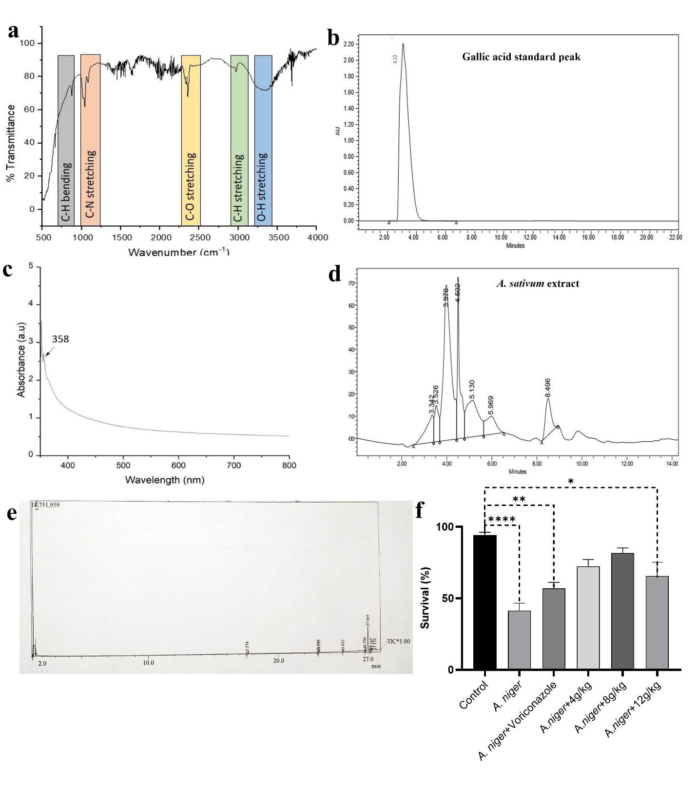
The biochemical composition of A. sativum extract was analyzed using FTIR spectroscopy, revealing the existance of various functional groups associated with its bioactive compounds (Table 1). The 870 cm-1 peaks represent the C-H bending mainly glycogen. The frequency range of 1044 cm-1 peaks represents the C-N stretching denoting amino acids. The frequency range from 2300 cm-1 peaks represents the C-O stretching which corresponds to proteins. The frequency range of 2970 cm-1 cm-1 peaks represents the C-H stretching which indicated presence of lipids. The frequency range of 3400 cm-1 peaks represents the O-H stretching vibration which indicates the presence of carbohydrate and amino acids (Nagarajan and Kumar 2017) (Figure 1a). The optical behavior of the ethanolic extract was studied by observing the UV-Vis spectrum. The UV-Vis spectra of A. sativum showed a broad absorption peak at 358 nm, which indicated the characteristic absorption of phenolic compounds (Figure 1b). HPLC analysis confirmed the presence of gallic acid peaks in the A. sativum extract at a retention time of 3.34 min, which is close to the standard peak observed at the retention time of 3.12 min (Figure 1c-d). GC-MS analysis revealed the presence of phytochemical compounds in A. sativum extract at different peaks and retention times (Figure 1e). The detected compounds are listed in table according to retention time with their molecular formula, molecular weight and chemical class (Table 2). Major detected compounds include, Cyclopropanedecanoic acid, 13- Docosenoic acid, 15- Tetracosenoic acid, 10-Octadecenoic acid, Oleic acid, 9-Octadecenoic acid, 1,2 Benzenedicarboxylic acid and Bis (2-ethylhexyl) phthalate. 2-Undecanol, Docosenoic acid, octadecanoic acid, Bis (2-ethylhexyl) phthalate are also identified by Alamu et al. (2024). Oleic acid, selenocoumarin and methyl esters exhibit antifungal properties, likely by disrupting fungal cell membranes. A variation in detection of various A. sativum compounds in different studies have been reported (Pârvu et al. 2019; Alamu et al. 2024; Bazaraliyeva et al. 2024; Sharma et al. 2018). The difference in the chemical compounds detected in different A. sativum samples might be due to difference in extraction methods, parts of the plant used, genotypic changes, environmental factors, harvesting season and the biomolecules detection method (El-Saadony et al. 2024; Bazaraliyeva et al. 2022).
_the_ft-ir_spectra_of_*a._sativum*_extract__(b)_the_uv-vis_spectra_of_*a._sativum*_extr.jpeg)
Pathological Scoring
Fish challenged with A. niger exhibited pronounced pathological symptoms at both 7 and 14 dpi, including eroded gills, damaged fins and body lesions with fins being the most affected area. The behavioral attributes of the challenged fish were also affected (Table 3) Fish treated with the positive control, Voriconazole exhibited mild behavioral and swimming abnormalities, less fin erosion and respiratory distress and moderate lethargy. Treatment with A. sativum extract resulted in healing of skin lesions, regeneration of scales and restoration of fin integrity, indicating the therapeutic potential of A. sativum in managing A. niger infection (Table 3). The survival of the juveniles was highest at the medium concentration of the extract (8g/kg) indicating an optimal dose response, whereas the highest concentration did not provide additional benefits (Figure 1f).
Hematological indices
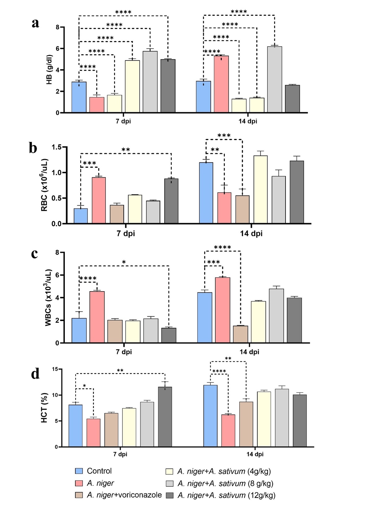
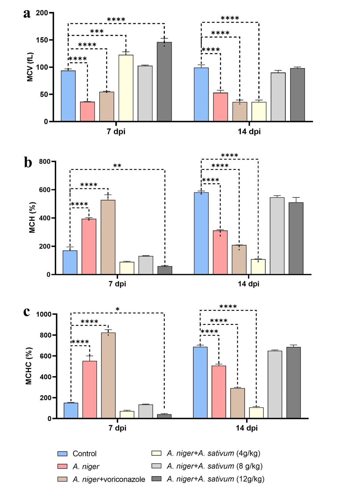
At 7 and 14 dpi, a significantly decreased level was observed in Hb, RBCs and Hct in A. niger challenged group as compared to the control. Treatment with Voriconazole did not result in noticeable recovery of Hb levels at either time point. Treatment with A. sativum extract resulted in improved Hb level at highest concentration (12g/kg) at 14 dpi (Figure 2a). When challenged fish treated with Voriconazole recovery effects were observed in RBC, WBC and HCT levels at 7 dpi. RBCs, WBCs and Hct level recovered to normal at all three A. sativum extract concentrations at 14 dpi (Figure 2b-d). MCV level displayed significant reduction at both time points in A. niger challenged group compared to the control. MCV level was decreased significantly in Voriconazole treated group and no noticeable recovery effects were observed at both points. Following treatment with A. sativum extract exhibited recovery effects in medium concentration at 7 dpi, while at 14 dpi medium and high concentrations were effective in recovering normal fish health (Figure 3a). MCH and MCHC level considerably increase at 7 dpi and decreased at 14 dpi in A. niger challenged group. Voriconazole treatment did not show any significant recovery in MCH and MCHC at both time points. A. sativum treatment recovered these effects at medium and high concentrations at 14 dpi, values reaching comparable to the control group (Figure 3b-c).
_hematological_indices_of_*a._sativum*_treated_groups_following_*a._niger*_challenge_.jpeg)
_hematological_indices_of_*a._sativum*_treated_groups_following_*a._niger*_challengea.jpeg)
Biochemical observations
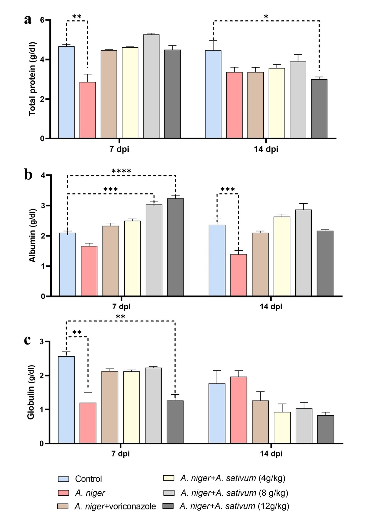
Total serum protein serves as a vital biomarker for evaluating the nutritional and health status of fish (Patriche et al. 2011). Albumin and globulin concentrations are pivotal indicators in evaluating the nutritional and immunological status of fish (Kaleeswaran et al. 2012). Total protein level was significantly reduced in the A. niger-challenged group compared to the control at 7 dpi and level was restored in Voriconazole and all A. sativum extract treated groups (Figure 4a). Albumin level significantly decreased A. niger challenged group at 14 dpi with subsequent marked improvement in all treated groups (Figure 4b). Globulin level was reduced in A. niger challenged group compared to the control group and restored level was observed at 7 dpi in the groups treated with Voriconazole, low and medium concentrations of A. sativum extract (Figure 4c).
_biochemical_analysis_of_*a._sativum*_treated_groups_following_*a._niger*_challenge_a.jpeg)
Enzyme analysis
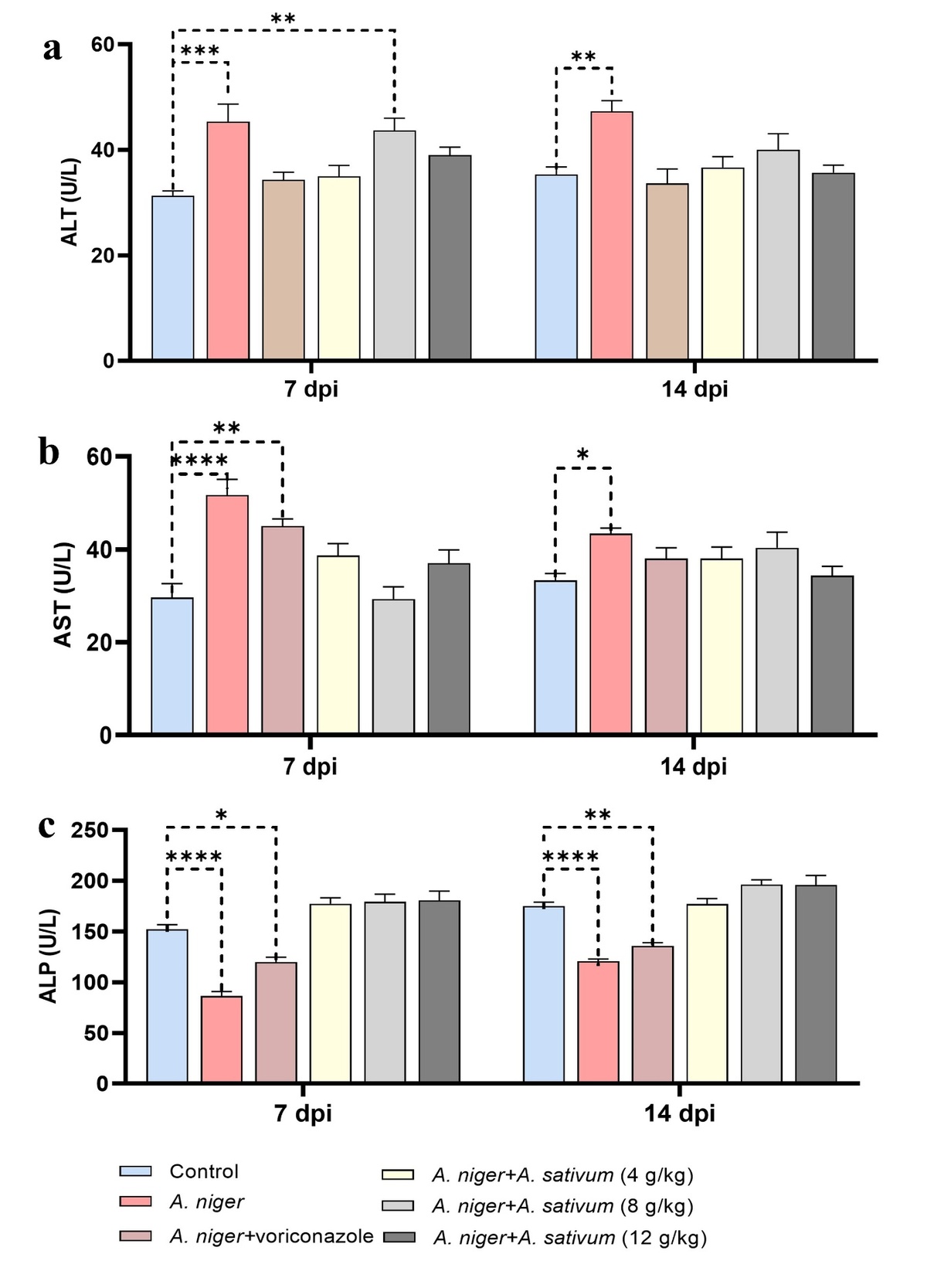
ALT and AST levels increased significantly at 7 and 14 dpi in A. niger challenged groups compared to the control. At 7 dpi, notable improvement in ALT level was observed in the Voriconazole-treated group however, treatment with A. sativum extract was effective at low and high concentrations of A. sativum extract. By 14 dpi, ALT levels were restored to normal across all treatment groups, comparable to the control (Figure 5a). At 7 dpi, AST activity did not recover in Voriconazole treated group, while Voriconazole was effective in reducing infection at 14 dpi. AST activity improved markedly at all concentrations of extract at both time points, reaching values similar to the control group (Figure 5b). ALP levels were significantly reduced in A. niger challenged fish at 7 and 14 dpi compared to the control. A. sativum extract resulted in noticeable recovery of ALP levels at both time points in all three extract concentrations (Figure 5c). The group treated with sole A. sativum extract did not show any signs of liver toxicity evident by comparable values of control (untreated) and A. sativum extract only treated groups (Supplementary data).
_enzyme_analysis_of_*a._sativum*_treated_groups_following_*a._niger*_challenge_assay_.jpeg)
Immune responses
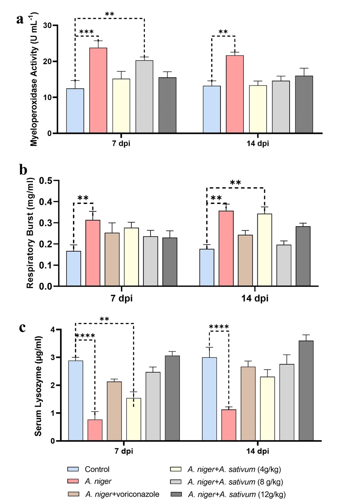
Myeloperoxidase activity was significantly higher in A. niger challenged group as compared to the control group at both time points. At 7 dpi, A. sativum-treated groups (low and high concentrations) returned to normal levels. By 14 dpi, all treated groups exhibited marked improvement and were comparable to the control unchallenged group (Figure 6a). Respiratory burst significantly increased in A. niger challenged group as compared to the control group at both time points. Respiratory burst restored to normal following treatment with Voriconazole at both time points. Treatment with A. sativum extract was effective at all concentrations at 7 dpi. At 14 dpi, medium and high concentrations showed improved Respiratory burst (Figure 6b). Serum lysozyme significantly decreased in A. niger challenged group as compared to the control group at both time points. When challenged fish treated with Voriconazole, it showed recovery at both time points. Maximum recovery was obtained at 14 dpi in all A. sativum treated groups comparable to the control group (Figure 6c).
_immune_responses_in_*a._sativum*_treated_groups_following_*a._niger*_challenge_assay.jpeg)
Discussion
A. sativum has been broadly studied in fish demonstrating its immunostimulatory and curative effects against microbial challenges (Nya and Austin 2009; Mosaddad et al. 2023; Siddique et al. 2026). The present study was conducted to evaluate the immunomodulatory roles of A. sativum on L. rohita challenged with A. niger.
Blood serves as a patho-physiological indicator of the whole body and haematological indices in blood reflects the health status of fish by detecting any disruption occurring due to the use of immunostimulants (Alsafah and Al-Faragi 2017). The results of the present study indicated that A. sativum extract supplementation restored the altered hematological parameters in A. niger infected L. rohita. The pathological symptoms were mitigated in the extract treated fish and survival rate was higher than the non-treated counterparts suggesting that A. sativum extract bolsters the fish’s immune response and resilience against A. niger. Similarly, Nwakpa and Ikwor (2024) demonstrated that A. sativum extract plays a vital role in improving hematological indices. Onomu (2019) also reported that dietary supplementation with garlic (0.5%) enhanced hematological parameters in Clarias gariepinus fingerlings, further supporting the beneficial effects of A. sativum inclusion in fish diets. The dietary inclusion of Thymus vulgaris (thyme) extract for Cyprinus carpio infected with Saprolegnia spp. resulted in improved hematological responses (Alsafah and Al-Faragi 2017).
Our results demostrated that serum biochemical parameters were altered upon A. niger infection and A. sativum extract supplementation normalised most of the blood parameter at both 7 and 14 dpis. Thyme and fennel supplementation improved serum protein levels in in rainbow trout challenged with Yersinia ruckeri (Gulec et al. 2013). The level of plasma protein have been reported to rise in in Nile tilapia given garlic supplemented diets (Aly et al. 2025).
Pathogenic infections affect the liver and its functions, including the release of liver enzymes (ALT and AST) into the blood. A substantial increase in ALT and AST activity is a chief indicator of hepatic dysfunction (Nazir et al. 2024). Our results showed increased AST and ALT activities upon A. niger infection which was normalised by A. sativum extract supplementation. The elevated levels of these enzymes are likely due to infection-induced cellular damage and disruption of cell membrane integrity, which results in the leakage of these enzymes into the bloodstream (El-Demerdash et al. 2022). The ALP level was significantly decreased in A. niger challenged group, while improved level was recorded in 4, 8 and 12g/kg A. sativum treatment groups. ALP activity was stimulated by administration of leaf extract of Aegle marmelos 10g/kg in Cyprinus carpio infected with the bacterial pathogen A. hydrophila (Pratheepa et al. 2010).
Myeloperoxidase plays an important role in leucocyte-mediated vascular injury responses in inflammatory vascular diseases (Chiu et al. 2007). The respiratory burst activity and serum lysozyme have been used as an indicator of nonspecific immunity in fish (Biller-Takahashi et al. 2013; Hardi et al. 2019). Our findings show that myeloperoxidase, respiratory burst and serum lysozyme levels were altered upon A. niger challenge indicating a triggered inflammatory reaction in the host. . The comparable levels of myeloperoxidase, respiratory burst and serum lysozyme markers in the A. sativum treated groups to the control group showed its regulatory effects on the immune response. Herbal extracts have been reported to improve the levels of these inflammatory markers in Cirrhina mrigala, against fungal pathogen, Aphanomyces invadans (Harikrishnan et al. 2009). Dietary supplementation of A. sativum extract at a concentration of 100 mg/kg feed for an extended feeding period of 8 weeks improved immune responses and disease resistance against Photobacterium damselae infection in Mugil cephalus larvae (Zorriehzahra et al. 2021). The difference in the optimal dosage in other studies and our work with partially high concentration might be due to variations in total duration of dietary administration, host species, extract formulations, and the infection model. Biological efficacy of A. sativum at higher dietary inclusion (10 g/kg feed) has been reported previously supporting elevated dietary inclusion levels (Sahu et al. 2007). Diets supplemented with P guajava plant extract significantly improved the respiratory burst and serum lysozyme of Nile tilapia after bacterial challenge (Kamble et al. 2024). Moreover, dietary supplementation with Flos populi extract (FPE) has been shown to boost the innate immune response and disease tollerance in gibel carp by mediating the myeloperoxidase activity (Zhang et al. 2022). Hydroxyl radicals and superoxide anions present in A. sativum can play major therapeutic roles serving as natural defense mechanisms against infection (Kim et al. 2001; Talpur and Ikhwanuddin 2012). All tested concentrations showed beneficial effects, but 8 g/kg feed consistently produced the optimal response in terms of all parameters, survival, and pathological outcomes. The lower concentration (4 g/kg) was less effective, likely due to insufficient bioactive compounds to elicit a full response, whereas the higher concentration (12 g/kg) did not produce further improvement and, in some parameters, showed slightly reduced efficacy possibly due to saturation of biological response or minor stress effects at higher concentrations (Talpur and Ikhwanuddin 2012; Zare et al. 2021).
The therapeutic effects of A. sativum can be attributed to the presence of various ester compounds in the extract. Esters are well known for their potent antimicrobial, antifungal, and antiviral properties (Williford et al. 2025; Lamba et al. 2023; López-Gresa et al. 2018). The principal mechanisms by which esters can hinder the microbial growth are disrupting cell membranes permeability and causing the release of essential cytoplasmic components, leading to pronounced cell death via generative oxygen species production. Another primary monounsaturated fatty acid molecule Oleic acid detected in the A. sativum extract, has been reported to perform antiinflammatory, antioxidant and anticancer activities (El-Saadony et al. 2024; Deng et al. 2023; Maccelli et al. 2020). Oleic acid exerts its effects primarily by disrupting cellular integrity, signal transduction pathways, nutrient and cofactor synthesis, cytoplasmic vacuolation, cell shrinkage, and membrane rupture ultimately triggering apoptosis. Different studies show some varations of the types of compounds detected in garlic which might be attributed to the extraction method, identification technique, parts of the plant used and harvesting time (Bazaraliyeva et al. 2022; El-Saadony et al. 2024; Pârvu et al. 2019). Further in vivo and in vitro studies to investigate the molecular interaction between A. sativum compounds and their targets of the fish immune system will greatly benefit our understanding to elevate the fish immune system to microbial pathogens. Overall, this study demonstrates that A. sativum extract exhibits antifungal activity against A. niger, suggesting that it serves as a natural and eco-friendly alternative to synthetic antifungal agents in aquaculture.
Conclusions
The findings of this study demonstrate that A. sativum supplementation can enhance immunomodulatory functions in L. rohita against A. niger challenge. A. sativum extract improved the serum hemato-biochemical and immune responses in the L. rohita juveniles, as well as increased the survival rate of the juveniles. The optimal responses were observed at the medium concentration (8g/kg) of the extract with maximum recovery points attained by this dose. Overall, we conclude that herbal extract of A. sativum can be used as a safe and potent alternative to synthetic drugs for controlling fungal infections in aquaculture.
Acknowledgements
Support provided by Department of Zoology, LCWU and Fisheries Research and Training Institute, Manawan are acknowledged.
Ethical Statement
This study was approved by the Ethical Committee of Department of Zoology, Lahore College for Women University, Lahore: RERC No. RER No/LCWU/Zoo/577c.
Author Contributions
Conceptualization: Z.K. and R.M.; Methodology: R.M., M.A.R., and Z.K.; Investigation: R.M., F.M., M.A., F.R., and M.A.R.; Resources: R.M., Z.K., and M.A.R.; Writing—original draft preparation: R.M. and Z.K.; Writing—review and editing: R.M. and Z.K.; Data curation and visualization: Z.K., R.M., and F.R.; Software: R.M.; Supervision: Z.K., F.M., and M.A.R.
Conflicts of Interest
The authors declare no conflicts of interest.
Data Availability Statement
All the related data will be provided on request.